
Hepatitis E virus vaccine and method

a technology for hepatitis e virus and vaccine, applied in the field of antigen and antibody vaccine compositions, can solve the problems of general unevenness of hev, and achieve the effect of neutralizing hepatitis e virus (hev) infection and preventing or treating hev infection

Inactive Publication Date: 2003-07-31
GENELABS TECH INC
View PDF7 Cites 2 Cited by
  • Summary
  • Abstract
  • Description
  • Claims
  • Application Information

AI Technical Summary

Benefits of technology

[0104] The recombinant or intact HEV capsid or capsid fragment peptides (HEV capsid antigens) described above are incorporated into a vaccine composition, according to known procedures, to enhance the antigenicity of the injected antigens.
[0114] At this stage, two of the four experimental animals received a third inoculation of non-adjuvanted, insoluble trpE-C2 peptide antigen. Four weeks later, these animals showed anti-HEV antibodies, as evidenced by Western blots. These results suggest that the trpE-C2 antigen may be more effective when administered in the absence of alum, possibly because of alum-denaturation of the antigen during the alum co-precipitation procedure.
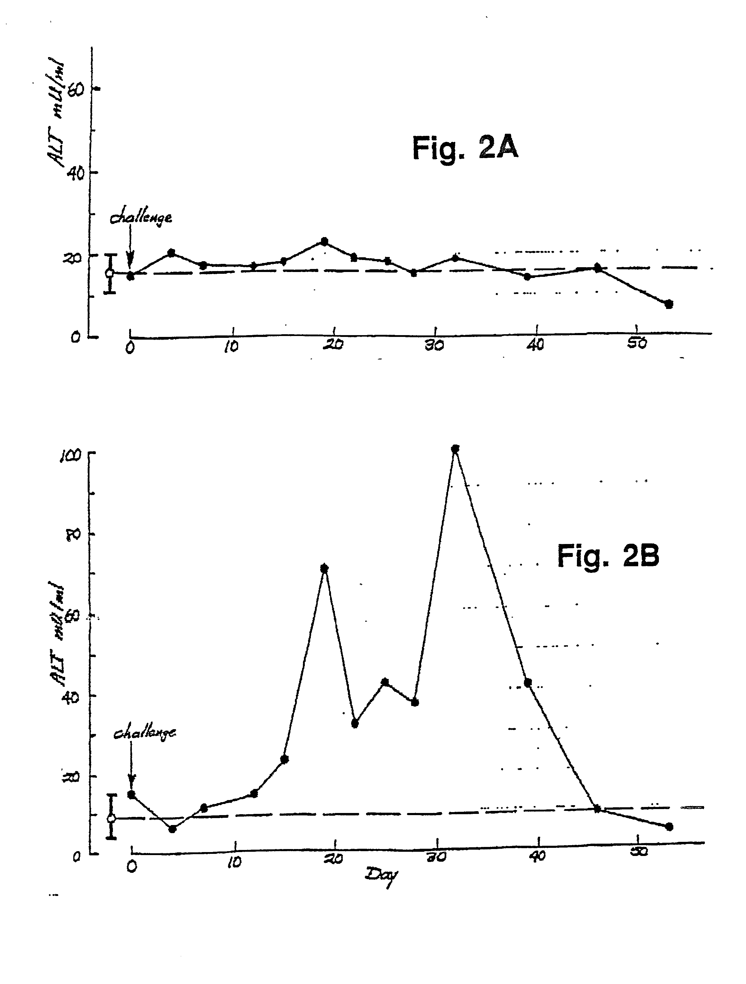
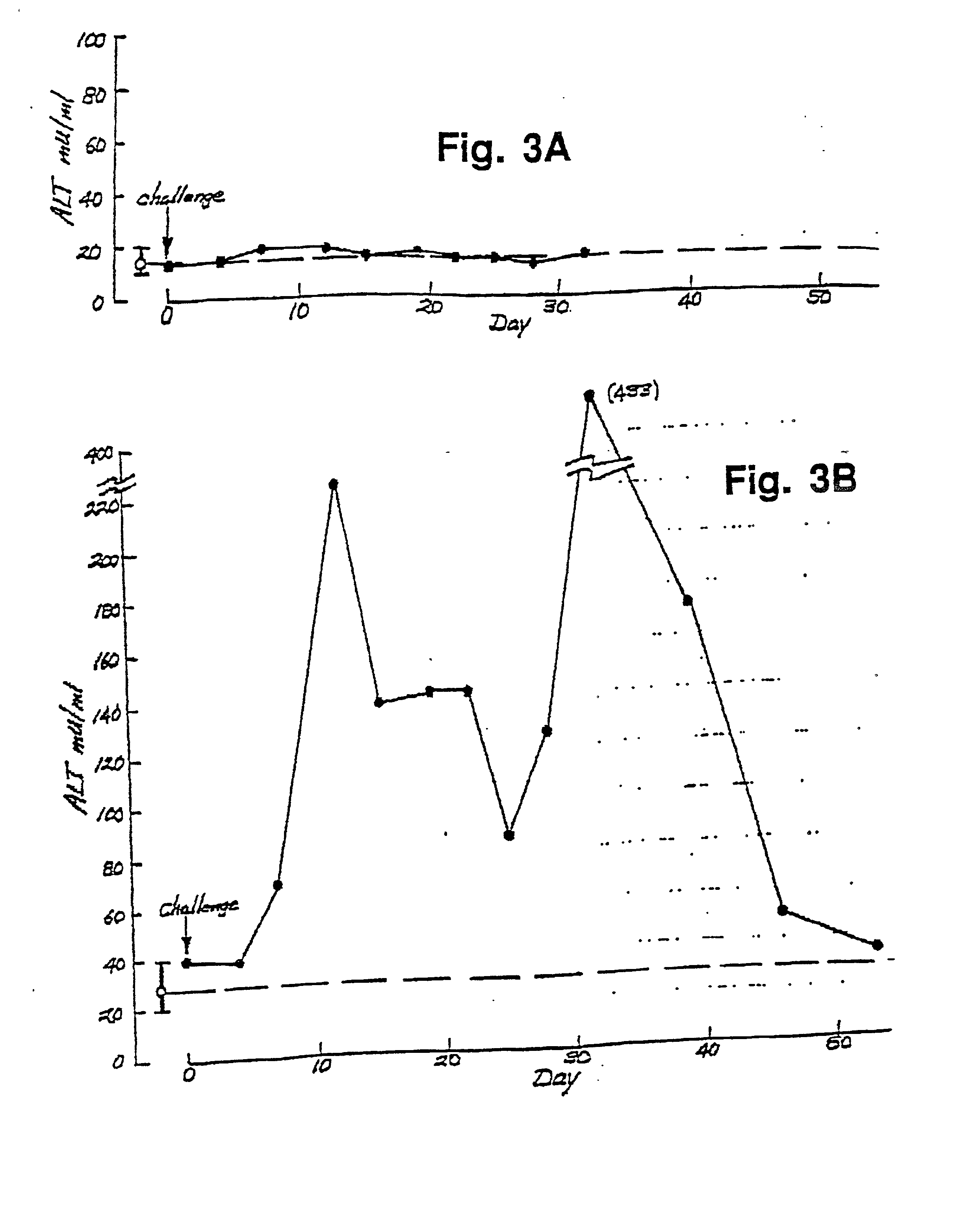
[0118] FIG. 3A shows the change in liver ALT levels following infection with Mexico-strain HEV virus, in one of the animals which received a third dose of trpE-C2. Again, there was no evidence of elevated ALT levels out to day 32 (The animal died of unrelated causes at day 32). The liver biopsy samples also showed minimal evidence of HEV antigen. This result demonstrates that an antigen vaccine directed against one HEV strain can provide protective immunity against other HEV strains.
[0123] The antibodies in the composition are preferably immunoreactive with a peptide containing one of the sequences: Sequence ID No. 13; Sequence ID No. 14, and internally consistent variations between Sequence ID Nos. 13 and 14. As will be seen below, antibodies prepared against the 406.3-2 antigen (M) are effective to block HEV infection in human primary hepatocytes.
[0132] The primary hepatocytes were prepared and cultured according to published procedures and as detailed in Example 1. The unique culture conditions allow for long-term cell growth in culture without loss of specialized hepatocyte function, as evidenced by the cells' continued ability to make and secrete liver-specific proteins, such as serum albumin, up to several months after initial culturing, as described in Example 1.

Problems solved by technology

The course of HEV is generally uneventful in healthy individuals, and the vast majority of those infected recover without the chronic sequelae seen with HCV.

Method used


Image

  • Hepatitis E virus vaccine and method
  • Hepatitis E virus vaccine and method
  • Hepatitis E virus vaccine and method

Examples


example 2

In vitro HEV Infection of Primary Human Hepatocytes

[0159] A. HEV Infection of Human Hepatocytes.

[0160] The HEV-infected cynomolgus monkey #73 stool pool (fourth passage) was used as an inoculum for infections of primary human hepatocytes. Various amounts of inoculum was diluted in 1 ml of serum-free medium (SFM) and applied to the culture during a 3 hr incubation period. This solution was then supplemented with 2 ml of fresh SFM and the entire mixture was incubated overnight. The next day, cell monolayers were washed with WME (10 mM HEPES, pH 7.4) for three times and changed to fresh SFM, which was changed at two day intervals thereafter.

[0161] B. Immunofluorescence Staining Assay.

[0162] Primary cynomolgus monkey hepatocytes were isolated and plated in tissue culture plates with collagen-coated coverslips as described. Cells on coverslips were infected with either the HEV-infected cynomolgus monkey #73 stool pool or the NIH normal human serum three days after initial plating. The in...

example 3

Preparation of 406.3-2 and 406.4-2 Antigens

[0169] A TZKF1 plasmid (ET1.1), ATCC deposit number 67717, was digested with EcoRI to release the 1.33 kb HEV insert which was purified from the linearized plasmid by gel electrophoresis. The purified fragment was suspended in a standard digest buffer (0.5M Tris HCl, pH 7.5; 1 mg / ml BSA; 10 mM MnC12) to a concentration of about 1 mg / ml and digested with DNAse I at room temperature for about 5 minutes. These reaction conditions were determined from a prior calibration study, in which the incubation time required to produce predominantly 100-300 basepair fragments was determined. The material was extracted with phenol / chloroform before ethanol precipitation.

[0170] The fragments in the digest mixture were blunt-ended and ligated with EcoRI linkers. The resultant fragments were analyzed by electrophoresis (5-10V / cm) on 1.2% agarose gel, using PhiX174 / HaeIII and lambda / HindIII size markers. The 100-300 bp fraction was eluted onto NA45 strips (Sc...

example 4

Neutralizing Activity of Anti-3.2(M) Antibody

[0190] A. In vitro Infection

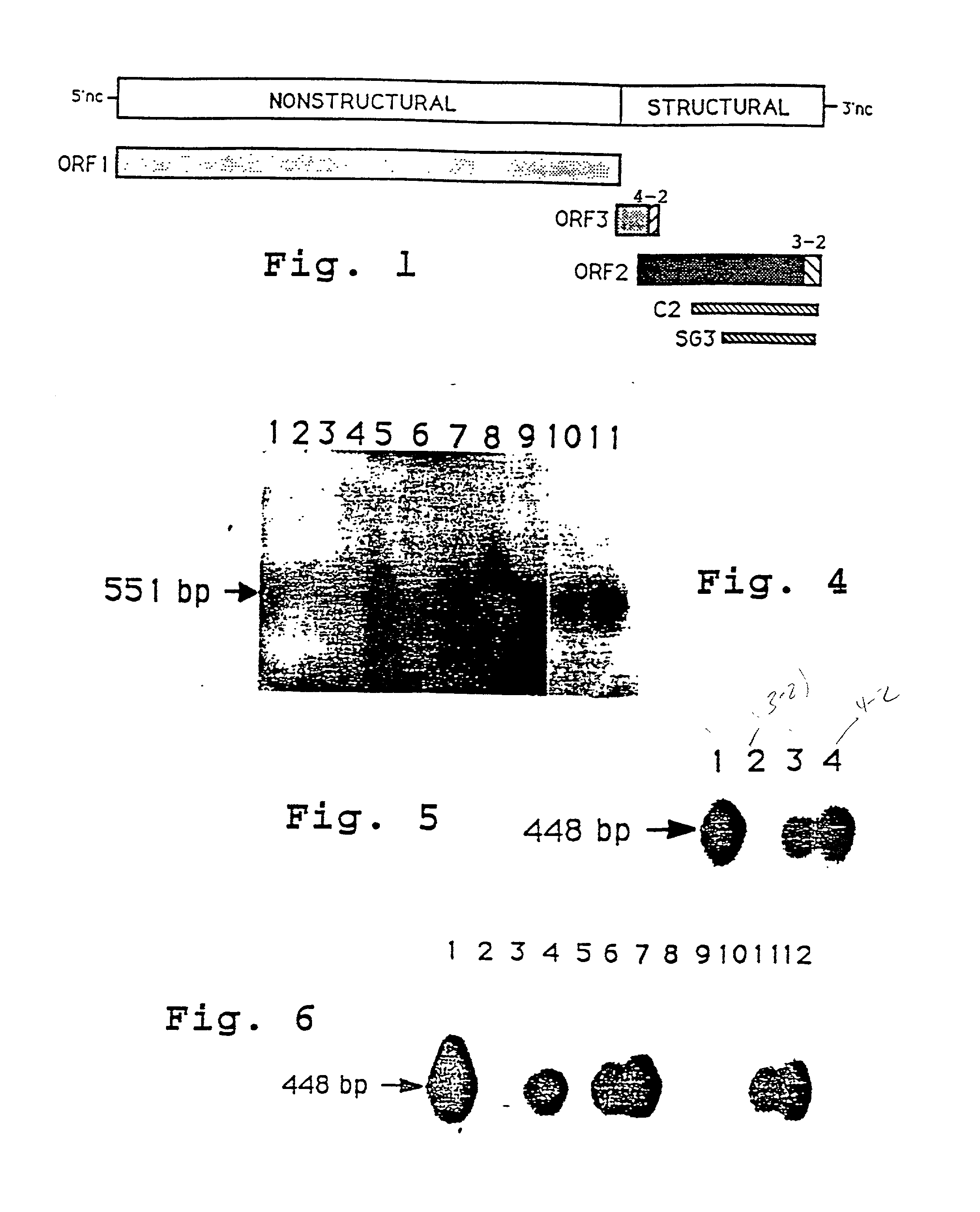
[0191] To prove that primary human hepatocytes were permissive for HEV infection and replication, cells were exposed to either normal human serum (NIH normal human serum pool) or HEV-infected cynomolgus macaque stool preparation (cyno#73). Fourteen days postinfection, total cellular RNAs were prepared for reverse-transcription (RT) / polymerase chain reaction (PCR) assays to evaluate the infectability of primary human hepatocytes with HEV. The results indicated that primary human hepatocytes were capable of supporting HEV propagation (FIG. 4).

[0192] Although quantitative PCR was not applied, total cellular RNA isolated from HEV-infected primary human hepatocytes would indicate a high level of virus replication as suggested by the extent of hybridization with the .alpha.-.sup.32P-dCTP labeled HEV-specific probe (lane 5). There was no evidence of HEV in total cellular RNA isolated from primary human hepatocytes tre...


PUM


Abstract

Antigen and antibody vaccine composition effective in preventing hepatitis E virus (HEV) infection are disclosed. The antigen composition includes a peptide corresponding to a carboxyl terminal end region of the capsid protein encoded by the second open reading frame 2 of the HEV genome. The composition is effective in preventing HEV infection after vaccination. The antibody composition contains an antibody effective to block HEV infection of human primary hepatocytes in culture.

Description

[0001] This application is a continuation-in-part of U.S. application Ser. No. 07 / 882,335, filed Jan. 17, 1992, which is a continuation-in-part of application Ser. No. 07 / 505,888, filed Apr. 5, 1990, which is a continuation-in-part of U.S. application Ser. No. 420,921, filed Oct. 13, 1989, which is a continuation-in-part of U.S. application Ser. No. 367,486, filed Jun. 16, 1989, which is a continuation-in-part of U.S. Application Serial No. 336,672, filed Apr. 11, 1989, which is a continuation-in-part of U.S. application Ser. No. 208,997, filed Jun. 17, 1988, all of which are herein incorporated by reference.1. FIELD OF INVENTION[0002] This invention relates to antigen and antibody vaccine compositions related to enterically transmitted nonA / nonB hepatitis viral agent, also referred to herein as hepatitis E virus (HEV), and to vaccine methods.2. REFERENCES[0003] Arankalle, V. A., et al., The Lancet, 550 (Mar. 12, 1988).[0004] Bradley, D. W., et al., J Gen. Virol., 69:1 (1988).[0005]...

Claims


Application Information

Patent Timeline
IPC IPC(8): A61K38/00C12N15/09A61K39/00A61K39/29A61K39/395A61P31/12C07K14/08C07K16/10C12N15/51C12P21/08C12Q1/70G01N33/576
CPCA61K38/00A61K39/00C07K14/005C07K16/10C07K2319/00G01N33/5767C07K2319/40C07K2319/61C12N2770/28022C12N2770/28122C07K2319/23A61P1/16A61P31/12A61P31/14A61P31/20
Inventor REYES, GREGORY R.BRADLEY, DANIEL W.TWU, JR-SHINPURDY, MICHAEL A.TAM, ALBERT W.KRAWCZYNSKI, KRZYSZTOF Z.YARBOUGH, PATRICE O.
Owner GENELABS TECH INC
Learn More