

Assay device and method for chemical or biological screening
a technology of assay device and method, applied in the direction of chemical analysis using titration, biochemistry apparatus, biochemistry apparatus and processes, etc., can solve the problems of inconvenient use, inability to efficiently or accurately perform large-scale cell sample processing, and inability to efficiently or accurately use microscopy-based methods or flow cytometric methods,
- Summary
- Abstract
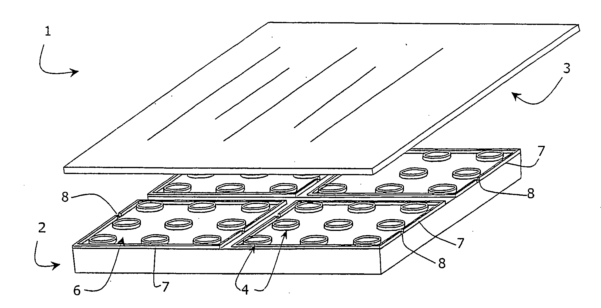
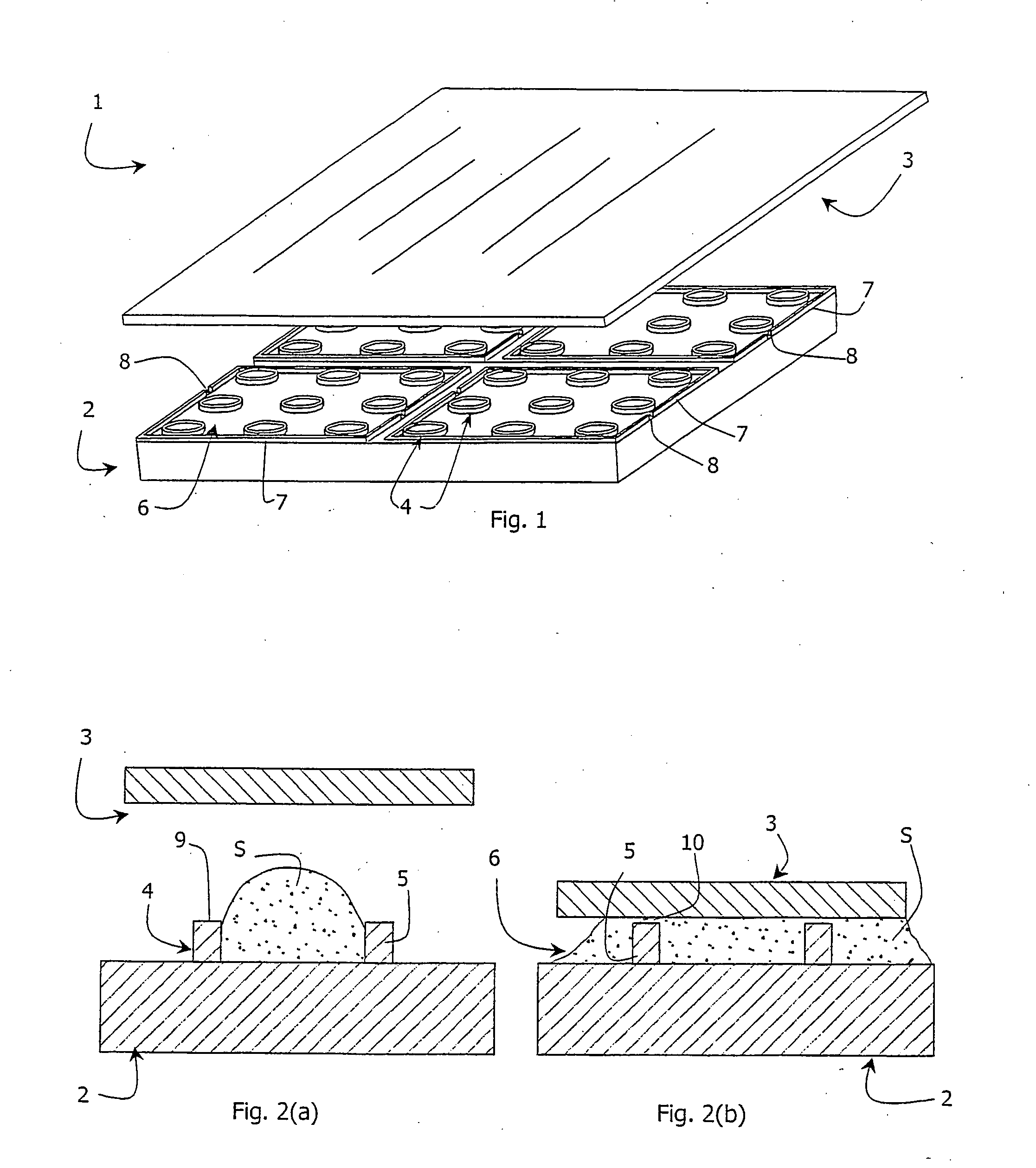
- Description
- Claims
- Application Information
AI Technical Summary
Problems solved by technology
Method used
Image

Examples
example 1
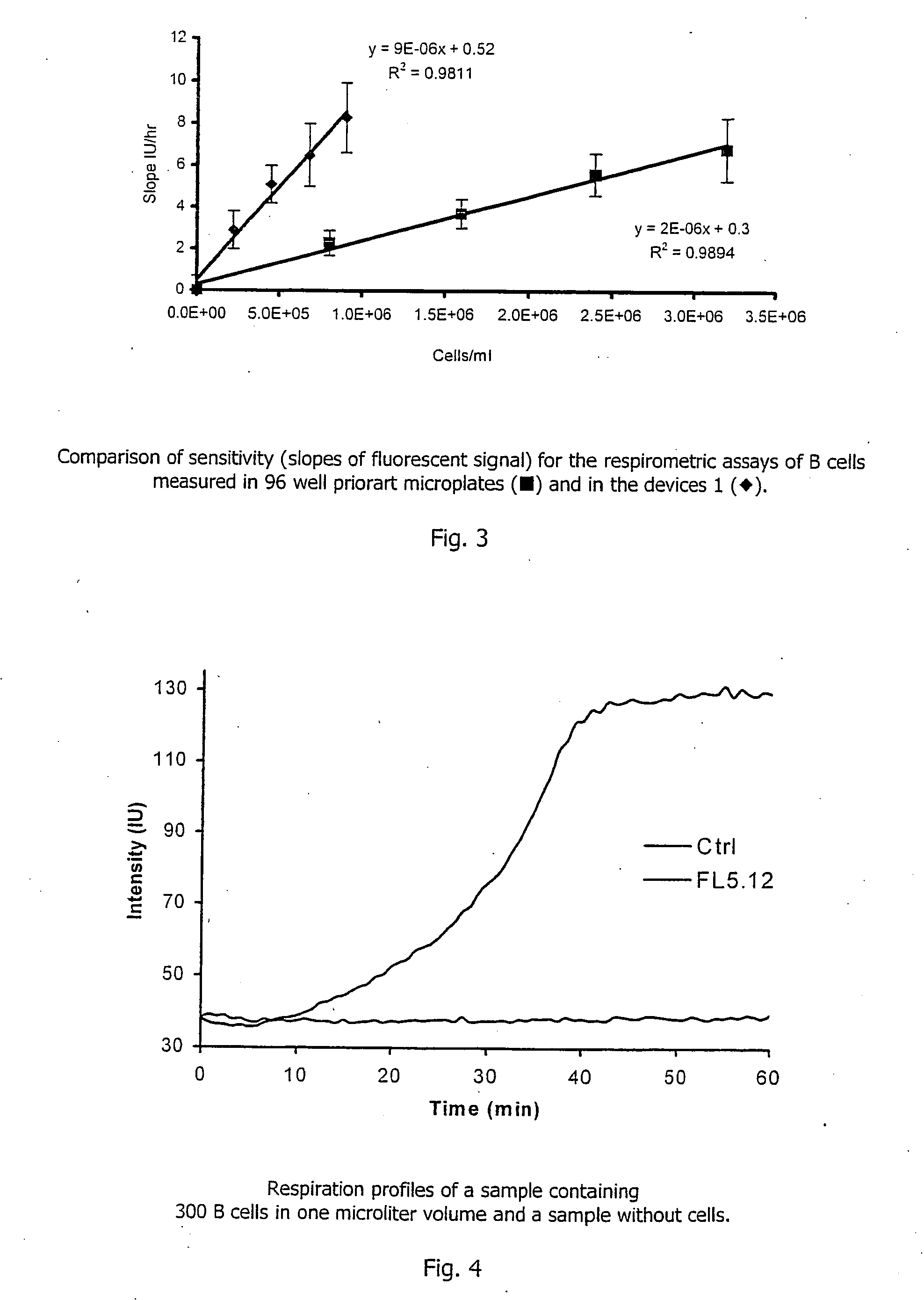
Using the Device for Monitoring Cellular Respiration
[0106] Three milligrams of platinum(II)-coproporphyrin-I (PtCP) was dissolved in 0.1 ml of dimethylformamide, mixed with 1 mg of 1-ethyl-3-(3-dimethylaminopropyl)carbodiimide in 0.1 ml dimethylformamide and incubated for 10 minutes at room temperature. The solution was added dropwise to 3 ml of solution of bovine serum albumin (BSA, 2 mg / ml) in 0.1M carbonate buffer, pH9.0 and incubated at room temperature to allow the dye derivative to react with protein amino groups. The covalent conjugate PtCP-BSA formed was purified from the excess of unbound dye by chromatography on a desalting column in phosphate buffer saline, collecting the fractions of PtCP-BSA conjugate. The concentration for the PtCP-BSA conjugate stock was determined spectrophotometrically. This stock solution of the oxygen probe was added to the samples to achieve the desired working concentration. Such samples can undergo respirometric measurements in the microwells o...
example 2
Using the Device for Measurement of Oxygen Uptake in an Enzymatic Reaction
[0108] Two microlitres of a solution containing 0.5 ug / ml of glucose oxidase enzyme, 10 mM of .beta.,D-glucose and 3*10.sup.-5M of PtCP-BSA probe prepared as described in Example 1 above in 0.1 M phosphate buffer, pH6.0 was aliquoted into each microwell of the device. The microwells were then sealed with the glass lid 3 placed in a plate reader SpectraMax Gemini (Molecular Devices), equilibrated at 37.degree. C. and the phosphorescent signal in each well was monitored. Phosphorescence was monitored at 650 nm using excitation at 380 nm. As in the Example 1, the device 1 demonstrates superior performance requiring less sample and showing greater sensitivity within a shorter time period, when compared to the assays performed in standard microplates.
example 3
Assessment of Viability of Cultured Yeast Cells B Treated with Toxicant
[0109] A solid-state oxygen probe comprising a thin film polymer coating of the phosphorescent dye platinum(II)-tetrakis(pentafluorophenyl)porphin-e (PtTFPP) and polycarbonate was used with the device 1 shown in FIG. 1. One milligram of PtTFPP is dissolved in 0.01% (w / v) solution of polycarbonate (M.ca. 60 000) in chloroform. One microlitre aliquotes of this solution were dispensed to the microwells and allowed to air dry for 10 minutes to produce thin film phosphorescent oxygen-sensitive coating inside the microwells.
[0110] S. pombe yeast cells were incubated with different concentrations of cadmium nitrate toxicant. Two .mu.l aliquots were taken from each sample at different time intervals, and placed in the wells of microwell chambers coated with a PtTFPP-polycarbonate solid-state oxygen probe. The respiration profiles of samples were monitored as described in Example 1 above, together with a control sample co...
PUM
 Abstract
Description
Claims
Application Information