
Cellular fibronectin as a diagnostic marker in stroke and methods of use thereof

a technology of fibronectin and diagnostic markers, applied in the field of cardiovascular disease and cerebral injury identification and use of diagnostic markers, can solve the problems of sudden weakness, loss of sensation, difficulty in speaking, seeing, walking, etc., and achieve the effects of rapid and accurate determination, maximum sensitivity, specificity, and predictive accuracy

Inactive Publication Date: 2005-06-16
PREDICTION SCI
View PDF2 Cites 157 Cited by
  • Summary
  • Abstract
  • Description
  • Claims
  • Application Information

AI Technical Summary

Benefits of technology

[0026] In one of its aspects, the invention discloses methods for determining a diagnosis or prognosis related to a cardiac event such as stroke, or for differentiating between stroke sub-type and / or determination of HT. The preferred method includes analyzing a fluid sample obtained from a person who has an unknown diagnosis for the levels of one or more markers specific to the damage caused by said cardiac event. In the case of stroke, these markers would be drawn from the group consisting of markers relating to vascular damage, glial activation, inflammatory mediation, thrombosis, cellular injury, apoptosis, myelin breakdown, and specific and non-specific markers of cerebral injury. The analysis of the preferred method thus more precisely includes identifying one or more markers the presence or amount of which is associated with the diagnosis, prognosis, or differentiation of stroke and / or determination of HT. Once such marker(s) are identified, the next in the method the level of such marker(s) in a sample obtained from a subject of interest can be measured. In certain embodiments of the preferred method, these markers can be compared to a level that is associated with the diagnosis, prognosis, or differentiation of stroke and / or determination of HT. By correlating the subject's marker level(s) to the diagnostic marker level(s), the presence or absence of stroke, and also the probability of future adverse outcomes, etc., in a patient may be rapidly and accurately determined.
[0027] In another of its aspects, the instant invention is embodied in methods for choosing one or more marker(s) for differentiation of stroke and / or determination of HT that together, and as a group, have maximal sensitivity, specificity, and predictive power. Said maximal sensitivity, specificity, and predictive power is in particular realized by choosing one or more markers as constitute a group by process of plotting receiver operator characteristic (ROC) curves for (1) the sensitivity of a particular combination of markers versus (2) specificity for said combination at various cutoff threshold levels. In addition, the instant invention further discloses methods to interpolate the nonlinear correlative effects of one or more markers chosen by any methodology to such that the interaction between markers of said combination of one or more markers promotes maximal sensitivity, specificity, and predictive accuracy in the prediction of any of the occurrence of stroke, identification of stroke subtype, or likelihood of HT.
[0032] Thus, in certain embodiments of the methods of the present invention, a plurality of markers are combined using an algorithm to increase the predictive value of the analysis in comparison to that obtained from the markers taken individually or in smaller groups. Most preferably, one or more markers for vascular damage, glial activation, inflammatory mediation, thrombosis, cellular injury, apoptosis, myelin breakdown, and specific and non-specific markers of cerebral injury are combined in a single assay to enhance the predictive value of the described methods. This assay is usefully predictive of multiple outcomes, for instance: determining whether or not a stroke occurred, then determining the sub-type of stroke, then further predicting stroke prognosis. Moreover, different marker combinations in the assay may be used for different indications. Correspondingly, different algorithms interpret the marker levels as indicated on the same assay for different indications.
[0040] The term “markers of thrombosis” as used in this specification refers to markers that indicate an coagulation event in ischaemic stroke. The blood clotting system is activated when blood vessels are damaged, exposing collagen, the major protein that connective tissue is made from. Platelets circulating in the blood adhere to exposed collagen on the cell wall of the blood vessel and secrete chemicals that start the clotting process as follows: Platelet aggregators cause platelets to clump together (aggregate). They also cause the blood vessels to contract (vasoconstrict), which reduces blood loss. Platelet aggregators include adenosine diphosphate (ADP), thromboxane A2, and serotonin (5-HT). Coagulants such as fibrin then bind the platelets together to form a permanent plug (clot) that seals the leak.
[0048] Macrophages and T lymphocytes kill target cells by inducing apoptosis, one of the potential mechanisms whereby the inflammatory cells invading the infarcted brain area participate in neuronal cell death. Stroke patients displayed an intrathecal production of proinflammatory cytokines, such as interleukin (IL)-1β, IL-6, IL-8, and granulocyte-macrophage colony-stimulating factor (GM-CSF), and of the anti-inflammatory cytokine IL-10 within the first 24 hours after the onset of symptoms, supporting the notion of localized immune response to the acute brain lesion in humans. Some of these cytokines (eg, IL-1β and IL-8) stimulate influx of leukocytes to the infarcted brain, a prerequisite for Fas / APO-1- and bcl-2-mediated apoptosis. TNF-α, a powerful cytokine inducing apoptosis in the extraneural compartment of the body, has been demonstrated to protect rat hippocampal, septal, and cortical cells against metabolic-excitotoxic insults and to facilitate regeneration of injured axons. More importantly, TNF-α and -β protect neurons against amyloid β-protein-triggered toxicity.
[0055] In yet another of its aspects, the present invention is embodied in methods for determining a treatment regimen for use in a patient diagnosed with stroke. The methods preferably comprise determining a level of one or more diagnostic or prognostic markers as described herein, and using the markers to determine a diagnosis for a patient. For example, a prognosis might include the development or predisposition to delayed neurologic deficits after stroke onset. One or more treatment regimens that improve the patient's prognosis by reducing the increased disposition for an adverse outcome associated with the diagnosis can then be used to treat the patient. Such methods may also be used to screen pharmacological compounds for agents capable of improving the patient's prognosis as above.

Problems solved by technology

A stroke may cause sudden weakness, loss of sensation, or difficulty with speaking, seeing, or walking.
Stroke or brain attack is a sudden problem affecting the blood vessels of the brain.
If an artery is blocked, the brain cells (neurons) cannot make enough energy and will eventually stop working.
If the artery remains blocked for more than a few minutes, the brain cells may die.
The most common problem is narrowing of the arteries in the neck or head.
If the arteries become too narrow, blood cells may collect and form blood clots.
These blood clots can block the artery where they are formed (thrombosis), or can dislodge and become trapped in arteries closer to the brain (embolism).
This naturally restricts the flow of blood to the brain and results in almost immediate physical and neurological deficits.
In addition, prior studies have found that the accuracy of stroke identification by medical personnel is modest and variable from one community to another.
Finally, any clinical neurological screening test will be limited by the training and experience of the examiner.
This insensitivity to acute stroke limits its use to post-stroke damage assessment.
However, this takes several hours, and thus is not used for rapid diagnosis of stroke.
However, as a practical issue, most hospitals do not have these specialized and highly expensive MRI services available in the acute setting.
Thus, without a practical and widely available radiological test, the diagnosis of stroke remains largely a clinical decision.
Thrombolytic therapy has been proven to be effective for the treatment of acute ischemic stroke, but the increased risk of hemorrhagic transformation (HT) associated with tissue plasminogen activator (tPA) administration is still of great clinical concern.
However, despite the available data, the underlying molecular mechanisms related to HT after thrombolytic treatment have yet to be fully elucidated.

Method used


Image

  • Cellular fibronectin as a diagnostic marker in stroke and methods of use thereof
  • Cellular fibronectin as a diagnostic marker in stroke and methods of use thereof
  • Cellular fibronectin as a diagnostic marker in stroke and methods of use thereof

Examples


example i

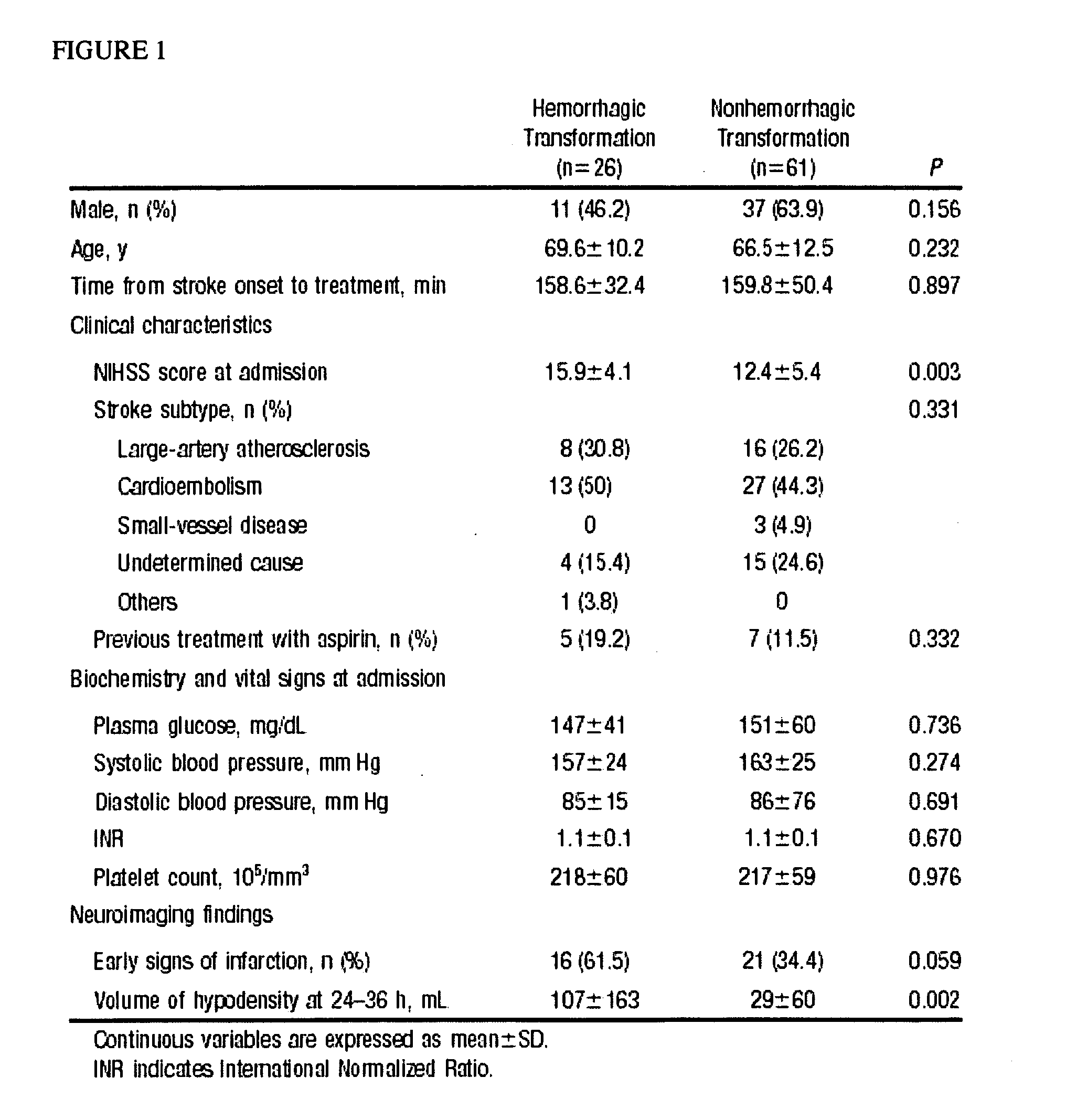
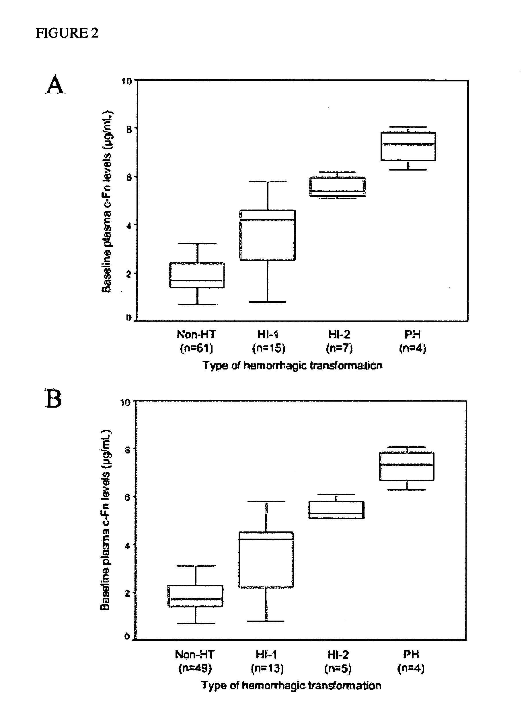
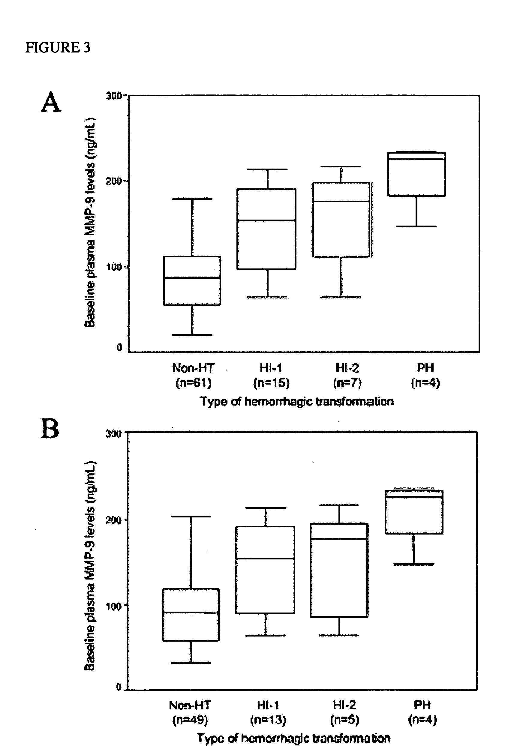
[0300] Prospectively studied patients (n=87, 59% men; mean age 67±12 years) received intravenous tPA following the European Cooperative Acute Stroke Study (ECASS) II criteria.12 Thrombolytic therapy was administered within 6 hours from the beginning of the symptoms at a dose of 0.9 mg / kg body weight, with an upper dose limit of 90 mg per patient. Ten percent of the total dose was given as a bolus over 1 to 2 minutes, followed by a 60-minute infusion of the remaining dose. The mean time to the infusion of the drug was 160—46 minutes. Seventy-one patients received the treatment within 3 hours from onset of symptoms, whereas 16 patients received tPA between 3 and 6 hours within onset of symptoms. Thirty healthy control subjects matched by age and sex (male: 57%; mean age: 63±9 years) and without history of neurological disorders or vascular risk factors were also included in the study. To determine the effect of stroke on the levels of the molecules, plasma c-Fn and MMP-9 concentration...


PUM

PropertyMeasurementUnit
molecular massaaaaaaaaaa
concentrationaaaaaaaaaa

Abstract

The present invention relates to methods for the diagnosis and evaluation of stroke and stroke sub-type. A variety of bio-markers are disclosed for assembling a panel for such diagnosis and evaluation. Methods are disclosed for selecting markers and correlating their combined levels with a clinical outcome of interest. In various aspects: the invention provides methods for early detection and differentiation of stroke subtypes, for determining the prognosis of a patient presenting with stroke symptoms, and identifying a patient at risk for hemorrhagic transformation after thrombolyic therapy. Methods are disclosed that provide rapid, sensitive and specific assays to greatly increase the number of patients that can receive beneficial stroke treatment and therapy, and reduce the costs associated with incorrect stroke diagnosis.

Description

RELATED APPLICATIONS [0001] The present application is descended from, and claims benefit of priority of, U.S. provisional patent application No. 60 / 505,606, the contents of which of which are hereby incorporated herein in their entirety, including all tables, figures, and claims. The present application is a continuation-in-part of U.S. utility patent application Ser. No. 10 / 948,834, which application is itself descended from U.S. provisional patent application 60 / 505,606 and 60 / 556,411FIELD OF THE INVENTION [0002] The present invention generally relates to the identification and use of diagnostic markers for cardiovascular disease and cerebral injury. In a various aspects, the present invention particularly relates to methods for (1) the early detection and differentiation of cardiovascular events stroke and transient ischemic attacks, and (2) the identification of individuals at risk for hemorrhagic transformation after both presentation with stroke symptoms and subsequent admini...

Claims


Application Information

Patent Timeline
Patent Type & Authority Applications(United States)
IPC IPC(8): G01N33/68G16B20/20G16B40/10G16B40/20
CPCG01N33/6848G01N33/6893G06F19/24G01N2800/2871G06F19/18G01N2333/78G16B20/00G16B40/00Y02A90/10G16B40/10G16B20/20G16B40/20
Inventor DAVALOS, ANTONICASTILLO, JOSECASTELLANOS, MARDIAMOND, CORNELIUS ALLEN
Owner PREDICTION SCI
Learn More