

Human tissue factor antibodies
a technology of human tissue factor and antibody, which is applied in the field of human tissue factor antibodies, can solve the problems of heavy bleeding, undesirable side effects of treatment with heparin and other anticoagulants, and difficulty in maintaining the proper dosage of heparin
- Summary
- Abstract
- Description
- Claims
- Application Information
AI Technical Summary
Benefits of technology
Problems solved by technology
Method used
Image

Examples
example 1
Preparation of Human Mabs Immunospecific for Human TF
Reagents.
[0194] Human TF can be isolated from human brain as described by Rao, L. V. M., Thrombosis Research, 51:373-384 1988.
[0195] Lipidated recombinant human TF (Dade Innovin, Baxter) can also be used as human thromboplastin reagent. Rat, rabbit, baboon, and pig thromboplastin are prepared from brain tissue. Two volumes of 45° C. 0.9% NaCl are added to the brain tissue, and the tissue is homogenized with a manual glass homogenisator. After 30 min incubation at 45° C. with occasional shaking, the samples are centrifuged 20 min at 2000×g. The precipitate are discarded, and the supernatant is aliquoted and stored at −80° C. until use.
[0196] Relipidated TF may be obtained by reconstitution of recombinant human full length TF (American Diagnostica #4500) into phospholipid vesicles (PC / PS 75 / 25).
[0197] Biotinylated human TF is produced as follows: Biotin-NHS (n-succinimido biotin, Sigma H-1759) is dissolved in DMF (dimethylform...
example 2
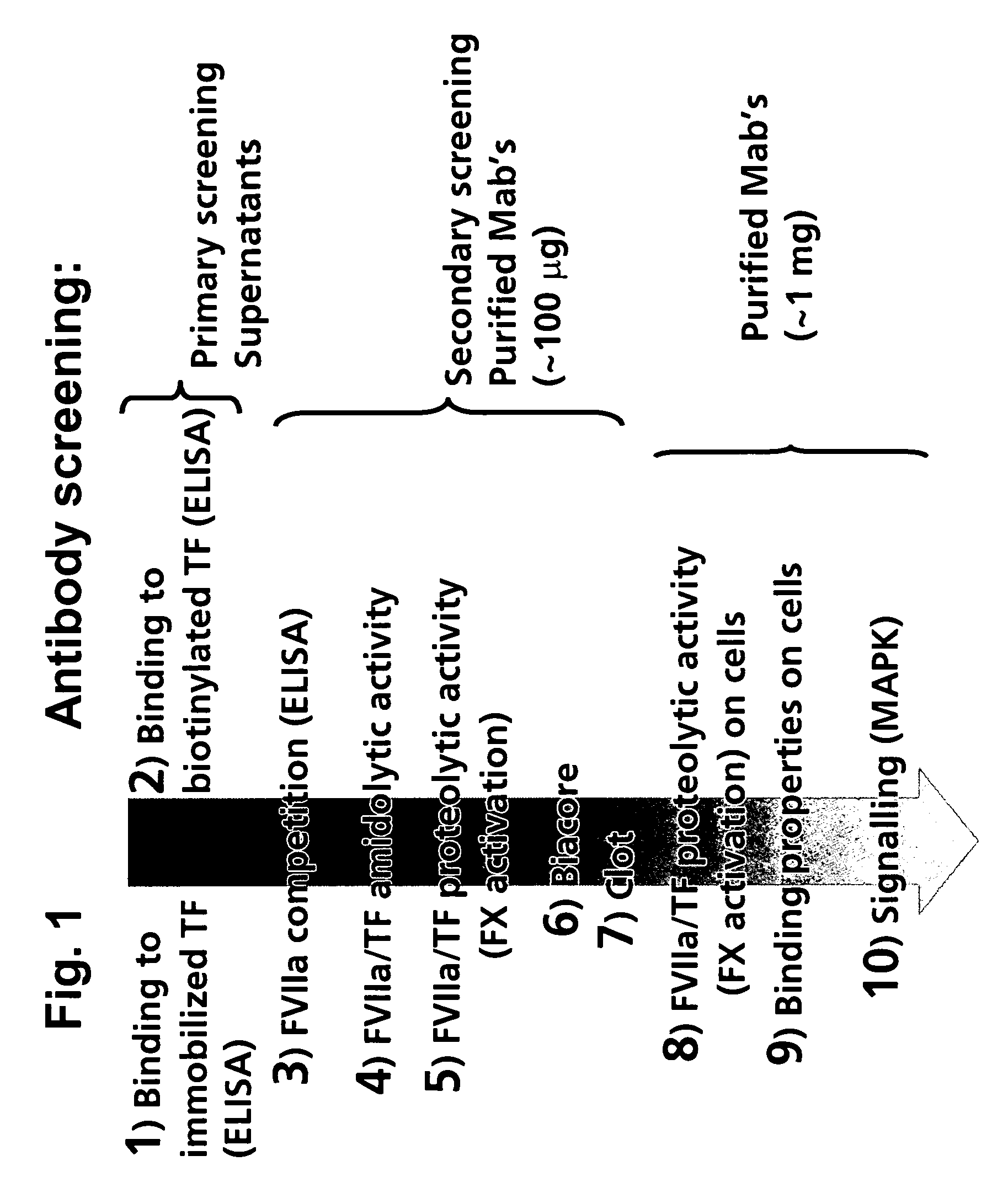
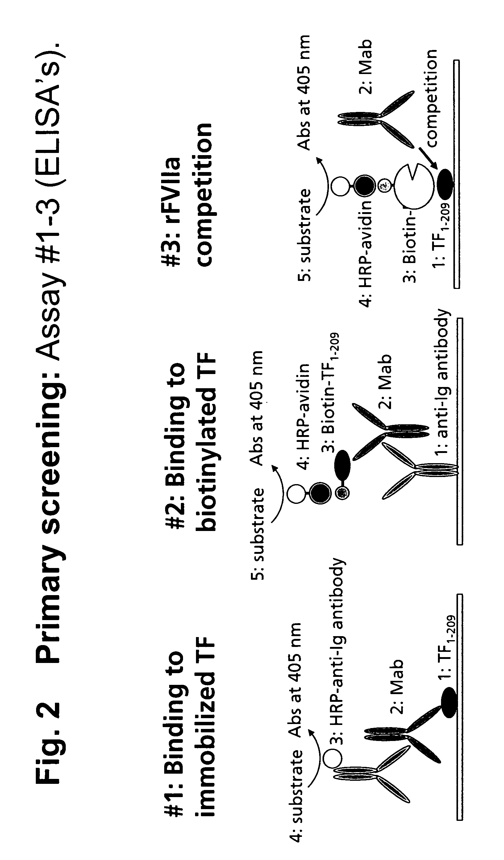
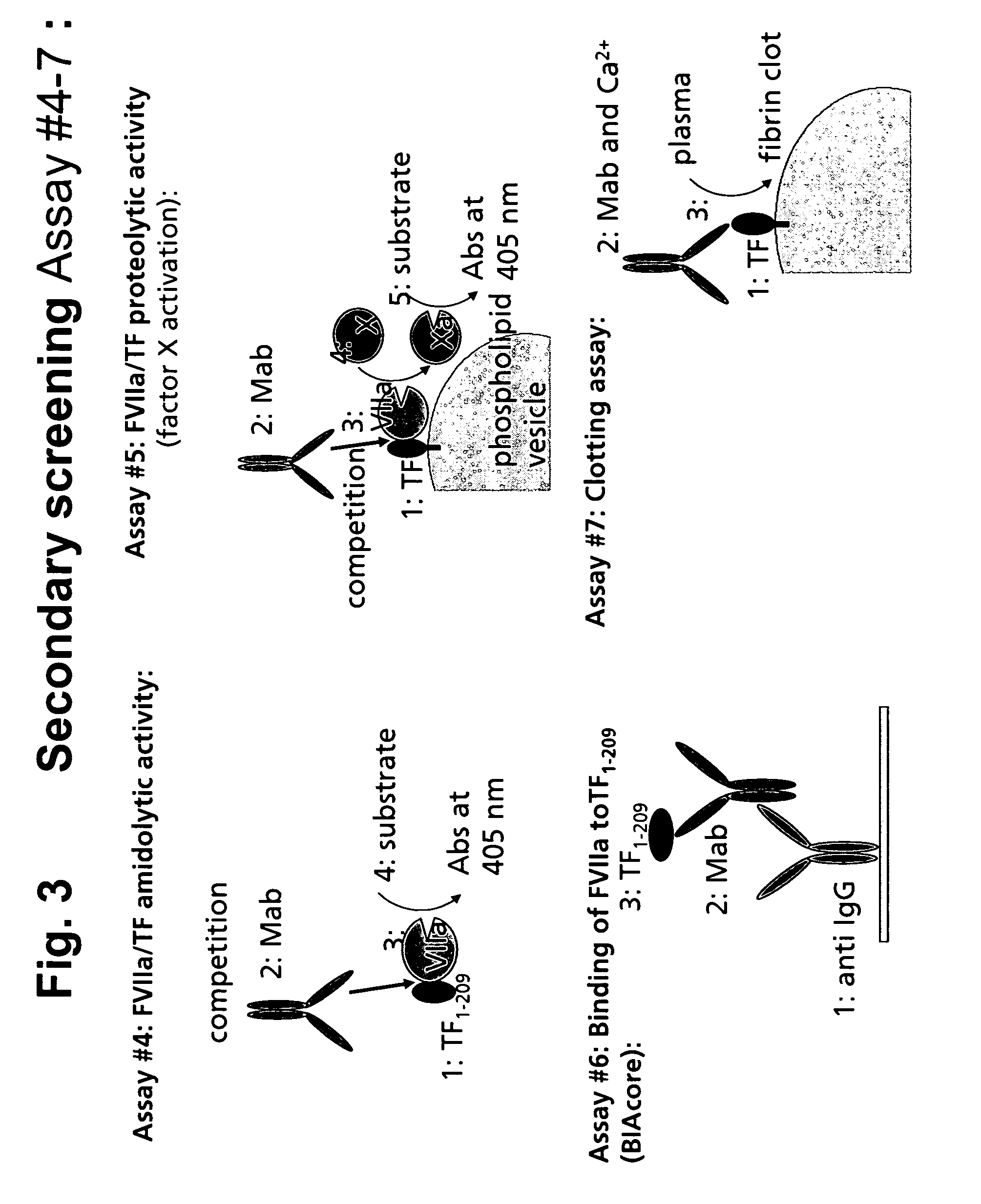
Screening.
[0210] The various assays used in the screening of serum and culture supernatants for specific selected antibodies are described in the following:
Direct TF ELISA Assay (Assay 1):
[0211] Nunc immunoplates are coated overnight at 4° C. with 1 μg / ml of human sTF in PBS. Plates are blocked with blocking buffer (TBS with 5 mM CaCl2 and 2% BSA) and washed in TBS+0.05% Tween 20, and the supernatants from the hybridoma cells are added. After incubation at room temperature for 1 hour, plates are washed and anti-human IgG labelled with horseradish peroxidase (HRPO) is added. After another hour of incubation, plates are washed and developed with TMB-substate (Kem-EN-Tec) as described by the manufactures. Absorbance at 450 nm is measured on an ELISA-reader.
Indirect TF ELISA Assay (Assay 2):
[0212] Nunc-immunoplates are coated with 0.5 μl / ml of goat anti-human IgG (Southern Biotechnology Associates, Cat-#2040-1) in PBS and incubated overnight at 4° C. Plates are blocked with bloc...
example 3
Human Cancer Assay. Investigating the Effects of Treatment with Human Anti-TF Mabs on Growth and Metastasis of Human Cancers in Mouse Models (Assay 13)
Treatment:
[0225] Human anti-TF Mabs given by bolus injection i.v.; 10 mg / kg=0.1 mg / 10 g; Injection-volume is 0.1 ml per 10 g mouse of either of three treatment solutions:
[0226] A. Vehicle control
[0227] B. 1 mg / ml Human FFR-rFVIIa
[0228] C. 1 mg / ml anti-TF Mabs
Description of Models:
I. Primary Growth and Liver Metastasis of Colon Cancer
[0229] Healthy female athymic mice (nu / nu) aged 7-8 weeks are used. To destroy the residual immunoresistance of the nude mice to the human cell implantation, the mice are routinely irradiated at 5 Gy 2 days before human tumor grafting (Vogel et al., 1997). Mice are challenged by tumor grafting of LS174T humancolon carcinoma cells (ATCC CCL 188) cultured in RPMI 1640 with 15% fetal calf serum (FCS) as described (Li et al., Human Gene Therapy 10: 3045-3053, 1999). In brief, the cells are harveste...
PUM
| Property | Measurement | Unit |
|---|---|---|
| Molar density | aaaaa | aaaaa |
| Nanoscale particle size | aaaaa | aaaaa |
Abstract
Description
Claims
Application Information