Selective detection of proteins that contain two or more alpha-helical transmembrane domains

a technology of alpha-helical transmembrane and protein, applied in the direction of fluid pressure measurement, liquid/fluent solid measurement, peptides, etc., can solve the problems of streaking in the isoelectric focusing dimension, lack of hydrophobic proteins in the proteomic profile generated from two-dimensional gel electrophoresis (2dge), and poor resolution of this class of proteins

Inactive Publication Date: 2006-02-09
LIFE TECH CORP
View PDF0 Cites 21 Cited by
  • Summary
  • Abstract
  • Description
  • Claims
  • Application Information

AI Technical Summary

Benefits of technology

[0008] Embodiments of the present invention provide staining solutions and method of using staining solutions for selectively detecting proteins that contain two or more α-helical transmembrane domains. The staining solutions of the present invention comprise at least one lipophilic dye and at least about 30% (v/v) hydrophobic solvent. The dyes of the present are represented by the general formula A-B-E wherein A is a nitrogen heterocycle, B is a bridge moiety

Problems solved by technology

However, proteomic profiles generated from two-dimensional gel electrophoresis (2DGE) are well known to lack highly hydrophobic proteins, particularly integral membrane proteins containing more than one α-helical transmembrane domain (Ito, K. and Akiyama, Y.
This is thought to be due to the very poor resolution of this class of proteins in the isoelectric focusing component of the procedure, arising from poor solubilization by nonionic detergents, even in the presence of high concentrations of urea.
Additionally, this class of proteins is known to display significant isoelectric point heterogeneity due to glycosylation, which results in streaking in the isoelectric focusing dimension.
However, selective methods for visualization of integral membrane proteins against a background

Method used


Image

  • Selective detection of proteins that contain two or more alpha-helical transmembrane domains
  • Selective detection of proteins that contain two or more alpha-helical transmembrane domains
  • Selective detection of proteins that contain two or more alpha-helical transmembrane domains

Examples


example 1

Selective Staining of Proteins Containing Two or More Alpha Helical Transmembrane Domains in the Protein Complex F1F0 ATPase After Separation by SDS Polyacrylamide Gel Electrophoresis

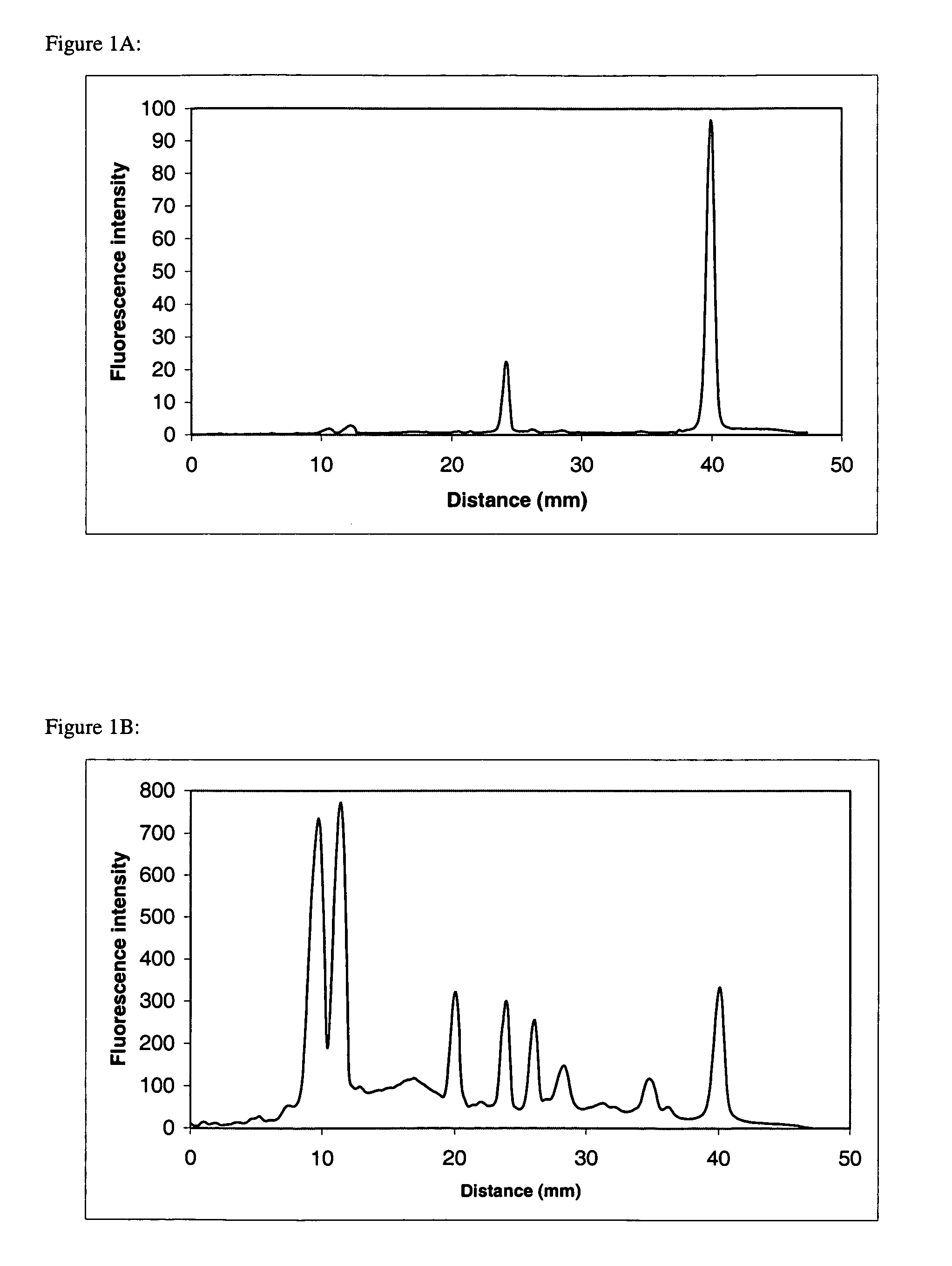
[0153] The protein complex F1F0 ATPase consists of a water-soluble F1-ATPase complex of 5 subunits (α, β, γ, δ, ε) and an insoluble membrane-embedded F0 complex of 3 subunits (a,b, c). F1 ATPase does not contain proteins having transmembrane domains, while F0 a, b, and c subunits contain a 5,1, and 2 α-helical transmembrane domains, respectively. Purified F1F0 ATPase was dissolved in 1× SDS sample buffer (50 mM Tris, 10% glycerol, 100 mM DTT, 2% SDS, 0.2% bromophenol blue, pH 6.8). Proteins were separated by SDS-polyacrylamide gel electrophoresis utilizing 10-20% acrylamide Tris-glycine precast Ready gels (Bio-Rad Laboratories, Hercules, Calif.). The 1 mm thick, 8×8 cm gels were subjected to electrophoresis using the Bio-Rad Mini Protean III system according to standard procedures. Following separation...

example 2

Selective Staining of Proteins Containing Two or More Alpha Helical Transmembrane Domains After Separation by SDS Polyacrylamide Gel Electrophoresis

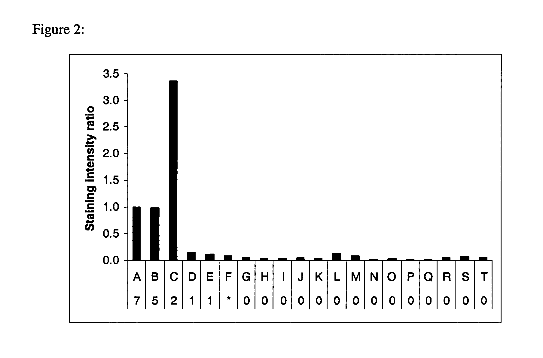
[0155] Representative examples of hydrophobic proteins containing no transmembrane domains, α helical transmembrane domains and a β sheet transmembrane domain along with water soluble proteins were prepared in 1×SDS sample buffer, separated by SDS-polyacrylamide gel electrophoresis, stained with Compound 4, imaged, stained for total proteins with SYPRO Ruby dye, and imaged again as described in Example 1. The intensity of the signals from Compound 4 and SYPRO Ruby stain was measured using ImageGauge software (Fuji Photo, Tokyo, Japan) for each protein and the ratio of the two signals was calculated and normalized for bacteriorhodopsin, a seven a helical transmembrane domain protein (FIG. 2). Proteins containing two or more a helical transmembrane domains were preferentially stained.

example 3

Selective Staining of Proteins Containing Two or More Alpha Helical Transmembrane Domains in the Protein Complex F1F0 ATPase After Separation by SDS Polyacrylamide Gel Electrophoresis

[0156] Bacteriorhodopsin, a seven α-helical transmembrane domain-containing protein, F1F0 ATPase complex, and a protein marker mixture containing myosin, β-galactosidase, phosphorylase b, BSA, ovalbumin, carbonic anhydrase, soybean trypsin inhibitor, lysozyme, and aprotinin (all non-transmembrane domain proteins) were prepared in 1× LDS sample buffer (50 mM Tris, 10% glycerol, 50 mM DTT, 2% LDS, 0.015% EDTA, 0.1% Serva Blue G250, and 0.1% Phenol Red, pH 8.5). Proteins were separated by SDS-polyacrylamide gel electrophoresis utilizing 4-12% acrylamide Bis-Tris NuPAGE precast gels (Invitrogen, Carlsbad, Calif.). The 1 mm thick, 8×8 cm gels were subjected to electrophoresis using the NuPAGE XCell Mini-Cell system according to standard procedures. Following separation, the gels were fixed 20 minutes in 100...


PUM

PropertyMeasurementUnit
Fractionaaaaaaaaaa
Fractionaaaaaaaaaa
Fractionaaaaaaaaaa

Abstract

Embodiments of the present invention provide a staining solution and of method of using the staining solution for selectively detecting proteins that contain two or more α-helical transmembrane domains. The staining solution comprises a lipophilic dyes and at least about a 30% hydrophobic solvent. The dyes of the present are represented by the general formula A-B-E wherein A is a nitrogen heterocycle, B is a bridge moiety and E is an electron pair accepting moiety that comprises either a carbonyl or nitrogen atom. In one embodiment these lipophilic dyes are merocyanine dye, a cyanine dye, a styryl dye or a carbazolylvinyl dye.

Description

CROSS-REFERENCE TO RELATED APPLICATIONS [0001] This application claims priority to U.S. Provisional Patent Application Ser. No. 60 / 599,339, filed Aug. 6, 2004, which disclosure is herein incorporated by reference.STATEMENT REGARDING FEDERALLY SPONSORED RESEARCH [0002] This invention was made in part with government support under grant number R33 CA093292-01, awarded by the National Cancer Institute. The United States Government may have certain rights in this invention.FIELD OF THE INVENTION [0003] Lipophilic fluorescent dyes, and to their use in selective detection of proteins that contain two or more α-helical transmembrane domains are disclosed. The dyes and their use have applications in the fields of cell biology, neurology, nutrition, immunology, proteomics and cancer biology. BACKGROUND OF THE INVENTION [0004] Integral membrane proteins typically contain one or more hydrophobic, transmembrane domains that intermingle with the hydrophobic portion of lipid bilayer membranes. Gi...

Claims


Application Information

Patent Timeline
IPC IPC(8): C07K1/26
CPCC07K1/26
Inventor PATTON, WAYNE F.HART, COURTENAY R.
Owner LIFE TECH CORP
Why Eureka
Try Eureka