Microchip-based system for hiv diagnostics

a technology of hiv diagnostics and microchips, applied in biochemistry apparatuses, instruments, peptides, etc., can solve the problems of requiring more cumbersome processing methods and detection equipment, and achieve the effect of enhancing detection of small molecules, reducing blood sample volume, reagent and power requirements, and enhancing detection

Inactive Publication Date: 2006-10-19
BOARD OF RGT THE UNIV OF TEXAS SYST +1
View PDF24 Cites 24 Cited by
  • Summary
  • Abstract
  • Description
  • Claims
  • Application Information

AI Technical Summary

Benefits of technology

[0034] Current methods of analyte screening often employ flow cytometry. Flow cytometry involves processing of a dynamic fluid stream that contains analytes of interest. Methods of the present invention pass analytes of interest through a unique flow cell comprising one or more cavities in which a capture agent is positioned to capture, and thereby separate, analytes from other aspects of whole blood and / or plasma. The captured analytes are then imaged optically, using a light source, one or more dichroic filters, and a detector capable of imaging the immobilized analytes (i.e., “static imaging”). Thus, methods of the present invention provide for efficient capture and static imaging of small molecule analytes of interest.
[0035] In one embodiment, CCD digital imaging is used for static imaging. Detection of small molecules is enhanced through a combination of efficient capture, static imaging and stable fluorescence signaling. Because the small molecules are readily captured and detected, blood sample volume, reagent and power requirements are significantly reduced. Flow cytometry, by contrast depends upon capture of signals from analytes moving through a flowing stream, thereby requiring more cumbersome processing methods and detection equipment.
[0049] b) forming a binding complex between the HIV-RNA and the complementary nucleotide sequence on the filter, whereby the signal emitted from the fluorescence-emitting compound is enhanced by formation of the binding complex;
[0060] b) forming a binding complex between the HIV-RNA and the complementary nucleotide sequence on the bead, whereby the signal emitted from the fluorescence-emitting compound is enhanced by binding of the HIV-RNA to the complementary nucleotide sequence;

Problems solved by technology

Flow cytometry, by contrast depends upon capture of signals from analytes moving through a flowing stream, thereby requiring more cumbersome processing methods and detection equipment.

Method used


Image

  • Microchip-based system for hiv diagnostics
  • Microchip-based system for hiv diagnostics
  • Microchip-based system for hiv diagnostics

Examples


example 1

Microchip-Based Assay for Measurement and Quantification of CD4+ T Cells

Design and Development of Flow Cell Analysis Chamber

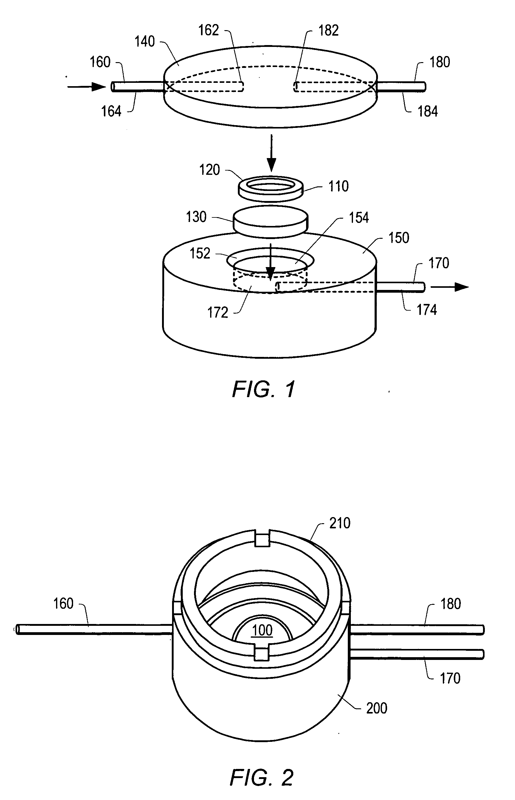
[0142] A chip-based sensor array composed of individually addressable microwells on a single silicon or plastic microchip has been developed. Microwells were created using molded-plastic methods, optimized, and the resultant structures, made with an anisotropic etch, served as miniaturized reaction vessels and analysis chambers. Each microwell has a volume of approximately 30 nanoliters (nL); a single microliter (μL) of fluid provides sufficient sample to complete multiple assays. Microwells possess pyramidal pit shapes, with openings that allow for both fluid flows through the analysis chambers as well as optical analysis of reactions occurring on the floor of the microwell, or on microbeads or a membrane filter resting within the microwell. Current chips contain up to 100 microwells on a single, dime-sized chip (FIGS. 7 and 8).

[0143] The microchip is anch...

example 2

Measurement of CD4:CD8 Ratios and CD4+ Percentages from Whole Blood

[0152] A rapid, whole blood assay (without red blood cell lysis, extra buffers or additional sample processing) for detecting CD4+ and CD8+ cells and establishing CD4:CD8 ratios was developed, based on the microchip discussed in Example 1. Adjustment of the flow parameters and dilution of the sample revealed an optimal flow rate of 0.8 mL / minute and a dilution of whole blood of 1:20.

[0153] The blood was obtained by venipuncture from healthy volunteers or HIV-infected subjects at the Massachusetts General Hospital and the Botswana-Harvard Laboratory in Gabarone, Bostwana. For the measurement of CD4:CD8 ratios and CD4+ percentages, 33 microliters of whole blood was mixed with 3 microliters of fluorophore-conjugated antibodies to CD3, CD4 or CD8 for staining. An optimized staining protocol was established for each of the antibodies utilized in this study by performing tube reactions and on-slide observation of the res...

example 3

Microbead Immunoassay

[0160] Agarose microbeads were coated with antigens specific to a variety of pathogens, including HIV gp41 / gp120, HIVp24 and hepatitis B surface antigen. Beads coated with antibody to specific antigens were placed in the wells of a silicon microchip and serum samples containing known titers of antibodies were then run through the microchip system, and detected by fluorescence microscopy using Cy2-labelled secondary antibodies. FIG. 13 shows results of one such experiment; with serum containing antibodies against HIV and hepatitis B easily detected using the microchip approach.

[0161]FIG. 15 shows the results of another microbead experiment where the HIV p24 antigen was detected in human serum containing 100 pg / ml of HIV p24 antigen. The same principle can be applied to any immunoassay, including liver enzymes.


PUM

PropertyMeasurementUnit
volumeaaaaaaaaaa
dead volumesaaaaaaaaaa
thicknessaaaaaaaaaa

Abstract

The invention relates to microchip-based assays to measure HIV-associated analytes of interest (e.g., CD4 lymphocytes, HIV RNA and liver enzymes) in a sample from a subject infected with the HIV virus. Methods of the present invention are optimal for use in monitoring HIV disease in resource-poor settings.

Description

RELATED APPLICATIONS [0001] This application claims the benefit of priority from U.S. application Ser. No. 60 / 445,143, filed on Feb. 5, 2003, U.S. application Ser. No. 60 / 447,070, filed on Feb. 13, 2003 and International Application No. PCT / US03 / 23131, filed Jul. 24, 2003. [0002] Each document cited or referenced in each of the foregoing applications, and any manufacturer's instructions or catalogues for any products cited or mentioned in each of the foregoing applications and in any of the cited documents, are hereby incorporated herein by reference. Furthermore, all documents cited in this text, all documents cited or referenced in documents cited in this text, and any manufacturer's instructions or catalogues for any products cited or mentioned in this text or in any document incorporated into this text, are incorporated herein by reference. Documents incorporated by reference into this text or any teachings therein can be used in the practice of this invention. Documents incorpo...

Claims


Application Information

Patent Timeline
Patent Type & Authority Applications(United States)
IPC IPC(8): C12Q1/70C07KC12Q1/52G01N33/537G01N33/543G01N33/569G01N33/58
CPCG01N33/537G01N33/56988G01N33/56972C12Q1/52G01N33/54366G01N33/582
Inventor WALKER, BRUCE D.RODRIGUEZ, WILLIAM R.MCDEVITT, JOHN C.
Owner BOARD OF RGT THE UNIV OF TEXAS SYST
Why Eureka
Try Eureka