Breast diagnostic apparatus for fused SPECT, PET, x-ray CT, and optical surface imaging of breast cancer

Inactive Publication Date: 2006-10-26
FUSED MULTIMODALITY IMAGING
View PDF31 Cites 32 Cited by
  • Summary
  • Abstract
  • Description
  • Claims
  • Application Information

AI Technical Summary

Benefits of technology

[0008] The present invention solves the problems that exist in prior art imaging systems and other problems by providing higher spatial resolution radioisotope imaging via breast anatomic specific imaging. The solution uniquely combines breast imaging with high resolution radioisotope imaging called micro single photon emission tomography (micro SPECT), high resolution positron emission tomography, micro positron emission tomography (micro PET), micro x-ray computed tomography (micro CT), and optical surface views. The term “micro” is used to describe the higher resolution capability of the system to image smaller details as compared to tr

Problems solved by technology

The radiologists reading these mammograms have difficulty reading the dense breast x-ray mammograms.
Once this exam has been completed and a dense breast indication has been found, t

Method used


Image

  • Breast diagnostic apparatus for fused SPECT, PET, x-ray CT, and optical surface imaging of breast cancer
  • Breast diagnostic apparatus for fused SPECT, PET, x-ray CT, and optical surface imaging of breast cancer
  • Breast diagnostic apparatus for fused SPECT, PET, x-ray CT, and optical surface imaging of breast cancer

Examples


Embodiment Construction

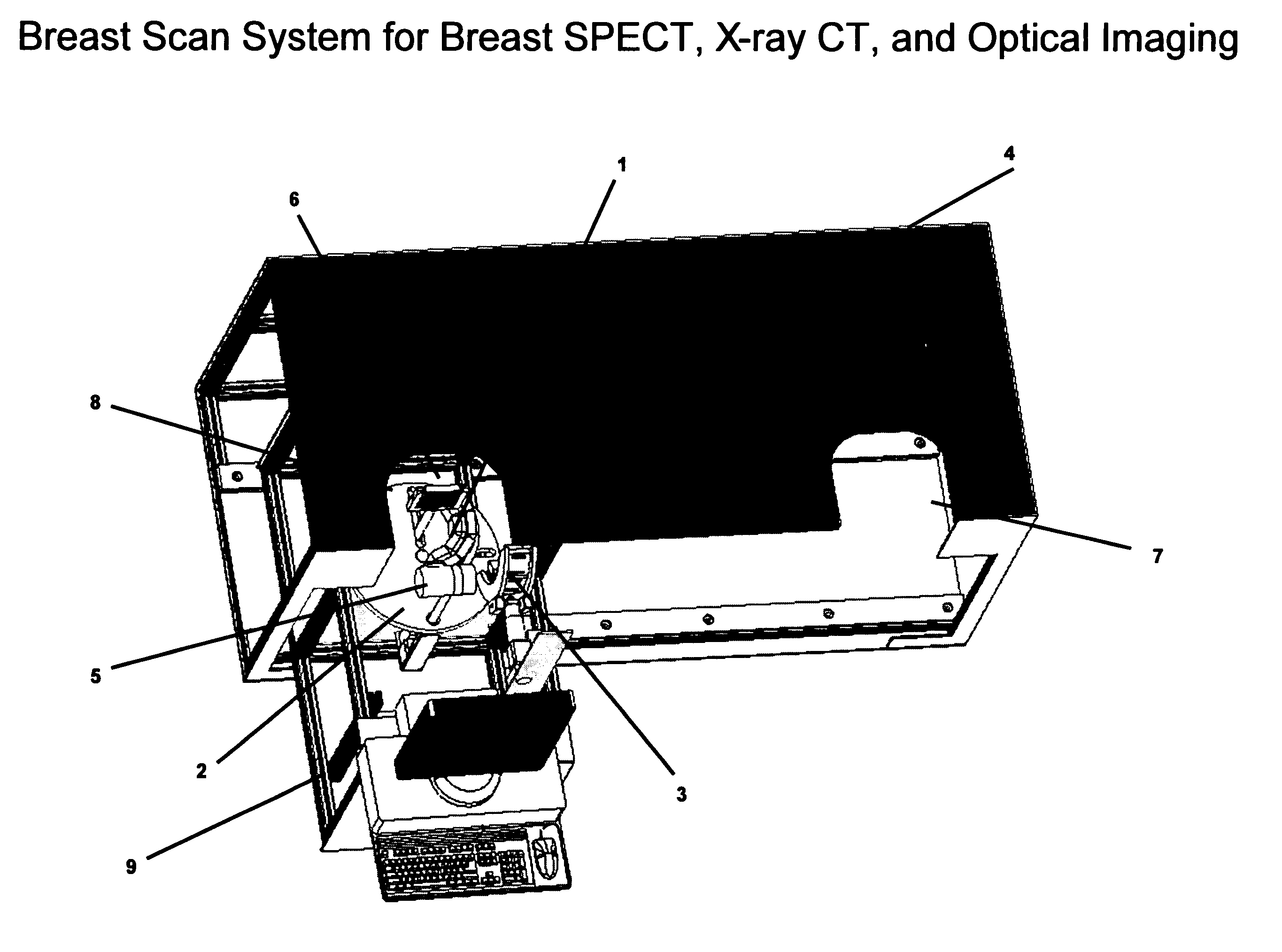
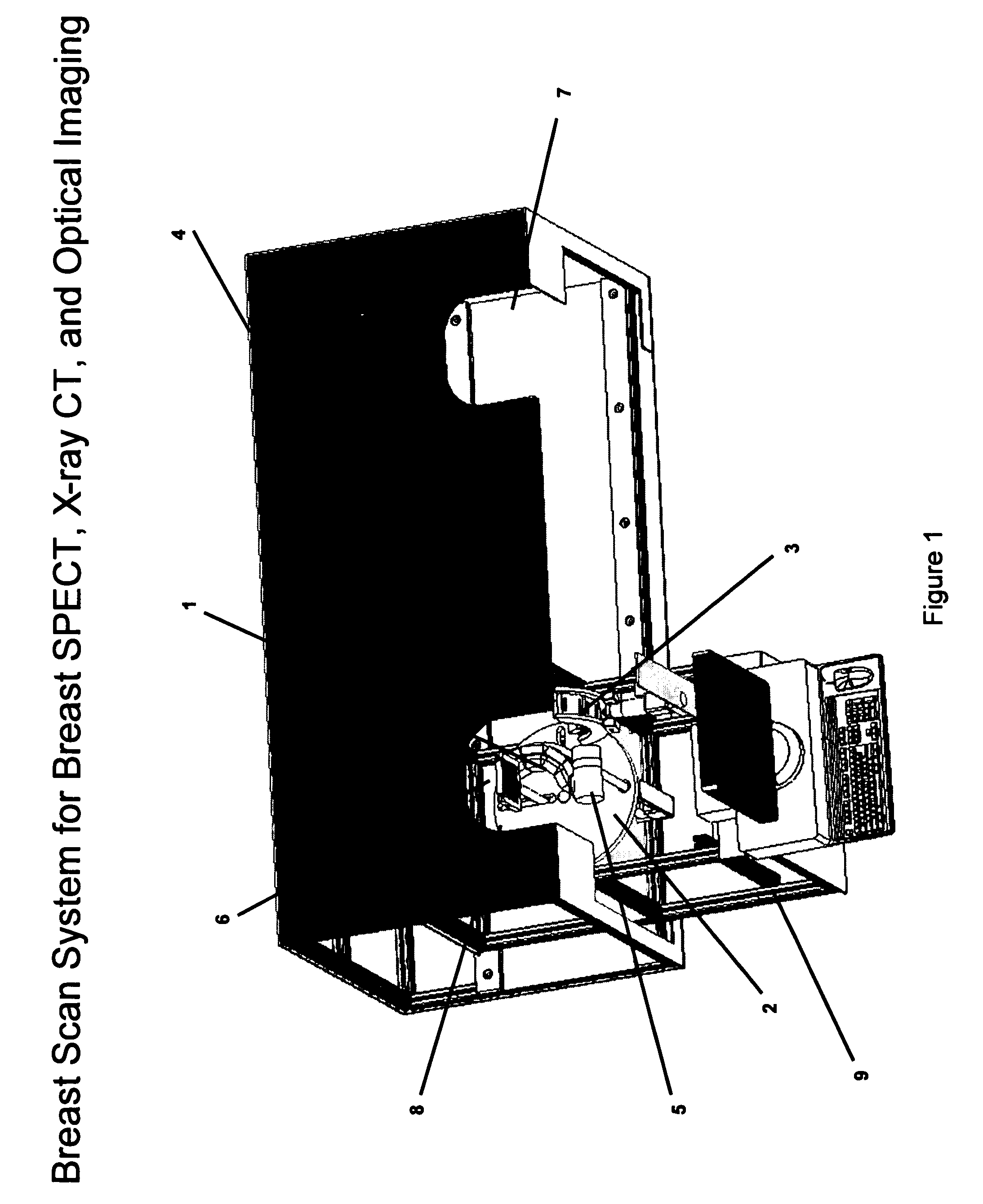
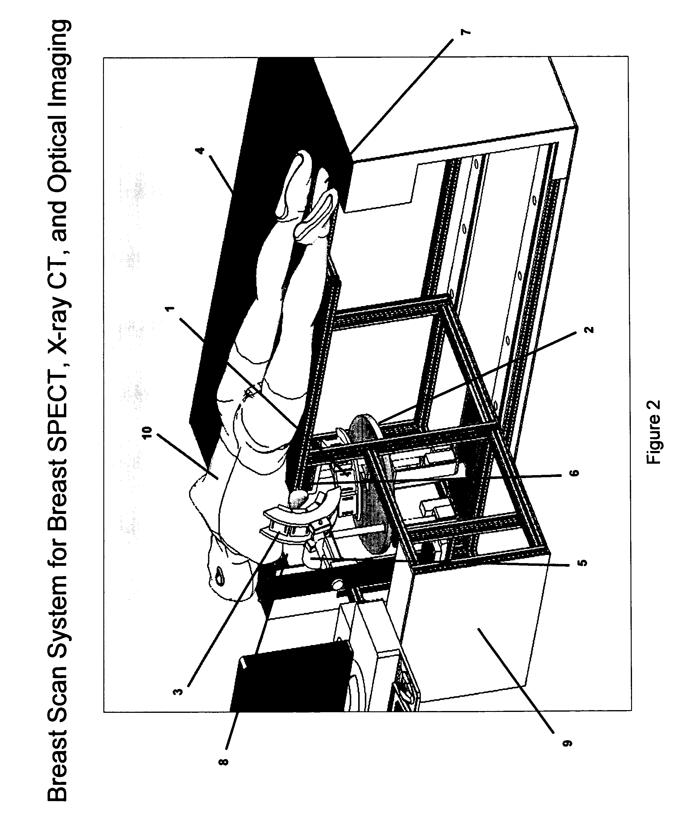
[0041] Referring now to the Figures where the illustrations are for the purpose of describing the preferred embodiment of the present invention and are not intended to limit the invention disclosed herein, FIG. 1 is a top frontal view of the apparatus utilized by the breast scan system of the present invention. As shown in FIG. 2, the patient 10 lies prone and slightly tilted to one side to allow full extension of the breast through a left breast hole 8 or right breast hole 7. The breast is scanned with an anatomic specific imaging central breast curved gamma detector 1 for single photon emission computed tomography (SPECT). Radioisotopes are injected into the patient 10 and emitted radiation is detected by the central breast curved gamma detector 1. The breast scan system also has an x-ray source 5 and an x-ray detector 6. The x-ray source 5 transmits x-rays through the breast of the patient 10 which are detected by the x-ray detector 6. The x-ray source 5 and x-ray detector 6 are ...


PUM


Abstract

A new method of breast imaging to improve the detection of cancer during early stages of development is disclosed. The system combines molecular images of radioisotope uptake in cancerous cells with three dimensional high resolution single photon emission computed tomography (SPECT), positron emission tomography (PET), x-ray computed tomography (CT) and optical reflectance and emission (ORE) images of the breast. The system acquires data from nuclear isotopes within the breast and processes the data into three dimensional molecular tomographic images of cancerous cellular activity, morphological three dimensional x-ray density tomographic images and three dimensional optical surface images. These three sets of images or data are then combined to provide information as to the sensitivity and specificity as to the type of cancer present, three dimensional information as to the physical location of the cancer and reference information for radiologists, surgeons, oncologists and patients in order to plan stereo-tactic biopsy, minimally invasive surgery and image guided therapy, if necessary.

Description

TECHNICAL FIELD [0001] The present invention relates, in general, to gamma ray and x-ray detector systems and signal processing for nuclear medicine gamma cameras, single photon emission tomography (SPECT), positron emission tomography (PET), x-ray computed tomography (CT), digital radiology, x-ray mammography, optical imaging, optical fluorescence imaging, and other limited field of view gamma ray and x-ray detection and signal processing instrumentation. BACKGROUND ART [0002] This invention applies to gamma ray imaging, nuclear SPECT imaging, PET imaging, x-ray CT imaging, digital radiography (DR) imaging, x-ray mammography, optical imaging, optical fluorescence imaging, small field of view imaging detectors and probes, and fused multimodality imaging. [0003] In breast imaging and screening, x-ray mammography is being used as a screening tool for women over the age of 40 years. During the screening process, 40% of women have dense breast or suspicious breast indications for cancer...

Claims


Application Information

Patent Timeline
IPC IPC(8): A61B6/04G01N23/04A61B5/05
CPCA61B5/0059A61B5/415A61B5/418A61B6/0435A61B6/502A61B6/037A61B6/4291A61B6/4417A61B6/5247A61B6/032G01T1/1603A61B5/0064
Inventor MCCROSKEY, WILLIAM K.DICKINSON, WILLIAM D.LEMASTER, WILLIAM S.SUMMERHILL, WALTER A.DOBOS, ALAN M.MILLIFF, MICHAEL E.
Owner FUSED MULTIMODALITY IMAGING
Why Eureka
Try Eureka