
Production of tissue engineered heart valves

Active Publication Date: 2006-11-09
WAKE FOREST UNIV HEALTH SCI INC
View PDF43 Cites 57 Cited by
  • Summary
  • Abstract
  • Description
  • Claims
  • Application Information

AI Technical Summary

Benefits of technology

[0006] The present invention provides methods and compositions for preparing tissue engineered heart valves. The tissue engineered valve has a viable cellular component, phenotypically compatible with the patient's own cardiac tissue. The replacement valve is produced from endothelial cells and muscle cells, such as smooth muscle cells. Endothelial cell coverage of the valve surface prevents the occurrence of thrombosis formation. In the present invention, circulating progenitor cells can be isolated from the peripheral blood of patients with vascular disease. These isolated progenitor cells can be grown and differentiated into endothelial and muscle cells that can be used to seed matrices. These cell seeded matrices can be exposed to fluid conditions that mimic blood flow through the heart, resulting in tissue engineered heart valves that are preconditioned to be analogous to native heart valves. Thus, the engineered valves can be designed to retain more of their natural flexibility and thus function more efficiently like their in vivo counterpart.

Problems solved by technology

Heart valve replacements are limited because donor tissues is in short supply.
While this approach is successful in limiting acute rejection of these grafts, there are serious limitations.
For example, leaflet flexibility is compromised, with the bending stiffness of the leaflets being significantly increased.
Also, glutaraldehyde fixation increases the incidence and extent of leaflet and aortic conduit calcification.
These factors combine to make bioprosthetic valves less durable and more prone to failure than natural valves.
Another major problem with replacement heart valves generally is the formation of blood clots and the risk of thrombosis.
Thrombosis alters the mechanical fluid flow property of the valves and reduces their functionality as well as increasing the risk of heart failure in the patient.

Method used


Image

  • Production of tissue engineered heart valves
  • Production of tissue engineered heart valves
  • Production of tissue engineered heart valves

Examples


example 1

Methods and Materials

Materials

[0186] Unless otherwise stated, all chemicals were purchased from Sigma (St. Louis, Mo.). ECL buffers and 125I-Sodium were purchased from PerkinElmer NEN (Boston, Mass.). Collagenase type II (1 mg / ml) was purchased from Boehringer Mannheim (Mannheim, Germany). Endothelial medium (EBM-2) was purchased from Cambrex Bio Science (Walkersville, Md.). PCR reagents and primers, M199 medium, fetal bovine serum (FBS) and penicillin were purchased from Life Technologies (Gaithersburg, Md.). Basic Fibroblast Growth Factor (bFGF) was a gift from Judith Abraham, (Scios Nova, Calif.). Anti human CD31, anti vonWillebrand factor (vWF) and anfi smooth muscle actin antibodies were purchased from DACO (Glostrup, Denmark). Anti-CD105 antibodies were purchased from BD Pharmingen (San Diego, Calif.). Anti Flk-1 antibodies were purchased from Santa Cruz (Santa Cruz, Calif.). FITC-conjugated avidin was purchased from Vector Laboratories (Burlingame, Calif.). Athymic mice we...

example 2

Electrospun Matrices

[0206] An electrospun matrix was formed using the methods outlined in Example 1. A solution of collagen type I, elastin, and PLGA, were used. The collagen type I, elastin, and PLGA were mixed at a relative concentration by weight of 45% collagen, 40% PLGA, and 15% elastin.

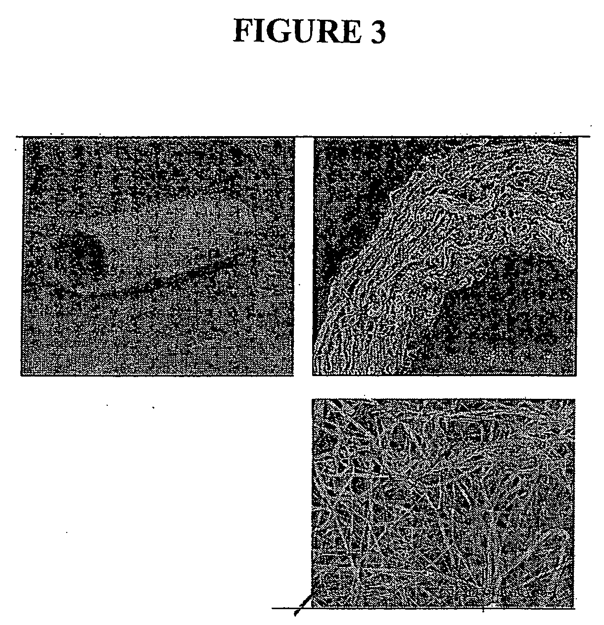
[0207] The resulting fibrous scaffold had a length of 12 cm with a thickness of 1 mm. A 2 cm representative sample is depicted in FIG. 3. This demonstrates the feasibility of spinning Type I Collagen and elastin into fibers from nanometer to micrometer diameter using concentrations from 3% to 8% by weight in solution. These results also show that by adding PLGA (Mw 110,000) to the mixture, solutions with higher viscosity and improved spinning characteristics could attained. By increasing the solution concentration to 15%, thicker, stronger scaffolds were able to be built while maintaining the collagen and elastin components.

[0208] Collagen type I stained positively on the decellularized scaff...

example 3

Cross-Linking of Electrospun Matrices

[0215] This example demonstrates how to increase the strength and stability of the electrospun scaffold by chemical cross-linking. The scaffolds were soaked in 20% dextran solution in phosphate buffered saline prior to crosslinking to reduce hydration-induced swelling and contraction of the scaffold. The scaffolds were crosslinked by immersion in EDC / NHS in MES / EtOH solution for 2 hours at room temperature. Scanning electron micrographs of the resulting fibers showed fiber diameters of 500 nm or less and a random orientation of fibers. Atomic force microscopy of the scaffold and a confocal image of nanofibers with an adhering endothelial cell demonstrate the scaffold structure. These data show that it is possible to fabricate vascular scaffolds from biological polymers with mechanics and structure similar to decellularized scaffolds and native arteries.


PUM


Abstract

The invention is directed to methods for preparing artificial heart valves by preconditioning a matrix seeded with endothelial cells and smooth muscle cells differentiated from isolated progenitor cells. These cell seeded matrices are exposed to fluid conditions that mimic blood flow through the heart to produce tissue engineered heart valves that are analogous to native heart valves.

Description

RELATED APPLICATIONS [0001] The present application claims priority to U.S. Provisional Application No. 60 / 660,832 filed Mar. 11, 2005 and U.S. Provisional Application Ser. No. 60 / 686,316 filed Jun. 1, 2005, the contents of which are hereby incorporated by reference in their entirety.BACKGROUND OF THE INVENTION [0002] The technical field of this invention relates to tissue engineered heart valves. Heart valve replacements are limited because donor tissues is in short supply. There are several types of valves that have been used for heart transplantations such as mechanical heart valves and bioprosthetic valves. The bioprosthetic valves are typically those obtained from other species and are known as xenografts. Porcine valves are the most common xenograft. [0003] Unlike mechanical heart valves, bioprosthetics have a natural trileaflet structure and should have similar flow characteristics as human valves. However, in order to prevent immune responses to the cellular components of th...

Claims


Application Information

Patent Timeline
IPC IPC(8): A61F2/24C12N5/08
CPCA61F2/2415A61L27/3604A61L27/3641A61L27/3808A61L27/3826B82Y10/00A61L27/3839A61L27/3886A61L27/507B82Y5/00A61L27/383C12N5/0068C12N5/069C12N2533/90
Inventor ATALA, ANTHONYYOO, JAMES J.
Owner WAKE FOREST UNIV HEALTH SCI INC
Learn More