
Targeted nanoparticles for magnetic resonance imaging

Inactive Publication Date: 2007-06-21
GENERAL ELECTRIC CO
View PDF15 Cites 40 Cited by
  • Summary
  • Abstract
  • Description
  • Claims
  • Application Information

AI Technical Summary

Benefits of technology

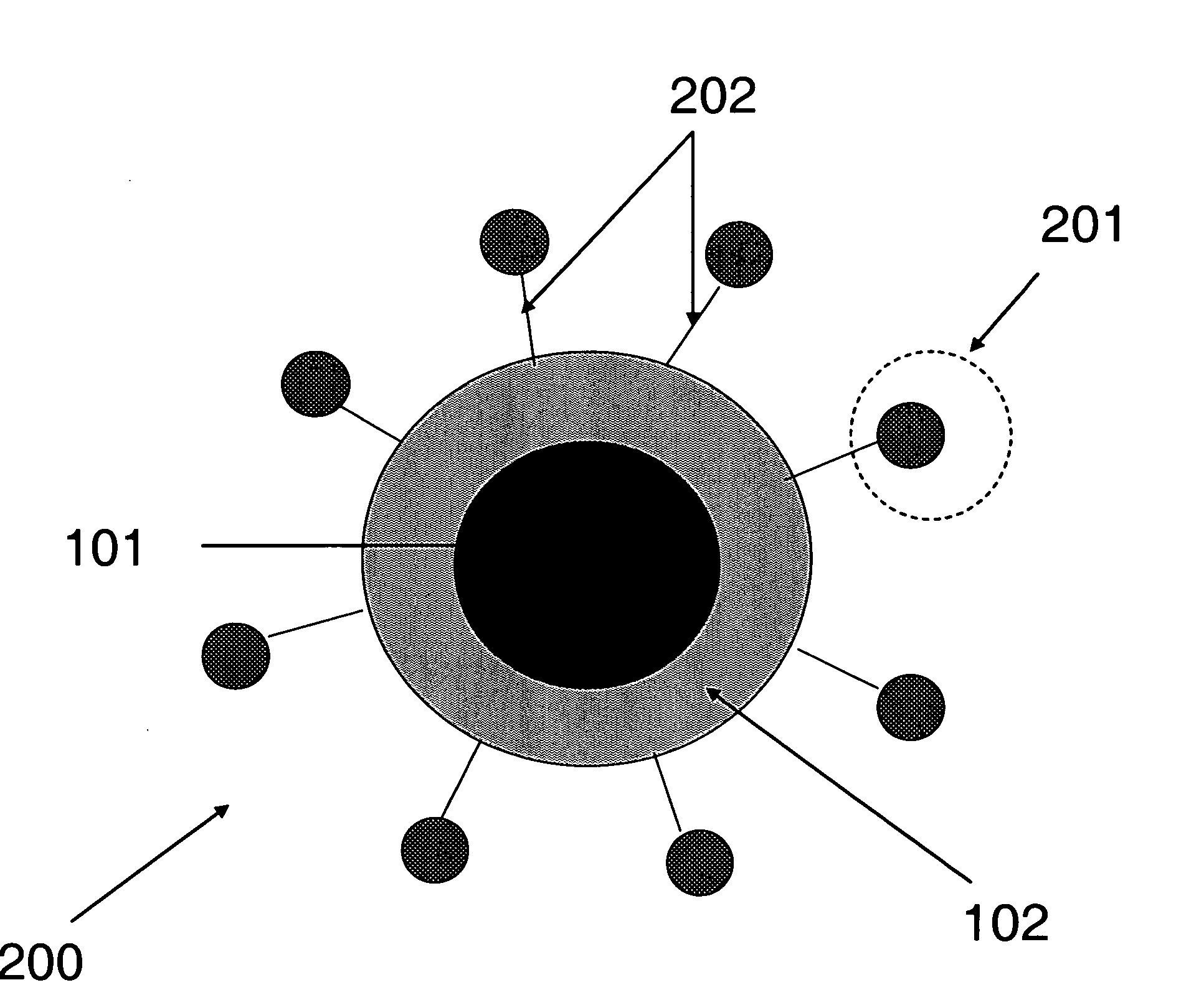
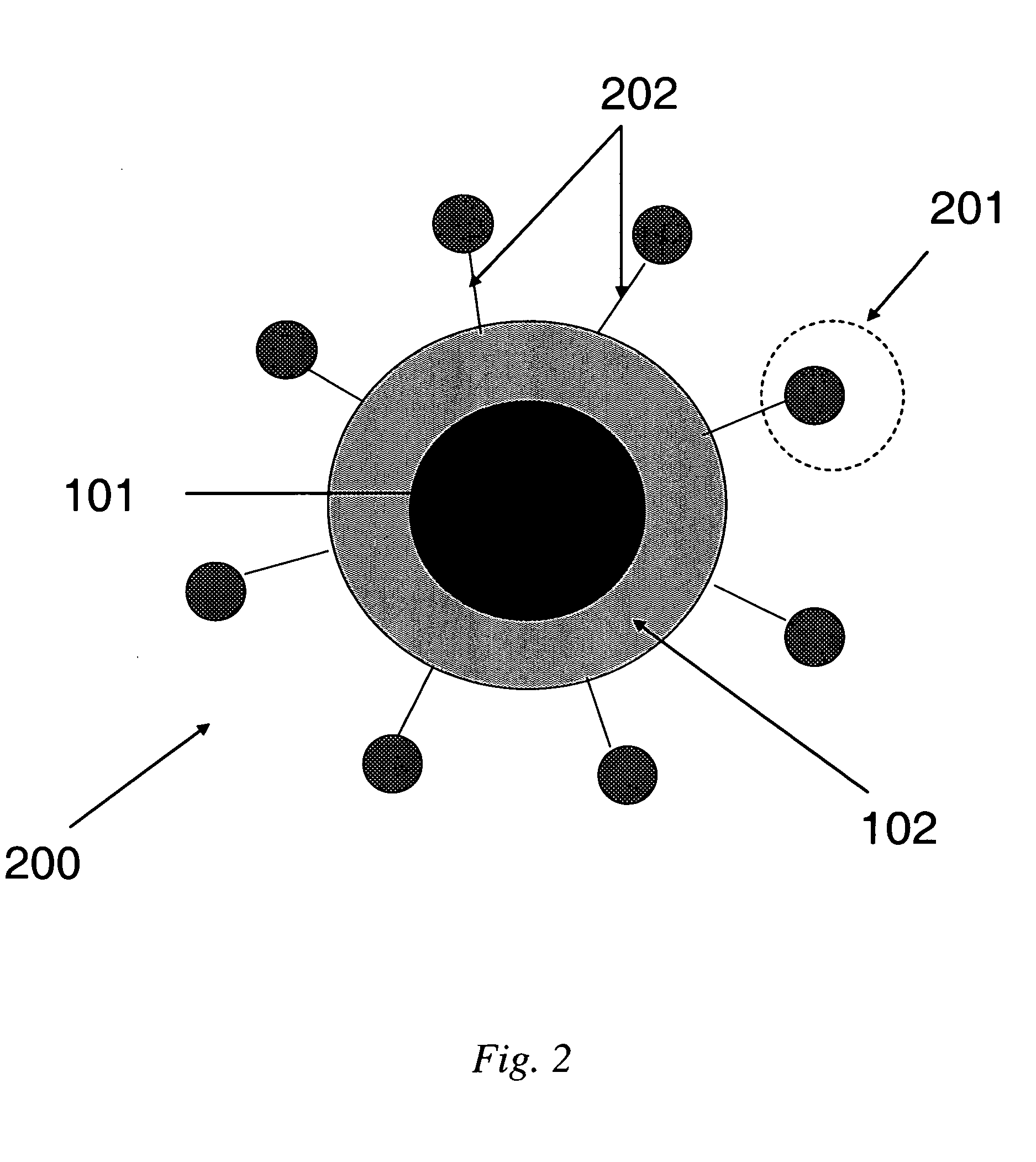
[0016] In some embodiments, the present invention is directed to novel targeted contrast agents for magnetic resonance imaging (MRI). The present invention is also directed to methods of making such targeted MRI contrast agents, and to methods of using such MRI contrast agents. Typically, such targeted MRI contrast agents provide enhanced relaxivity, improved signal-to-noise, targeting ability, and resistance to agglomeration. Methods of making such MRI contrast agents typically afford better control over particle size, and methods of using such MRI contrast agents typically afford enhanced blood clearance rates and biodistribution.

Problems solved by technology

2 mm), however, it offers poor sensitivity when compared with other imaging techniques.
However, dextran coatings have been claimed to be unstable at the alkaline conditions of the particle synthesis, and their chemical composition has therefore been questioned.
Additionally, dextran-induced anaphylactic reactions present potential problems (R. Weissleder U.S. Pat. No. 5,492,814).
In addition, this method provides little control over the degree of coating leading to particles containing multiple iron oxide nanoparticles within a single agent.
Nanoparticles obtained using conventional methods also have a low level of crystallinity, which significantly impacts the sensitivity of the contrast agent.
Moreover, nanoparticles tend to agglomerate due to their high surface energy, which is a significant problem encountered during synthesis and purification steps.
Such agglomeration increases the size of the particle, resulting in rapid blood clearance as well as reducing targeting efficiency, and may result in a reduction in relaxivity.
When large particles are employed, only a few targeting ligands may be attached before the particles become large enough to activate the RES, resulting in almost instantaneous clearance from the blood and failure of the agent to reach the intended target.
When coatings are globular, reactive sites intended for ligand attachment are generally hindered, thereby reducing conjugation efficiency.
In addition, once bound, ligands may reside in the interior of globular coatings, preventing easy access to the biomarkers.
The plaque only becomes detectable once it reaches a critical size, resulting in blocked blood flow, or when it ruptures, which may lead to thrombus (blood clot) formation, resulting in acute myocardial infarction or death.
Contrast agents, however, have inherent problems that limit targeting efficiency, such as low sensitivity, low signal-to-noise ratio, large particle sizes, rapid blood clearance, low efficiency of ligand attachment and the accessibility of ligands to the biomarkers' targets.
The primary limitation of this technology is the measured Msat values of these agents is between 43-60 emu / g for 4-6 nm core nanoparticles.

Method used


Image

  • Targeted nanoparticles for magnetic resonance imaging
  • Targeted nanoparticles for magnetic resonance imaging
  • Targeted nanoparticles for magnetic resonance imaging

Examples


example 1

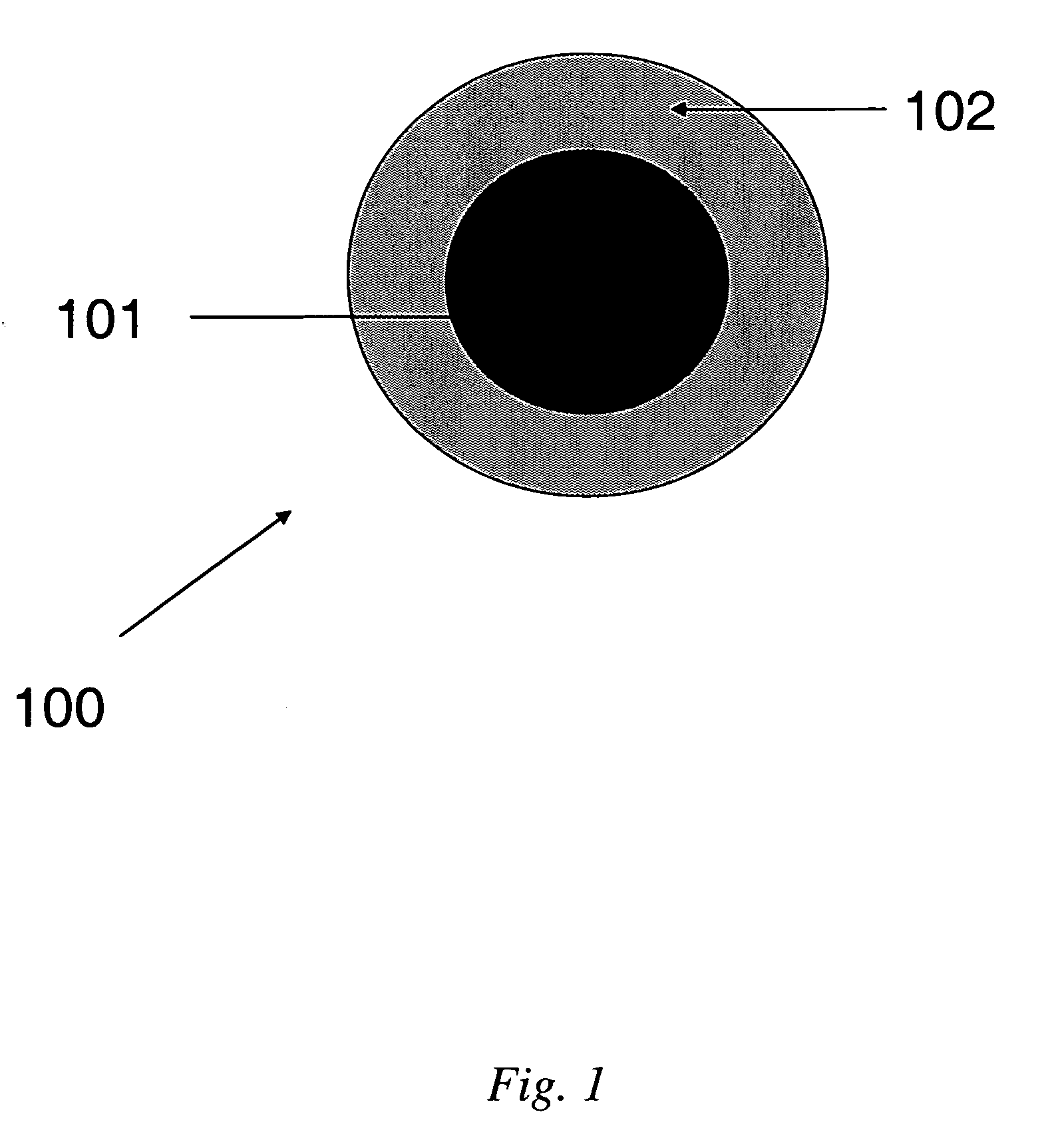
[0045] This Example illustrates the synthesis and characterization of SPIO nanoparticles and preparation of PEI-silane coated SPIO nanoparticles.

[0046] Synthesis of 5 nm SPIO nanoparticles. A 25 mL, 3-neck Schlenk flask was fitted with a condenser, stacked on top of a 130 mm Vigreux column, and a thermocouple. The condenser was fitted with a nitrogen inlet and nitrogen flowed through the system. The Schlenk flask and Vigreux column were insulated with glass wool. Trimethylamine-N-oxide (Aldrich, 0.570 g, 7.6 mmol) and oleic acid (Aldrich: 99+%, 0.565 g, 2.0 mmol) were dispersed in 10 mL of dioctylether (Aldrich: 99%). The dispersion was heated to 80° C. at a rate of about 20° C. / minutes. Once the mixture had reached ˜80° C., 265 μL of Fe(CO)5 (Aldrich: 99.999%, 2.0 mmol) was rapidly injected into the stirring solution through the Schlenk joint. The solution turned black instantaneously, with a violent production of a white “cloud.” The solution rapidly heated to ˜-120-140° C. Withi...

example 2

[0050] This Example illustrates attachment of peptides to PEI-coated siloxane core / shell nanoparticles. Polyethylene imine-coated siloxane core / shell naonoparticles are conjugated to N-acelyated peptides utilizing EDC. The reaction takes place in 0.1M MES, pH 4.5-5, as depicted in the synthetic scheme of FIG. 4. The polyethylene imine (PEI)-coated core / shell nanoparticles have numerous available secondary amines for coupling to N-acetylated peptides with the amount of conjugation controlled to achieve maximum binding efficiency to the biological target, as depicted in FIG. 5.

example 3

[0051] This Example is illustrative of cell uptake studies. NHS ester-Cypher5E dye was covalently bound to the PEI-coated nanoparticles. These amine-coupled dyes indicate the uptake of these nanoparticles into phagocytic cells and demonstrate the utility of the free amines of the PEI coating for attachment using NHS ester chemistry (similar to coupling chemistry for peptide, etc). Peptides can be coupled to these particles in a similar manner for uptake in non-phagocytic disease-specific cells expressing biomarkers of interest for diagnosis. FIG. 6 is a micrograph of MRI contrast agents comprising NHS ester-Cypher5E dye covalently bound to the PEI-coated nanoparticles and delivered to phagocytic cells stained with Cell Tracker Green dye, in accordance with some embodiments of the present invention.

[0052] Peptide-functionalized cationic nanoparticles could also deliver oligonucleotides to disease-specific sites for therapeutic or diagnostic purposes.


PUM

PropertyMeasurementUnit
Nanoscale particle sizeaaaaaaaaaa
Hydrodynamic diameteraaaaaaaaaa

Abstract

In some embodiments, the present invention is directed to novel targeted contrast agents for magnetic resonance imaging (MRI). The present invention is also directed to methods of making such targeted MRI contrast agents, and to methods of using such MRI contrast agents. Typically, such targeted MRI contrast agents provide enhanced relaxivity, improved signal-to-noise, targeting ability, and resistance to agglomeration. Methods of making such MRI contrast agents typically afford better control over particle size, and methods of using such MRI contrast agents typically afford enhanced blood clearance rates and biodistribution.

Description

TECHNICAL FIELD [0001] The present invention relates generally to nanoparticles for use in diagnostic imaging, and more specifically to nanoparticles functionalized with a targeting moiety for use as contrast agents in magnetic resonance imaging. BACKGROUND INFORMATION [0002] Diagnostic imaging procedures and contrast agents are used to study organs, tissues, and diseases in a body. One example of an imaging technique comprises magnetic resonance (MR), which is a technique that uses a powerful magnetic field and radio signals to create sophisticated vertical, cross-sectional and three-dimensional images of structures and organs inside a body. Unlike conventional radiography and computed tomographic (CT) imaging, which make use of potentially harmful radiation (X-rays), magnetic resonance imaging (MRI) is based on the magnetic properties of atoms. MRI is most effective at providing images of tissues and organs that contain water, such as the brain, internal organs, glands, blood vess...

Claims


Application Information

Patent Timeline
IPC IPC(8): A61K49/10
CPCA61K47/48192A61K47/48238A61K47/48861A61K47/48884A61K49/1848H01F1/0054A61K49/1866B82Y5/00B82Y25/00G01N33/552G01R33/5601A61K49/1857A61K47/59A61K47/62A61K47/6923A61K47/6929
Inventor TORRES, ANDREW SOLIZSYUD, FAISAL AHMEDWOOD, NICHOLE LEAKULKARNI, AMIT MOHANBAILLIE, MARK THOMASMOASSER, BAHRAMBALES, BRIAN CHRISTOPHERBELETSKII, ANTONBONITATEBUS, PETER JOHN JR.
Owner GENERAL ELECTRIC CO
Learn More