Method for culturing stem cells

a stem cell and culturing technology, applied in the field of culturing stem cells, can solve the problems of inconvenient long-term hesc culture, lack of simple methods for producing ebs of consistent and desired size, and lack of 2-d sam monolayers, etc., to achieve the effect of ensuring the cell differentiation profile of ebs and facilitating the control of the cell differentiation profil

Pending Publication Date: 2008-01-31
WISCONSIN ALUMNI RES FOUND
View PDF3 Cites 19 Cited by
  • Summary
  • Abstract
  • Description
  • Claims
  • Application Information

AI Technical Summary

Benefits of technology

[0015] In a second aspect, the invention is summarized in that a method for forming EBs having a narrow size distribution includes the steps of harvesting substantially undifferentiated ESCs from the microwells and culturing the harvested ESCs under differentiating culture conditions until the culture contains differentiated cells. Because the undifferentiated ESCs for use in the EB-forming method can be obtained from dimension-constrained microwells having uniform dimensions, supra, aggregates having a narrow size distribution can be harvested, thereby avoiding a shortcoming of existing EB-forming methods, namely that clumps of ESCs from which EBs are now derived can vary widely in size, volume and cell number. The harvesting step can include harvesting entire colonies or harvesting cell aggregates anchored to colonies in the microwells but unattached to the coating. Colonies can be released by enzymatic treatment. Aggregates can be released from the colonies by gentle shearing without dislodging the colonies, which can be cultured again to yield more cell aggregates. Aggregates released by gentle shearing have a narrow size distribution and yield cultured EBs also having a narrow size distribution. Where the entire culture in inadvertently harvested during the shearing step, the resulting EB is substantially larger than the majority of the EBs and can be discarded or ignored. Advantageously, the cell differentiation profile of EBs can be controlled by controlling the size, shape and volume of the undifferentiated cell cultures that give rise to the EBs.

Problems solved by technology

Unfortunately, the art lacks simple methods for producing EBs of consistent and desired size from ESCs.
Unfortunately, 2-D SAM monolayers are of limited utility for culturing primate ESCs because of the cell's growth nature.
Likewise, initial efforts at 2-D micro-contact printing of Matrigel® on SAM surfaces indicated that this method was not suitable for long-term hESC culture because of substrate instability and because growing colonies could span across unpatterned regions.
With few exceptions, current literature regarding patterned 2-D monolayers focuses primarily on cell attachment and replication to generate confluent monolayers in patterned regions, but does not investigate effects of three-dimensional confined geometries on long term health and stability of cell lines that are not strictly contact dependent.
Because hESC differentiation does not occur immediately, short-term analysis may not accurately represent hESC response to confinement.
These cells, however, were incubated for only four hours to investigate initial cell attachment and spreading, rather than long-term behavior in microwells.
Unfortunately, cell viability after two days was determined solely by visual cell replication [29].
These studies demonstrated the possibility of cell attachment in microwells, but did not show a marked improvement over prior patterned microwells that also constrained cells for at least two days.

Method used


Image

  • Method for culturing stem cells
  • Method for culturing stem cells
  • Method for culturing stem cells

Examples


example 1

Microwells for hESCs Culture and Embryoid Body Generation

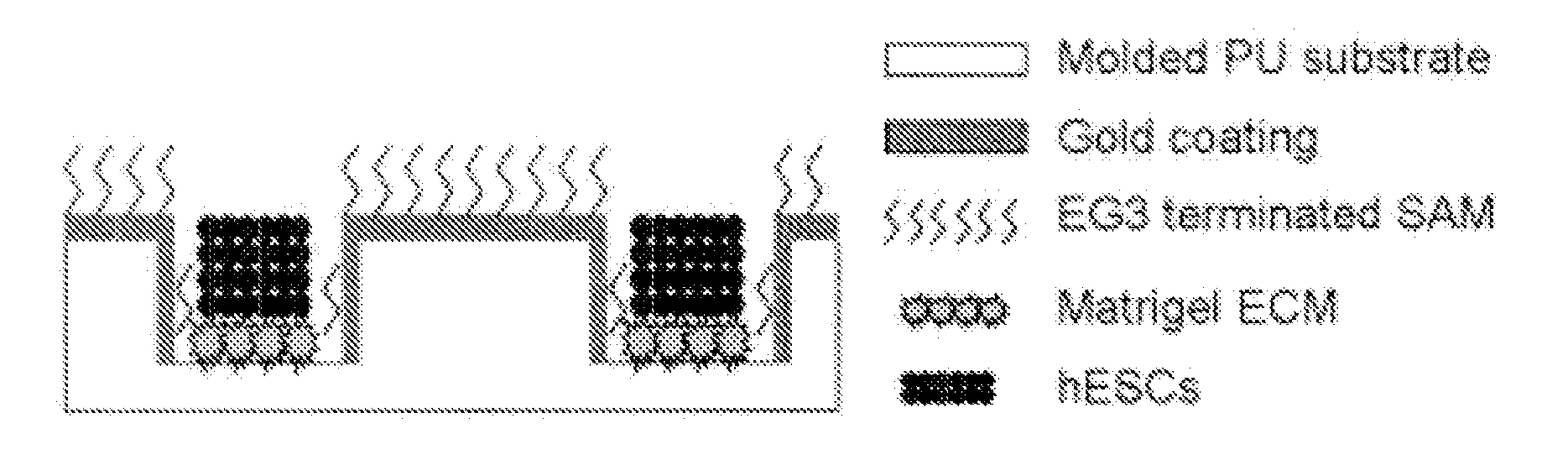
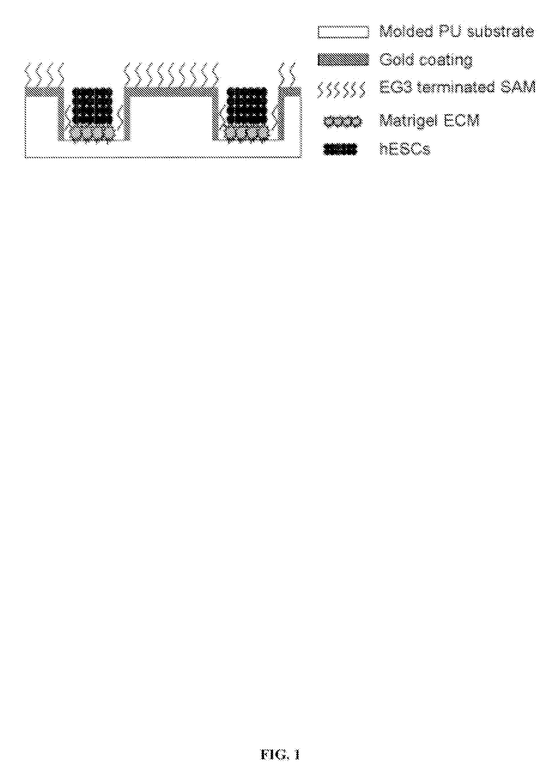
[0027] Reference is made to FIG. 1. Microscope slides having formed thereupon a homogeneous distribution of wells of identical size and shape were constructed in three steps using a polydimethylsiloxane (PDMS) stamp to shape a surface of a UV-crosslinkable polyurethane polymer substrate. First, silicon masters, each having desired microwell patterns formed into a surface thereof, were prepared using photolithography and plasma etching techniques similar to those described by Chen et.al. [22], incorporated herein by reference as if set forth in its entirety. The surfaces were passivated by fluorination with (tridecafluoro-1,1,2,2,-tetrahydrooctyl)-1trichlorosilane vapor. Second, a mixture of PDMS elastomer pre-polymer with curing agent (10:1) (Sylgard 184 Silicon Elastomer; Dow Corning, Midland, Mich.) was poured over silicon masters to form PDMS stamps. The mixture was degassed under vacuum and incubated overnight at 70° C. t...

example 2

Directed Differentiation of hESCs from Microwell Cultures

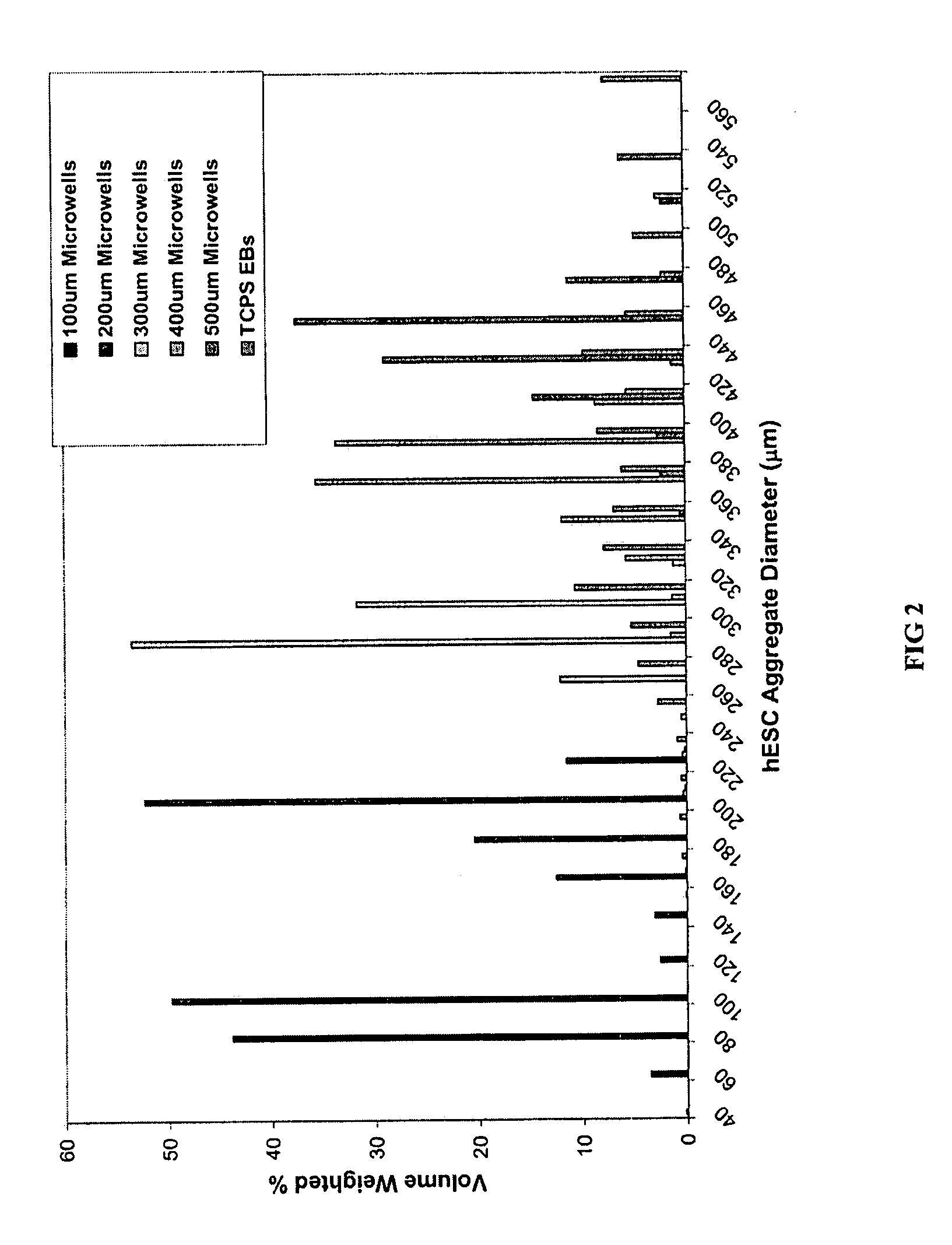
[0046] hESCs were cultured either in microwells having depths of 50 μm-120 μm and lateral dimensions of 50 μm-500 μm or in TCPS plates as described above. Briefly, samples were incubated for 30 minutes at 37° C. to allow hESCs to settle into the microwells before adding 1.5 ml / well CMF+ to the wells of a 6-well plate. The medium was changed daily thereafter and the cells typically reached confluence within a week. TCPS EBs and microwell-derived EBs were then formed as described above.

[0047] Keratinocytes differentiation was achieved by initially forming EBs from microwell-derived hESC aggregates as described above. EBs were grown in suspension for fourteen days in UMF-medium and then attached to gelatin-coated plates in Defined Keratinocyte Serum Free Medium (DSFM) (Invitrogen; Carlsbad, Calif.). Cells were cultured 2-3 weeks prior to analysis by immunocytochemistry and flow cytometry, as described for undifferentiated cells...

example 3

Microwells for hESCs Culture and Cryopreservation

[0051] hESCs were cultured in microwells having dimensions of 50 μm deep with 50 μm-400 μm lateral dimensions as described above. Briefly, samples were incubated for 30 minutes at 37° C. to allow hESCs to settle into the microwells before adding 1.5 ml / well CMF+ to the wells of a 6-well plate. The medium was changed daily thereafter and the cells typically reached confluence within a week. Alternatively, hESCs were cultured in TCPS dishes, as described above.

[0052] After 7 days of culture, the hESCs in microwells, suspensions and TCPS plates were place in freezers at −80° C. for up to 4 weeks. Following cryopreservation, cells frozen in suspension were thawed by immersion of the cryovial in a 37° C. waterbath with agitation. Cells were immediately diluted in 10 ml CMF (i.e. hESC medium conditioned on mouse embryonic fibroblasts with bFGF) and centrifuged. hESCs were then diluted in 2 ml CMF and plated to 1 well of a 6-well plated pr...


PUM

PropertyMeasurementUnit
depthaaaaaaaaaa
depthaaaaaaaaaa
depthaaaaaaaaaa

Abstract

A three-dimensional microwell system that supports long term embryonic stem cell (ESCs) culture and formation of homogeneous embryoid bodies (EBs) is described. Microwell-cultured ESCs remain viable and undifferentiated for several weeks in culture and maintain undifferentiated replication when passaged to Matrigel®-coated, tissue culture-treated polystyrene dishes. Microwell-cultured ESCs maintain pluripotency, differentiating to each of the three embryonic germ layers. ESC aggregates released from microwells can be passaged for undifferentiated replication or differentiated to monodisperse EBs. The ability to constrain ESC growth in three dimensions advantageously provides for more efficient, reproducible culture of undifferentiated cells, high-throughput screening, and the ability to direct ESC differentiation by generating monodisperse EBs of a desired size and shape.

Description

CROSS-REFERENCE TO RELATED APPLICATIONS [0001] This application claims the benefit of U.S. Provisional Patent Application No. 60 / 814 / 975, filed Jun. 20, 2006, incorporated herein by reference as if set forth in it entirety.STATEMENT REGARDING FEDERALLY SPONSORED RESEARCH OR DEVELOPMENT [0002] This invention was made with United States government support awarded by the following agency: NSF DMR-0079983. The United States government has certain rights in this invention.BACKGROUND [0003] Embryonic stem cells (ESCs) can proliferate without limit and can differentiate into each of the three embryonic germ layers [1-3]. To facilitate self-renewal, primate (including human) ESCs are typically co-cultured with mouse embryonic fibroblast (MEF) feeder cells, or cultured in MEF-conditioned medium (MEF-CM) on a Matrigel® extracellular matrix. [0004] Cell microenvironment influences embryonic stem cell (ESC) differentiation [4,5]. For example, spontaneous differentiation of ESC cultures occurs a...

Claims


Application Information

Patent Timeline
Patent Type & Authority Applications(United States)
IPC IPC(8): C12N5/02C12M1/16C12N5/08C12N5/073
CPCC12N5/0603C12N2501/115C12N2502/13C12N2533/90C12N2535/10C12N2533/30
Inventor PALECEK, SEAN P.MOHR, JEFFREY C.DE PABLO, JUAN J.
Owner WISCONSIN ALUMNI RES FOUND
Why Eureka
Try Eureka