Support Accumulating In Injured Part In Vascular Channel

a vascular channel and support technology, applied in the direction of antinoxious agents, drug compositions, extracellular fluid disorders, etc., can solve the problems of hemostatic reaction, morbidity and mortality, and the circulatory system

Inactive Publication Date: 2008-03-13
KEIO UNIV
View PDF3 Cites 2 Cited by
  • Summary
  • Abstract
  • Description
  • Claims
  • Application Information

AI Technical Summary

Benefits of technology

[0008]The present inventors devoted themselves to keen studies to solve the problems mentioned above. As a result, they succeeded in observing behavior of a carri

Problems solved by technology

Thrombus formation, however, is a main cause of circulatory system diseases due to its properties of narrowing a vascular caliber and interrupting a blood flow.
Narrowing of a blood vessel caused by the thrombus and the resultant ischemia could be a main cause of morbidity and mortality.
Furthermore, a hemostatic reaction by thrombus formation is deteriorated by thrombocytopenia associated with the use of cancer drugs or blood disorders such as leukemia or by deterioration of blood coagulation factor associated with liver disorder and results in diapedetic hemorrhage from a space between vascular endothelial cells, under such circumstance bleeding in the brain blood vessels

Method used


Image

  • Support Accumulating In Injured Part In Vascular Channel
  • Support Accumulating In Injured Part In Vascular Channel
  • Support Accumulating In Injured Part In Vascular Channel

Examples


example 1

Preparation of Liposome

[0125]One g of mixed lipid powder (DPPC (dipalmitoylphosphatidylcholine) / cholesterol / DPEA / PEG-DSP (N-(monomethoxypolyethyleneglycol-carbamyl)distearoylphosphatidyl ethanolamine)=5 / 5 / 10 / 0.033 (molar ratio)) was dispersed in 25 mL of saline and agitated at room temperature for 10 hours. The resultant was passed through a filter with a pore diameter of 0.22 μm twice in an extruder to obtain liposome dispersion with an average particle size of 263±43 nm. Lipid concentration in saline was 3 g / dL. To 20 ml of this dispersion, separately prepared Octadecyl Rhodamine B (from Molecular Probe) in ethanol solution (1 mg / 1 mL) was dropped for 600 μL while agitating, and further subjected to 2 hours of agitation with light being shielded. Liposomes were precipitated by ultracentrifugation (100,000 g, 60 min.+150,000 g, 30 min.). A slight amount of unintroduced dye and ethanol remaining in the supernatant were removed. Saline was added to redisperse the vesicles and the res...

example 2

Analysis of Liposome Behavior by Image Analysis Method

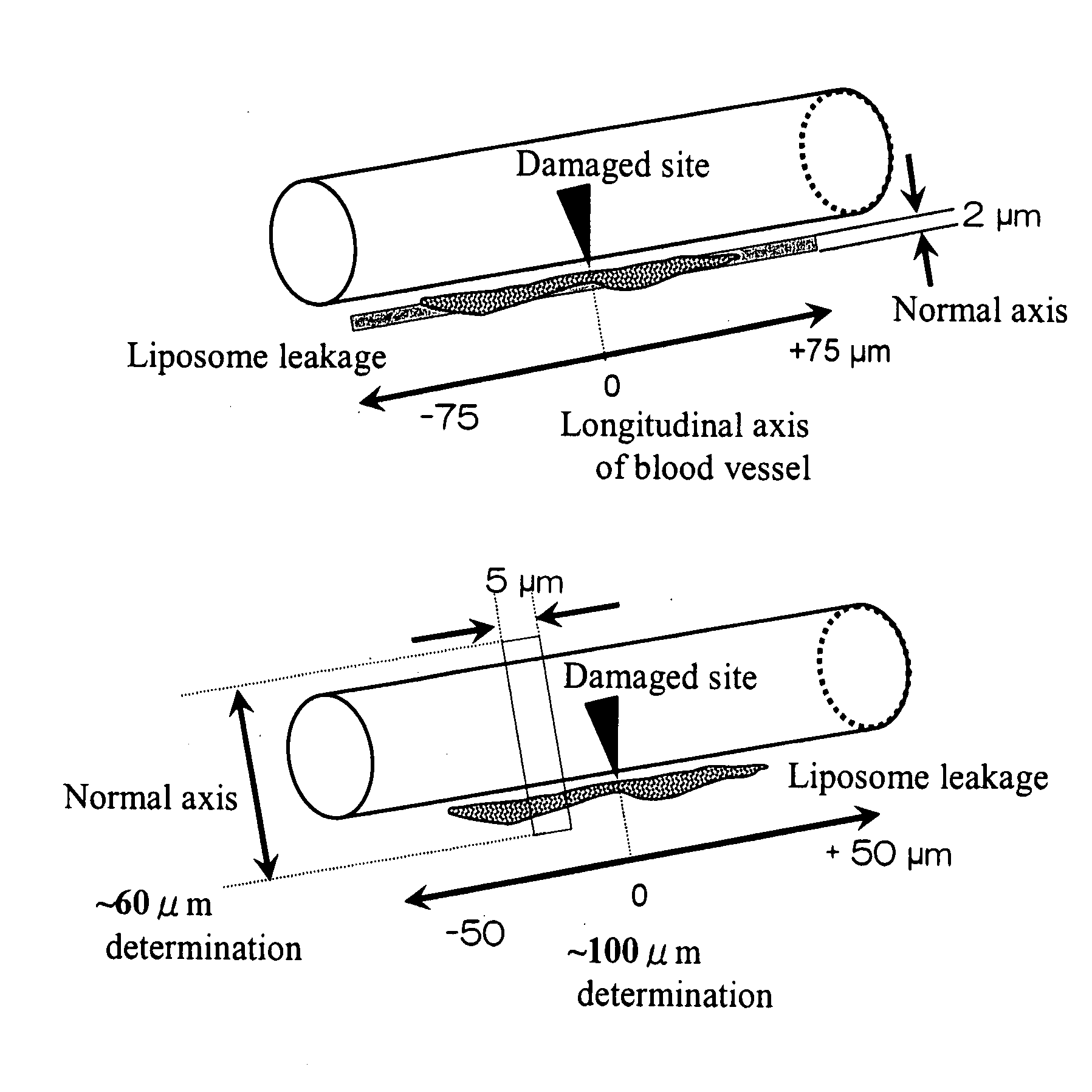
[0127]1. Biomicroscopic System and Microcirculation Observing / Recording System

[0128]Rats were bred under the control of the Laboratory Animal Center, School of Medicine, Keio University. According to the method of Suematsu et al. (Suematsu M, et al. Lab Invest 1994), male Wistar rats (200-250 g; Clea Japan, Tokyo) were anesthetized by intramuscular injection of pentobarbital sodium at 50 mg / kg, and a catheter was inserted into a femoral vein to administer carboxyfluorescein diacetate succinimidyl ester (CFDA-SE) at 1 mg / kg for biostaining of the platelets in the body.

[0129]The mesenterium at the ileocecal region was opened extraperitoneally to observe the microcirculatory system with an erecting-type confocal laser microscope.

[0130]A 60× objective lens was used. Within the light path of the microscope are implemented an argon laser capable of laser output at 488 nm that allows imaging of the CFSE fluorescence and laser output at ...

example 3

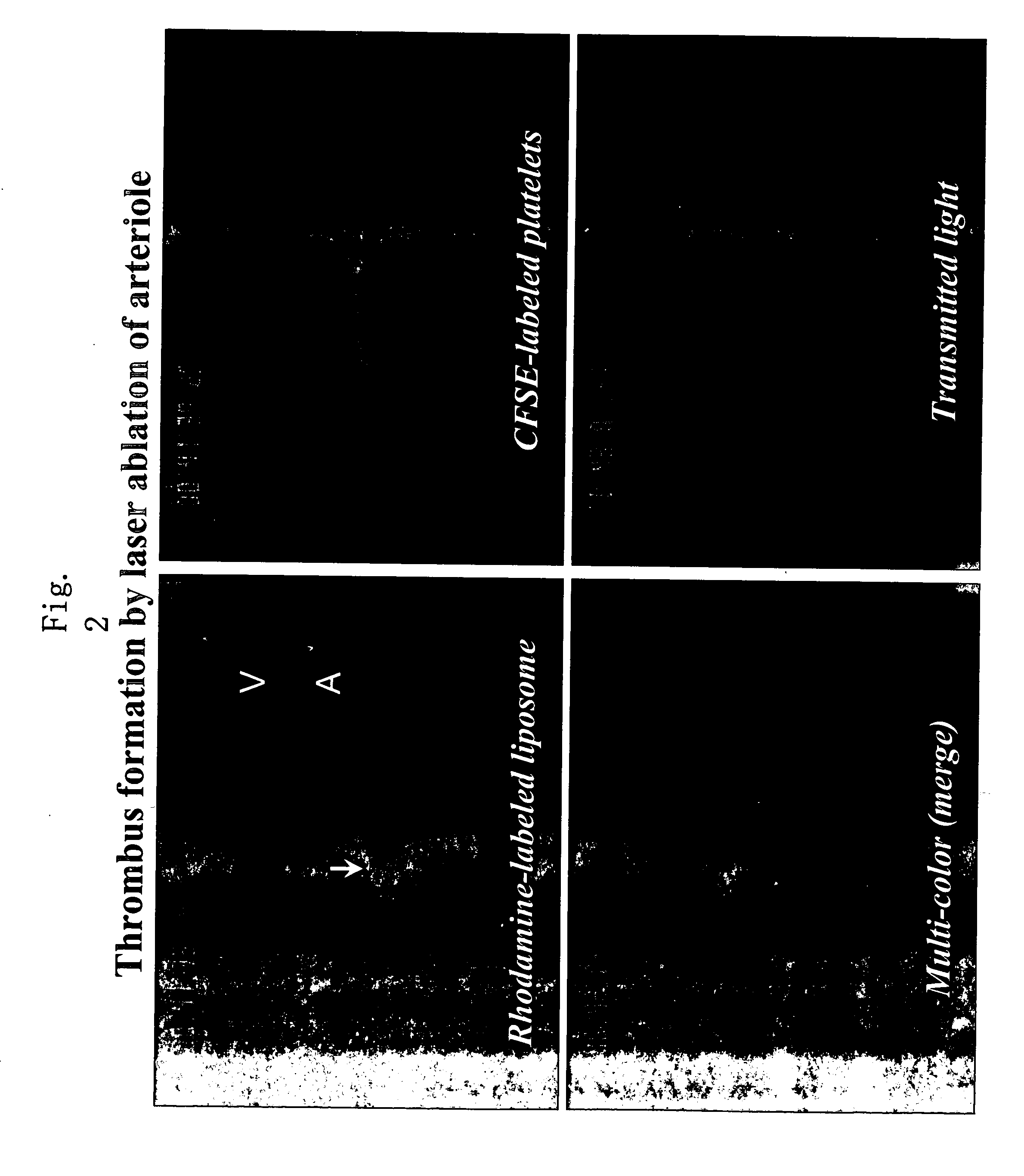
Liposome Behavior under Inflammation Stimulation

[0145]The present example aims at revealing liposome behavior upon stimulation of mesenterium with a general inflammation substance instead of mechanical stimulation. Histamine was used as the inflammation stimulating substance. In the same manner as the method of Example 2, male Wistar rats (200 to 250 g) were anesthetized by intramuscular injection of pentobarbital sodium at 50 mg / kg, and a catheter was inserted into a femoral artery. The mesenterium at the ileocecal region was opened extraperitoneally to observe the microcirculatory system with an erecting-type confocal laser microscope. A 60× objective lens was used.

[0146]The liposome dispersion prepared according to the method in Example 1 was infused into the femoral vein via the catheter for 250 μl per rat weighing 100 g (250 μl / 100 g rat, where proportion of liposome in the dispersion was 35% by volume (estimated number of liposomes in the sample being 4.8×10¹² / ml)).

[0147]Ten m...


PUM


Abstract

The present invention provides a carrier that accumulates on a damaged tissue and that functions as a drug for controlling a platelet function, a drug delivery method using the carrier and a pharmaceutical composition comprising the carrier. The present invention also provides a carrier with a non-cationic surface, a drug transporter and a pharmaceutical composition comprising the carrier.

Description

FIELD OF THE INVENTION[0001]The present invention relates to a non-cationic carrier that attaches to and accumulates on a damaged tissue and fills in the damaged site, to a pharmaceutical composition comprising such carrier incorporating or carrying a drug, and to a drug delivery method.BACKGROUND OF THE INVENTION[0002]Thrombus formation is an essential function in an organism in that it closes wound and stops bleeding. Thrombus formation, however, is a main cause of circulatory system diseases due to its properties of narrowing a vascular caliber and interrupting a blood flow. For example, when the outer wall of the blood vessel is damaged, thrombus formation plays a role in a biological defense function for maintaining the perfusion pressure necessary for blood circulation. On the other hand, damage to vascular endothelial cells caused, for example, by peroxidative response causes arteriosclerosis, and significant development of such arteriosclerosis induces thrombus formation. Na...

Claims


Application Information

Patent Timeline
IPC IPC(8): A61K9/127
CPCA61K9/1272A61P29/00A61P31/00A61P35/00A61P39/06A61P7/00A61P7/02A61P7/04A61P7/10A61P9/10A61K47/24A61K9/127
Inventor SUEMATSU, MAKOTOTAKEOKA, SHINJI
Owner KEIO UNIV
Why Eureka
Try Eureka