
Devices containing DNA encoding neurotrophic agents and related compositions and methods

a neurotrophic agent and dna technology, applied in the field of neurotrophic agent treatment, can solve the problems of preventing reconnection of neural pathways, affecting axonal regrowth following ns injury, and threatening neuronal survival, so as to promote neuronal survival, promote neuronal survival, and effectively transfer

Inactive Publication Date: 2008-06-26
TISSUE REPAIR CO +2
View PDF99 Cites 3 Cited by
  • Summary
  • Abstract
  • Description
  • Claims
  • Application Information

AI Technical Summary

Benefits of technology

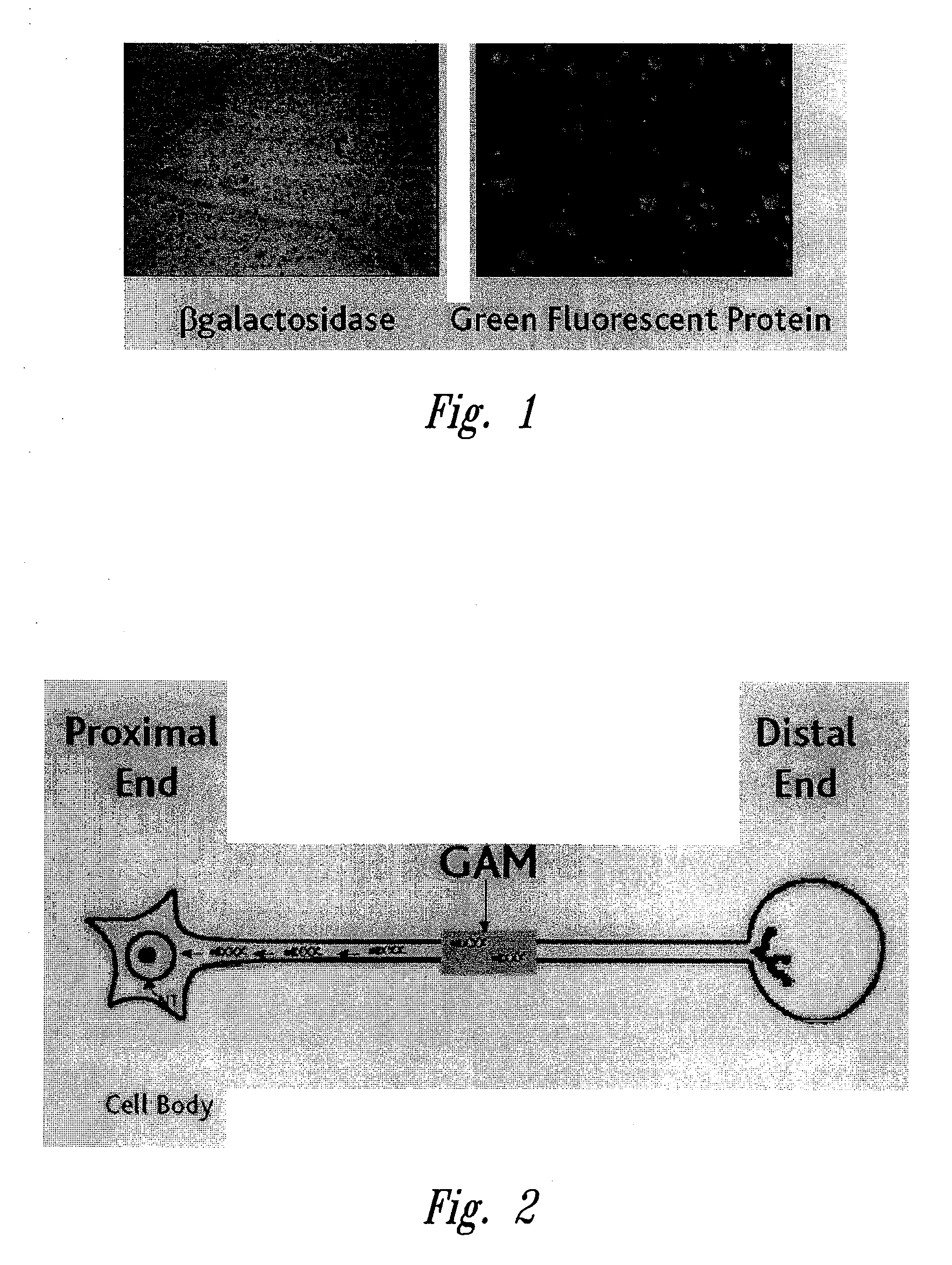
[0012]Interruption of neural connections may be a consequence of acute or chronic NS injury leading to physical severance of axons that threatens neuronal survival, as described above. Accordingly, the compositions and methods of the present invention may delay cell degeneration and cell death by restoring the continuous retrograde flow of neurotrophic factors, from distal neuronal targets to neuronal perikarya, that is essential for maintenance of neural networks.
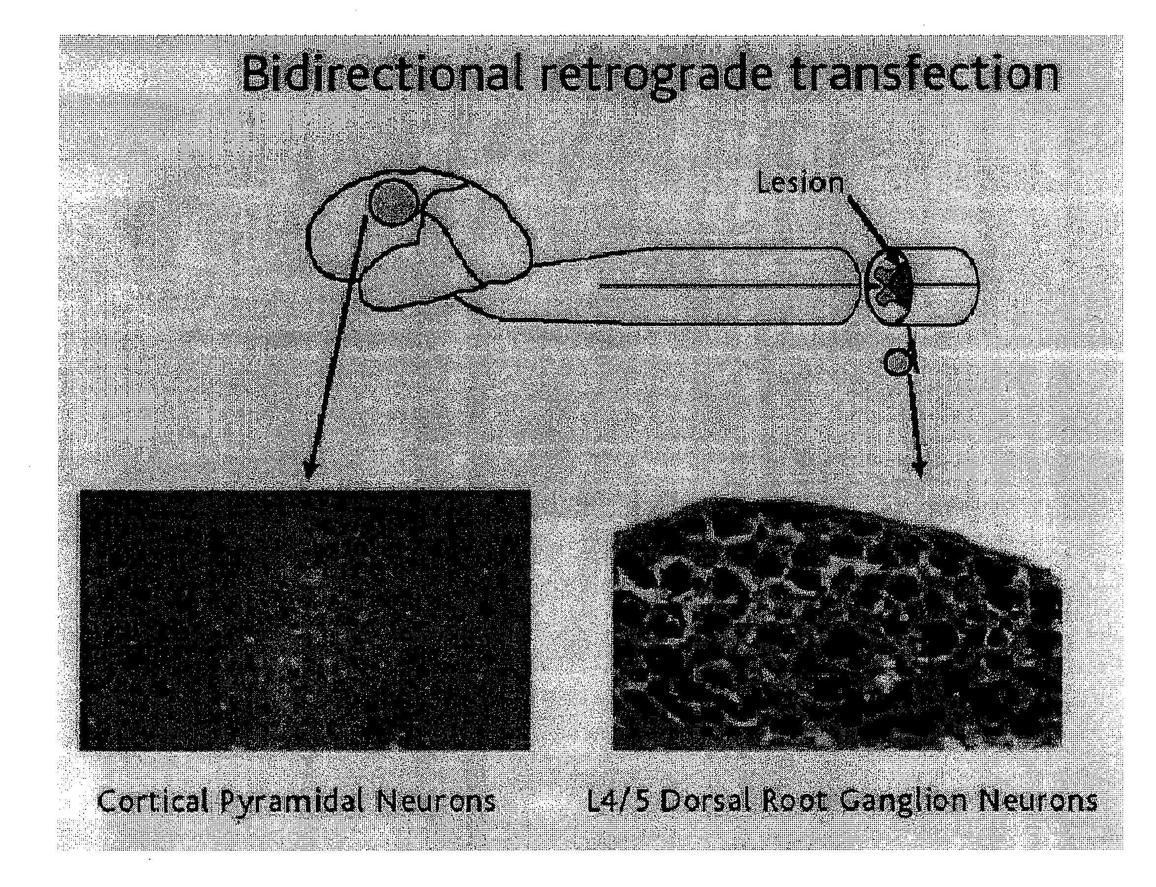
[0024]Turning to another aspect of the invention, a method is provided for transferring a neuronal therapeutic encoding agent into a neuronal cell, comprising contacting a neuronal cell with any one of the devices just described to effectively transfer the neuronal therapeutic encoding agent into the neuronal cell. In one embodiment, transfer of the neuronal therapeutic encoding agent comprises retrograde axonal transport of the neuronal therapeutic encoding agent. In another embodiment the method further comprises expression of the neuronal therapeutic encoding agent at a neuronal cellular site distinct from a site of contact between the device and the neuronal cell. In another embodiment, the device is contacted with a neuronal cell at a neuronal injury site. In another embodiment, the device is contacted with a neuronal cell in a manner such that axonal generation or regeneration occurs. In a further embodiment, axonal regeneration occurs without axonal entrapment. In another embodiment, the device is contacted with a neuronal cell in a manner that promotes neuronal survival. In a further embodiment, neuronal survival is promoted without axonal entrapment. In certain further embodiments a neural connection is established or reestablished.

Problems solved by technology

Interruption of neural connections by physical severance of axons disconnects neuron from target and threatens neuronal survival.
Because of the spatiotemporal regulation of cues, including neurotrophic factors, essential for the maintenance of neural networks, axonal regrowth following NS injury is impaired by the absence of one or more appropriate stimuli in the vicinity of the damaged neuron.
Consequently, reconnection of neural pathways is prevented and functional recovery may be compromised.
These methods often produce localized sinks of high neurotrophin concentration at the lesion site in which axons may become entrapped.
Thus, axonal extension beyond the lesion and along the damaged projection tracts may be impossible.
Failure to re-establish neural connections and the ensuring neuronal atrophy may result in complete loss of function.
Depending on the viral vector construct and delivery vehicle used, such approaches may under certain circumstances, (i) elicit inappropriate antiviral immune responses, (ii) promote undesirable viral toxic effects, (iii) have limited efficacy due, for example, to inefficiency of genetically altered viral gene promoter sequences, (iv) be tumorigenic and / or (v) lack specificity regarding the cell type to which therapeutic genes are delivered.
Poor targeting of such recombinant viral vectors to specific cell types, for example, may limit the value of such an approach and may establish localized accumulations of therapeutic gene products at the site of vector delivery, giving rise to the problems associated with localized growth factor sinks and axonal entrapment.

Method used


Image

  • Devices containing DNA encoding neurotrophic agents and related compositions and methods
  • Devices containing DNA encoding neurotrophic agents and related compositions and methods
  • Devices containing DNA encoding neurotrophic agents and related compositions and methods

Examples


example 1

Preparation of Gene Activated Matrix Containing FGF2-Poly-L-Lysine Complexed with a Plasmid Encoding GFP Protein (GFP) Reporter Gene Under Promoter Regulation

[0276]Plasmid isolation, production of competent cells, transformation and manipulations using the M13 cloning vectors are performed as described (Sambrook et al., Molecular Cloning, a Laboratory Manual, Cold Spring Harbor Laboratory Press, Cold Spring Harbor, N.Y., 1989). DNA fragments are purified using the Geneclean II kit, purchased from Bio 101 (La Jolla, Calif.). Recombinant DNA constructs are sequenced using the Sequenase kit (version 2.0, United States Biochemical, Cleveland, Ohio) according to the manufacturer's instructions. Conjugation of FGF2 to poly-L-lysine K84 homopolymer to produce FGF2-K is as described by Sosnowski et al. (1996 J. Biol. Chem. 271:33647-33653). Preparation of FGF2-poly-L-lysine complexed with a plasmid encoding green fluorescent protein (GFP) under CMV promoter regulation is also essentially as...

example 2

Preparation of DNA Construct Containing the Neuronal GAP43 Promoter

[0280]Plasmid isolation, production of competent cells, transformation and manipulations using the M13 cloning vectors are performed as described (Sambrook et al., Molecular Cloning, a Laboratory Manual, Cold Spring Harbor Laboratory Press, Cold Spring Harbor, N.Y., 1989). DNA fragments are purified using the Geneclean II kit, purchased from Bio 101 (La Jolla, Calif.). Recombinant DNA constructs are sequenced using the Sequenase kit (version 2.0, United States Biochemical, Cleveland, Ohio) according to the manufacturer's instructions. DNA containing the human GAP43 promoter sequence (Genbank accession number X840768) is obtained as described in de Groen et al. (J. Mol. Neurosci. 6:109-119, 1995) and incorporated into plasmids in operative linkage with reporter gene encoding or neuronal therapeutic agent encoding sequences.

example 3

Delivery and Expression of Targeted GFP Gene in Lesioned Rat Optic Nerve Repair Model

[0281]In this example, targeted delivery of the GFP reporter gene to rat optic nerve neurons is conducted using a ligand as a molecular targeting agent in an in vivo model of neuronal regeneration.

[0282]Transgene expression of neuronal cells in vivo following experimentally induced axonal lesion is monitored in the rat optic nerve repair model. See, e.g., Logan et al., Meth. Neurosci. 21:3-19, 1994, which is hereby incorporated by reference in its entirety. Adult rats are anesthetized by intraperitoneal injection of physiological saline solution containing ketamine (40 mg / kg), acepromazine (1.2 mg / kg) and xylazine (8 mg / kg). The optic nerve is accessed intraorbitally by a dorsolateral approach and severed by transection using manual pressure applied with surgical forceps. (Berry et al., J. Neurocytol. 25:147-170, 1996). Care is taken to avoid damaging the central retinal artery or the optic nerve sh...


PUM

PropertyMeasurementUnit
lengthaaaaaaaaaa
inner diameteraaaaaaaaaa
diameteraaaaaaaaaa

Abstract

Devices useful in the delivery of DNA encoding neurotrophic agents, anti-fibrotic agents, and related compositions are disclosed herein for use in the treatment of central and / or peripheral nervous system injury. Methods of making and using the disclosed devices and DNA are also described. In various embodiments, the invention also discloses compositions and devices that may further include a targeting agent, such as a polypeptide that is reactive with an FGF receptor (e.g., bFGF), or another ligand that binds to cell surface receptors on neuronal cells, or a support cell. The invention also discloses methods of promoting neuronal survival and regeneration via transfection of an axon as it grows through a device or composition of the present invention, or via transfection of a repair cell.

Description

CROSS-REFERENCE TO RELATED APPLICATIONS[0001]The application is a continuation-in-part of U.S. application Ser. No. 09 / 088,419, filed Jun. 1, 1998, which is hereby incorporated by reference.TECHNICAL FIELD[0002]The present invention relates generally to the treatment of neurons following NS injury that may result from surgery, trauma, compression, contusion, transection or other physical injury, from vascular pharmacologic or other insults including hemorrhagic or ischemic damage or from neurodegenerative or other neurological diseases. More specifically, the invention relates to the preparation and use of devices for transferring neuronal therapeutic agents and / or DNA encoding neuronal therapeutic agents into the NS, including devices that are gene activated matrices, to alter the function, gene expression or viability of neuronal cells therapeutically. The invention further relates to administration of such devices, including administration of matrices containing useful genes.BACK...

Claims


Application Information

Patent Timeline
Patent Type & Authority Applications(United States)
IPC IPC(8): A61K9/00A61K35/12A61P25/00C12N15/09A61K35/15A61K35/30A61K38/00A61K38/16A61K38/17A61K38/18A61K38/45A61K41/00A61K47/48A61K48/00A61P43/00C07K14/415C07K14/475C07K14/50C07K14/52C12N15/62C12N15/87
CPCA61K9/0024A61K35/15A61K35/30A61K38/00A61K38/1841A61K38/185A61K41/0042A61K47/42A61K48/00A61K48/0008A61K48/0058A61K48/0075C07K14/415C07K14/475C07K14/503C07K14/52C07K2319/00C07K2319/03C07K2319/04C07K2319/09C07K2319/21C07K2319/50C07K2319/55C07K2319/75C07K2319/80C12N15/62C12N15/85C12N15/87C12N2799/026C12N2800/107C12N2810/851C12N2830/008A61K38/1709A61K38/179Y10S977/927Y10S977/912A61K47/642A61K47/6425A61P25/00A61P43/00
Inventor BAIRD, ANDREWGONZALEZ, ANA MARIABERRY, MARTINLOGAN, ANN
Owner TISSUE REPAIR CO
Learn More