
Drugs And Gene Carrier Particles That Rapidly Move Through Mucous Barriers

a gene carrier and drug technology, applied in the direction of peptides, drug compositions, aerosol delivery, etc., can solve the problems of impede the transport rate of large macromolecules and nanoparticles, and achieve the effect of promoting adhesion or complexation

Active Publication Date: 2008-07-10
THE JOHNS HOPKINS UNIVERSITY SCHOOL OF MEDICINE
View PDF48 Cites 205 Cited by
  • Summary
  • Abstract
  • Description
  • Claims
  • Application Information

AI Technical Summary

Benefits of technology

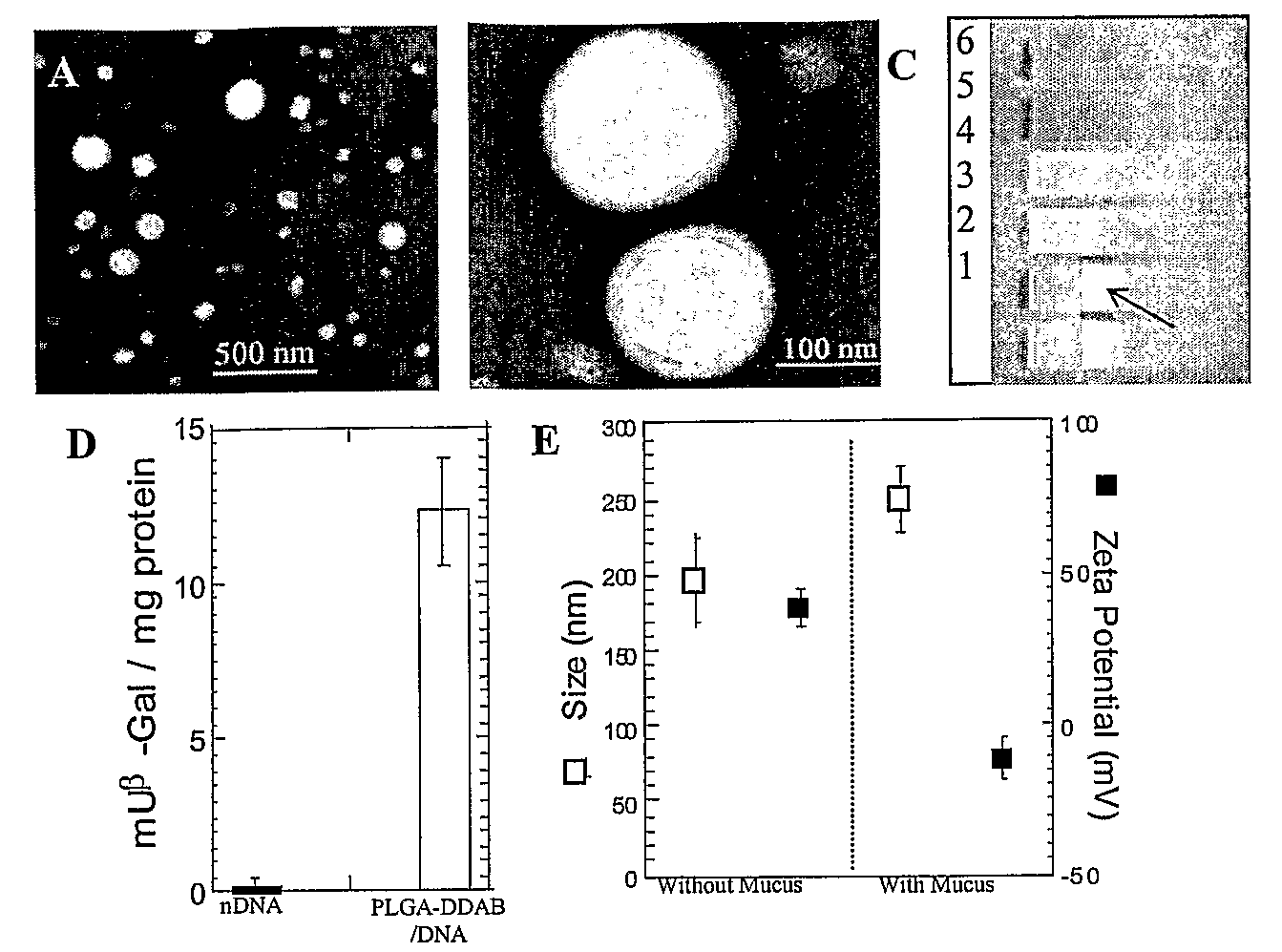
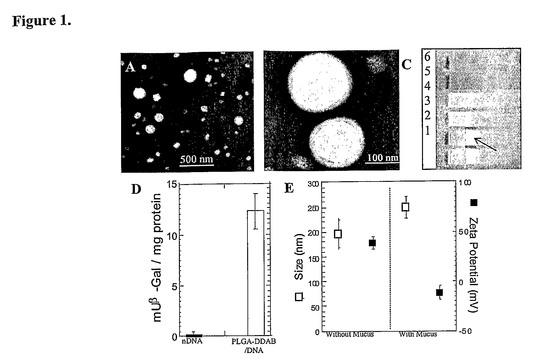
[0004]In one aspect, a polymeric particle comprises a pharmaceutically acceptable polymer core and a surface-altering agent that is embedded or enmeshed in the particle's surface or that is disposed (e.g., by coating, adsorption, covalent linkage, or other process) on the surface of the particle. The surface-altering agent may be a bioactive agent itself. For example, in certain embodiment, a polymeric particle may comprise a pharmaceutically acceptable polymer and a nucleic acid coating the surface of the particle. In such embodiments, the nucleic acid molecule may alter the surface of the particle and make it mucus resistant. In certain other embodiments, a polymeric particle comprises a pharmaceutically acceptable polymer and a protein (e.g., serum albumin) disposed on the surface of the particle. The protein may alter the surface of the particle and make it mucus resistant. In any of these embodiments, the polymeric particle may comprise an imaging agent, e.g., that may include a diagnostic agent and / or a detectable label. For example, the nucleic acid or the protein included in the polymeric particle may comprise an imaging agent itself, e.g., a detectable label can be attached to the DNA or the protein. Alternatively, the polymeric particle may comprise an imaging agent that is separate from the nucleic acid or the protein, e.g., encapsulated in the core or disposed on or coupled to the surface. Additionally, the polymeric particle may comprise a targeting moiety or molecule coupled to the polymeric particle and / or the protein or nucleic acid, and the targeting moiety can help deliver the nucleic acid, the protein, and / or the imaging or diagnostic agent to a targeted location in a patient.
[0006]Alternatively, a polymeric particle may comprise a pharmaceutically acceptable polymer core, a surface-altering agent, e.g., a surfactant, that is embedded or enmeshed in the particle's surface, or disposed (e.g., by coating, adsorption, covalent linkage, or other process) on the surface of the particle and that alters the surface of the particle, such as by making it mucus-resistant, and a bioactive agent disposed on the polymeric particle. The bioactive agent may be coated or otherwise disposed on the surface of the polymeric particle, or be coupled to the polymeric particle by, e.g., covalent linkage, complexation, or other process. In certain such embodiments, the surface-altering agent is selected to promote adhesion or complexation of the bioactive agent to the surface of the particle. In such embodiments, the surface-altering agent and / or the bioactive agent may contribute to mucus-resistant properties of the modified particles. The polymeric particles may comprise an imaging agent, such as a diagnostic agent and / or a detectable label. The bioactive agent coated or disposed on the surface of the particle or coupled to the particle may be or comprise an imaging agent itself, e.g., a detectable label can be attached to a therapeutic agent. Alternatively, the polymeric particle may comprise an imaging agent that is separate from the bioactive agent, e.g., encapsulated in the core or disposed on or coupled to its surface. Additionally, the polymeric particle may comprise a targeting moiety or molecule coupled to the polymeric particle, and the targeting moiety can help deliver the bioactive agent and / or the imaging agent to a targeted location in a patient.
[0009]The surface-altering agent may increase charge or hydrophilicity of the polymeric or liposomal particle, or otherwise decrease interactions between the particle and mucus, thereby promoting motility through mucus. The surface-altering agent may enhance the average rate at which the polymeric or liposomal particles, or a fraction of the particles, move in or through mucus. Examples of suitable surface-altering agents include but are not limited to anionic protein (e.g., serum albumin), nucleic acids, surfactants such as cationic surfactants (e.g., dimethyldioctadecyl-ammonium bromide), sugars or sugar derivatives (e.g., cyclodextrin), polyethylene glycol, mucolytic agents, or other non-mucoadhesive agents. Certain agents, e.g., cyclodextrin, may form inclusion complexes with other molecules and can be used to form attachments to additional moieties and facilitate the functionalization of the particle surface and / or the attached molecules or moieties.

Problems solved by technology

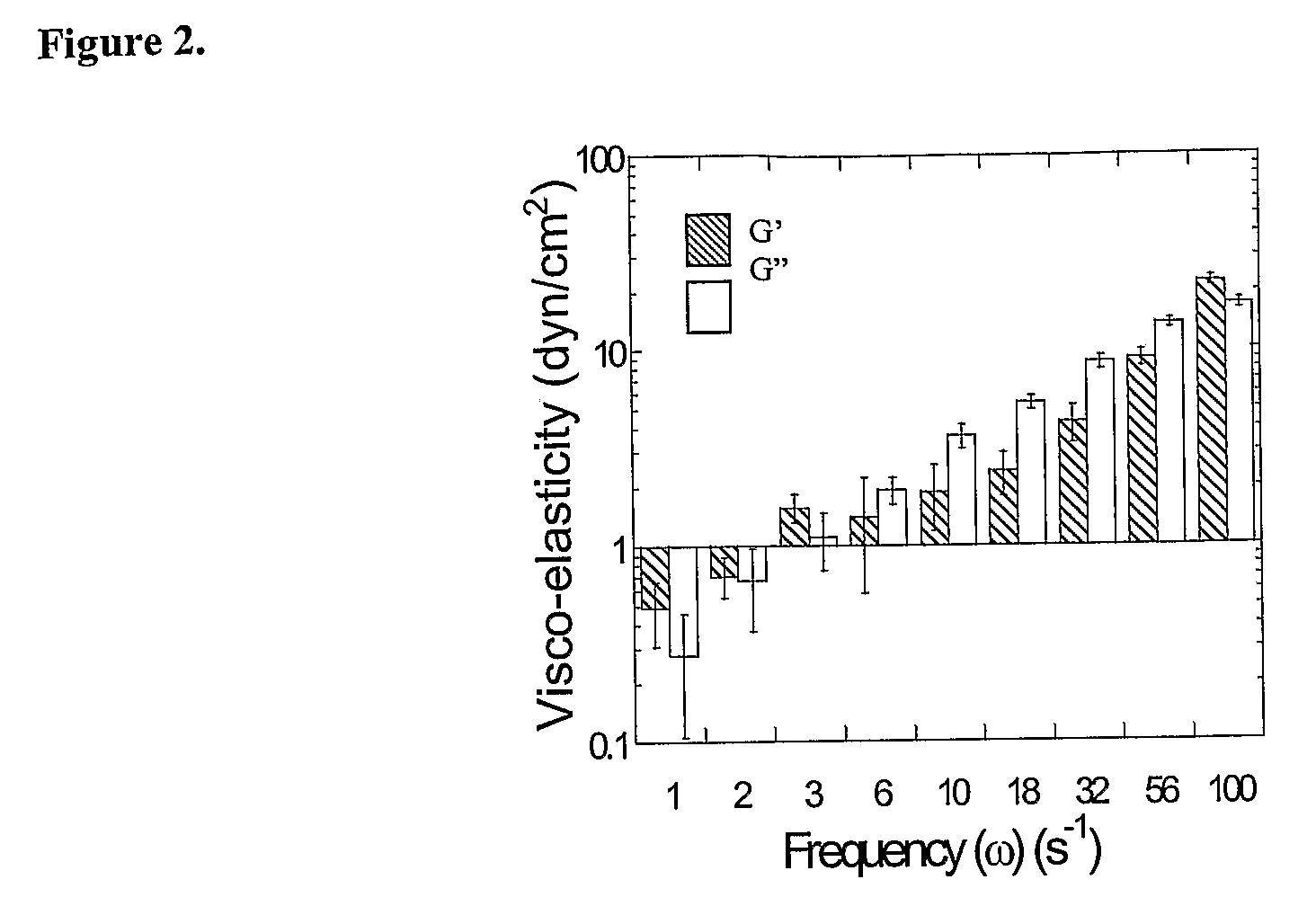
The condensed and complex microstructure of the mucus network gives rise to a highly viscoelastic gel, which significantly impedes the transport rates of large macromolecules and nanoparticles.

Method used


Image

  • Drugs And Gene Carrier Particles That Rapidly Move Through Mucous Barriers
  • Drugs And Gene Carrier Particles That Rapidly Move Through Mucous Barriers
  • Drugs And Gene Carrier Particles That Rapidly Move Through Mucous Barriers

Examples


Embodiment Construction

1. Overview

[0038]Cationic microparticles with DNA adsorbed to their surfaces have been shown to efficiently transfect cells in vitro. E.g., Singh et al., Proc Natl Acad Sci USA 2000, 97, 811-816; Singh et al., Pharm Res 2001, 18, 1476-1479; Denis-Mize et al., Gene Ther 2000, 7, 2105-2112. However, obtaining high transfection efficiencies in vivo is often limited by particle transport through extracellular barriers, including the mucosal barrier, which has been described as the foremost barrier to transfection in mucus-covered cells. See, e.g., Ferrari et al., Gene Ther 2001, 8, 1380-1386; Yonemitsu et al., Nat Biotechnol 2000, 18, 970-973.

[0039]High MW poly(ethylene glycol) (PEG) has been used as a mucoadhesive added to polymeric systems for its reported ability to interpenetrate into the mucus network (Bures et al., J. Controlled Release, (2001) 72:25-33; Huang et al., J. Controlled Release, (2000) 65:63-71; Peppas et al., J. Controlled Release, (1999) 62:81-87) and hydrogen bond t...


PUM

PropertyMeasurementUnit
diameteraaaaaaaaaa
diameteraaaaaaaaaa
diameteraaaaaaaaaa

Abstract

The invention generally relates to polymeric particles suitable for transporting bioactive agents across mucosal barriers. The invention also relates to methods of making and using those polymeric particles.

Description

GOVERNMENT FUNDING[0001]The work related to this application has been funded by the National Science Foundation (CTS0210718 and fellowship funding).BACKGROUND[0002]The primary component of mucus is higher molecular weight mucin glycoproteins, which form numerous covalent and noncovalent bonds with other mucin molecules and various constituents, including DNA, alginate, and hyaluronan. Hanes et al., Gene therapy in the lung. in Pharmaceutical Inhalation Aerosol Technology, 2d ed.; Marcel Dekker Inc.: New York, 2003; pp. 489-539. Reconstituted mucus formulated from pig gastric, human cervical, and tracheobronchial mucins display similar mucus structures, with large rod or fiberlike aggregates of 5 nm in diameter and 100-5000 nm in length. Khanvilkar et al., Adv. Drug Deliv. Rev. 2001 48, 173-193. The condensed and complex microstructure of the mucus network gives rise to a highly viscoelastic gel, which significantly impedes the transport rates of large macromolecules and nanoparticle...

Claims


Application Information

Patent Timeline
Patent Type & Authority Applications(United States)
IPC IPC(8): A61K9/14A61K31/7088A61K38/00C12N5/02A61M15/00A61K9/16A61K9/50A61K9/51A61K47/48
CPCA61K9/0073A61K9/167A61K9/5031A61K9/5123B82Y5/00A61K47/48046A61K47/48907A61K47/48915A61K9/5153A61K47/543A61K47/6935A61K47/6937A61K9/5146A61K48/0033A61K48/0041
Inventor HANES, JUSTINDAWSON, MICHELLE ROSEWIRTZ, DENISFU, JIEKRAULAND, ERIC MARK
Owner THE JOHNS HOPKINS UNIVERSITY SCHOOL OF MEDICINE
Learn More