Material and Methods for Nerve Grafting

a nerve grafting and material technology, applied in the field of nerve grafting materials and methods, can solve the problems of pain, neuronal death, muscle atrophy and permanent functional deficit, and the inability to achieve full restoration of function after nerve transection repair, so as to improve the ability of regenerating axons, promote the repair of damaged nerve tissue, and increase the number of axons

Inactive Publication Date: 2008-12-04
UNIV OF FLORIDA RES FOUNDATION INC
View PDF38 Cites 0 Cited by
  • Summary
  • Abstract
  • Description
  • Claims
  • Application Information

AI Technical Summary

Benefits of technology

[0020]The present invention also concerns methods to promote the repair of damaged nerve tissue in a human or animal. Methods of the present invention comprise administering one or more CSPG-degrading enzymes to a nerve repair, coaptation, graft, or damaged nerve tissue. The methods of the subject invention improve the ability of regenerating axons to traverse the nerve-nerve or nerve-graft interface and potentiates axonal growth within the basal lamina scaffold. The degradation of inhibitory CSPG creates a more permissive nerve substratum and allows axon sprouts greater access to Schwann cell basal lamina of the nerve, thereby increasing the number of axons that successfully penetrate damaged nerve tissue or implanted nerve grafts. The appropriate routing of the axon sprouts may also be enabled leading to further improvements in recovery of function.

Problems solved by technology

Poor management of nerve injuries is associated with muscle atrophy and can lead to painful neuroma when severed axons are unable to reestablish continuity with the distal nerve.
Regenerating axons that fail to traverse the gap or injury site and enter the basal lamina of the severed distal nerve segment will deteriorate, resulting in neuronal death, muscle atrophy and permanent functional deficit (Fawcett J W et al.
Full restoration of function after repair of nerve transection remains an unobtainable ideal because of the fine microstructure of nerves and an inability to achieve precise axon-to-axon coaptation, despite the current state of the art in microsurgical techniques.
Nerve grafting is warranted with nerve ablation but presents several practical challenges.
While the availability of donor grafts suffers the difficulties of other organ replacement strategies, the importance of viable cellular elements in nerve grafts may be far less important.
However, while normal (uninjured) nerve is rich in laminin, normal nerve remains inhibitive or refractory to axonal growth.
Loss of function associated with nerve injury results from axon disruption.
Axons are very thin and fragile and the slightest injury (including compression) can cause a severing response (axotomy).
Transection injuries are those in which both axons and the nerve sheaths are severed, disrupting the continuity of the nerve and the guidance mechanisms required for axon regeneration.
In addition, axonal regrowth after nerve transection and repair is further complicated by the misalignment of proximal and distal elements.
Even in the instances of clean transection by a sharp instrument, the entire nerve structure is disrupted.
Swelling and axoplasmic outflow from the cut ends causes a mushrooming effect which interferes with accurate coaptation and realignment of the basal lamina scaffolding.
Consequently, any misalignment of nerve microstructure (after injury and repair) forces regenerating axonal sprouts to negotiate nonpermissive tissues which may severely limit their access to basal laminae in the distal nerve.
However, the procedure for creating pre-degenerated nerves in humans is impractical (i.e., nerve injury followed by a period of survival in vivo to allow tissue degeneration).
However, in vivo studies testing predegenerated nerve grafts have produced conflicting results, especially when using cellular (live) nerve grafts (Gordon et al.
Although not beneficial for optimizing the growth-promoting potential of nerve grafts, continued cold storage does further decrease cell viability, immunogenicity and the concerns of immunorejection of allogenic nerve grafts (Evans et al.

Method used


Image

  • Material and Methods for Nerve Grafting
  • Material and Methods for Nerve Grafting
  • Material and Methods for Nerve Grafting

Examples


example 1

Degradation of CSPG by Treatment of Acellular Nerve Segments with Chondroitinase

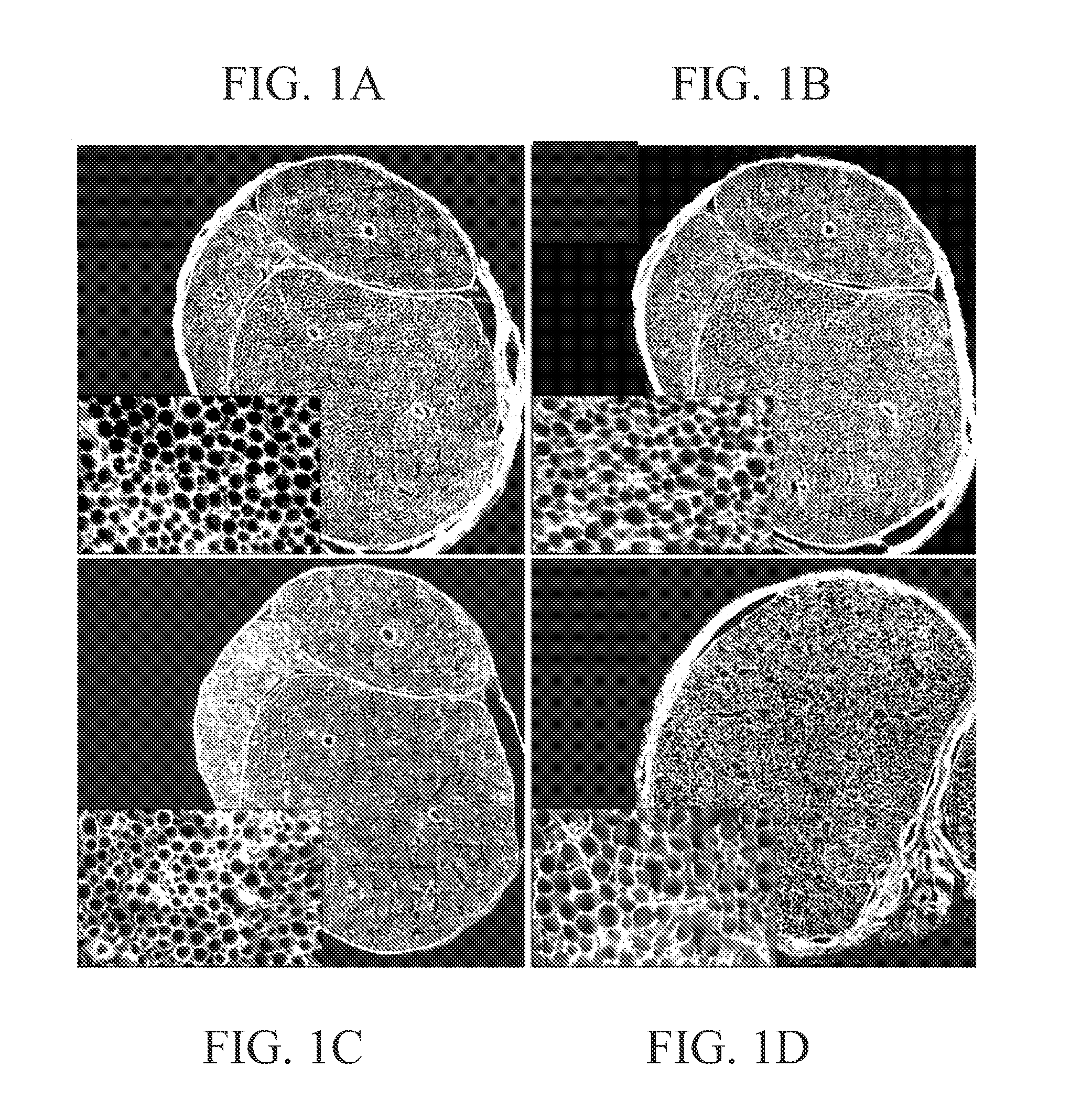
[0125]The purpose of this experiment was to determine if chondroitinase treatment effectively degraded CSPG throughout intact segments of acellular nerves. Segments of rat sciatic nerve (1.5 cm in length) were made acellular by repeated freeze-thaw cycles and then bathed en bloc in a chondroitinase ABC solution for 16 hours. CSPG degradation within the chondroitinase pretreated nerves was examined by immunolabeling with neoepitope antibody Ab1918. This antibody binds to an epitope created on the core protein after lysis of the chondroitin sulfate chains by chondroitinase ABC (Bertolotto et al. [1986] J. Neurol Sci, 73:233-244). Ab1918 immunostaining was intense throughout the entire pretreated nerve segment, as shown in FIG. 1A. Furthermore, the intensity of Ab1918 immunostaining was not increased by an additional post-treatment of the sections with chondroitinase, as shown in FIG. 1B. Ab1918 immunoreact...

example 2

Inactivation of Inhibitory CSPG by Treatment of Acellular Nerve Segments with Chondroitinase

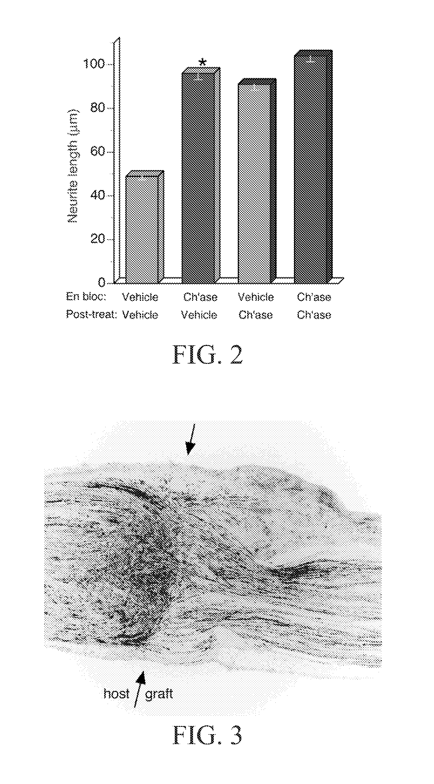
[0127]Inactivation of inhibitory CSPG in chondroitinase-treated acellular nerve was determined by cryoculture bioassay. Embryonic chick DRG neurons were seeded onto sections of prepared nerve segments and the neurite-promoting activity was assessed by scoring neurite growth. Results are shown in FIG. 2. On sections of acellular nerve pretreated en bloc with vehicle only the average neurite length was 49 μm. Neurite growth on acellular nerve pretreated en bloc with chondroitinase averaged 96 μm, representing a 95% increase compared to the control condition. To determine if the en bloc chondroitinase treatment was thorough, cryoculture assays were performed on nerve tissues treated with chondroitinase after sectioning (post-treatment). As expected, the neurite-promoting activity of acellular nerve treated en bloc with vehicle only was increased significantly (86%) by post-treatment with chondro...

example 3

Nerve Regeneration is Enhanced by Chondroitinase Treatment of Acellular Nerve Grafts

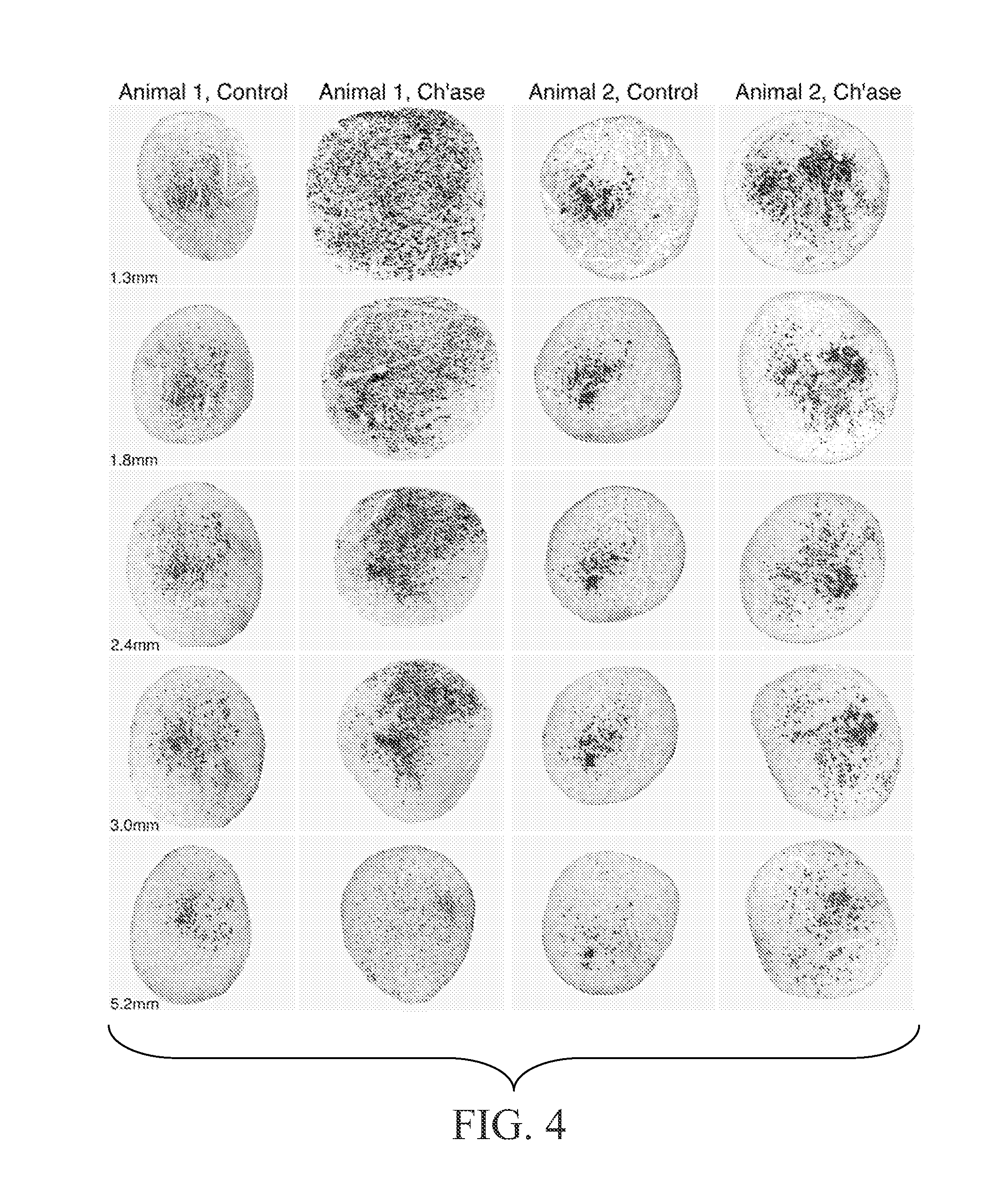
[0129]The following experiments tested the hypothesis that chondroitinase treatment improves nerve regeneration through acellular nerve syngrafts. As described in Example 2, acellular sciatic nerve segments were treated en bloc with vehicle or chondroitinase ABC. Ten-mm interpositional nerve grafts were joined to the host nerve by epineurial neurorrhaphy reinforced with fibrin glue. Each of nine host rats received bilateral grafts, one vehicle-treated and one chondroitinase-treated graft. Regeneration was initially examined after 8 days. First, the proximal and distal nerve-graft coaptations were examined in longitudinal section to assess the alignment of the surgical coaptation, as shown in FIG. 3. All of the grafts were in continuity and thus were included in the subsequent analysis. Scoring of regeneration was based on GAP-43-immunolabeling which intensely stained growing axons. Axon and Schwann c...


PUM

PropertyMeasurementUnit
lengthaaaaaaaaaa
lengthaaaaaaaaaa
lengthaaaaaaaaaa

Abstract

The subject invention pertains to compositions and methods for promoting repair of damaged nerve tissue using nerve grafts and preparation of nerve grafts. The compositions and methods of the subject invention can be employed to restore the continuity of nerve interrupted by disease, traumatic events or surgical procedures. Compositions of the subject invention comprise one or more chondroitin sulfate proteoglycan (CSPG)-degrading enzymes that promote axonal penetration into damaged nerve tissue and nerve graft. The invention also concerns methods for promoting repair of damaged nerve tissue using the present compositions and nerve tissue treated according to such methods. The invention also includes storage solutions for nerve tissue.

Description

CROSS-REFERENCE TO RELATED APPLICATION[0001]This application claims the benefit of provisional patent application Ser. No. 60 / 311,870, filed Aug. 13, 2001, which is hereby incorporated by reference in its entirety, including all figures, tables, and drawings.[0002]The subject invention was made with government support under a research project supported by National Institutes of Health Grant No. R01 NS37901. The government has certain rights in this invention.BACKGROUND OF THE INVENTION[0003]Peripheral nerve injuries are a major source of chronic disability. Poor management of nerve injuries is associated with muscle atrophy and can lead to painful neuroma when severed axons are unable to reestablish continuity with the distal nerve. Although nerves have the potential to regenerate after injury, this ability is strictly dependent upon the regenerating nerve fibers (and their axonal sprouts) making appropriate contact with the severed nerve segment (and the Schwann cell basal laminae ...

Claims


Application Information

Patent Timeline
Patent Type & Authority Applications(United States)
IPC IPC(8): A01N1/02A61K35/12A61K35/16C12N5/06A61K35/30A61K38/00A61K38/22A61K38/27A61K38/51A61K45/00A61K47/30A61K48/00A61L27/00A61L27/22A61L27/36A61L27/38A61L31/04A61P25/00A61P25/02A61P43/00C12N5/08
CPCA61K38/51A61L27/227A61K35/30A61L2430/32A61L31/047A61L27/3604A61L27/3675A61L27/3683A61L27/3687A61L27/383A61L27/3878A61K2300/00A61P25/00A61P25/02A61P43/00A61K38/43
Inventor MUIR, DAVID F.
Owner UNIV OF FLORIDA RES FOUNDATION INC
Why Eureka
Try Eureka