
Detection of Antibodies

a technology of antibodies and detection methods, applied in the field of detection of antibodies, to achieve the effect of high-efficiency methods

Inactive Publication Date: 2009-01-29
BEESON DAVID
View PDF1 Cites 10 Cited by
  • Summary
  • Abstract
  • Description
  • Claims
  • Application Information

AI Technical Summary

Benefits of technology

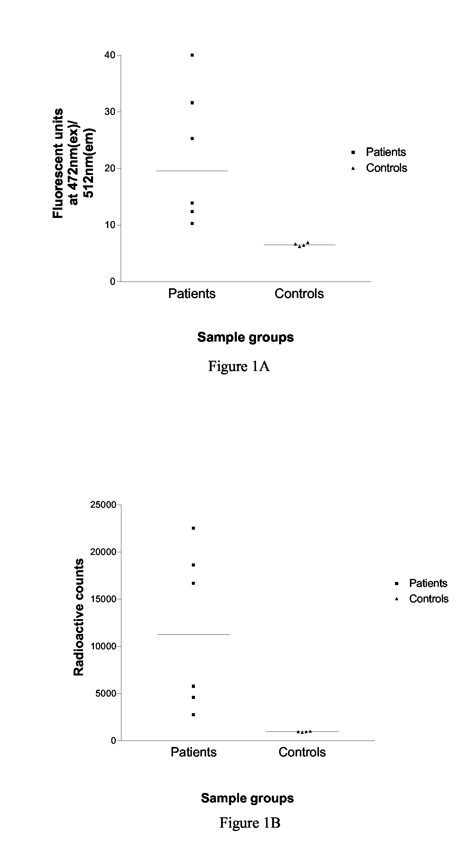
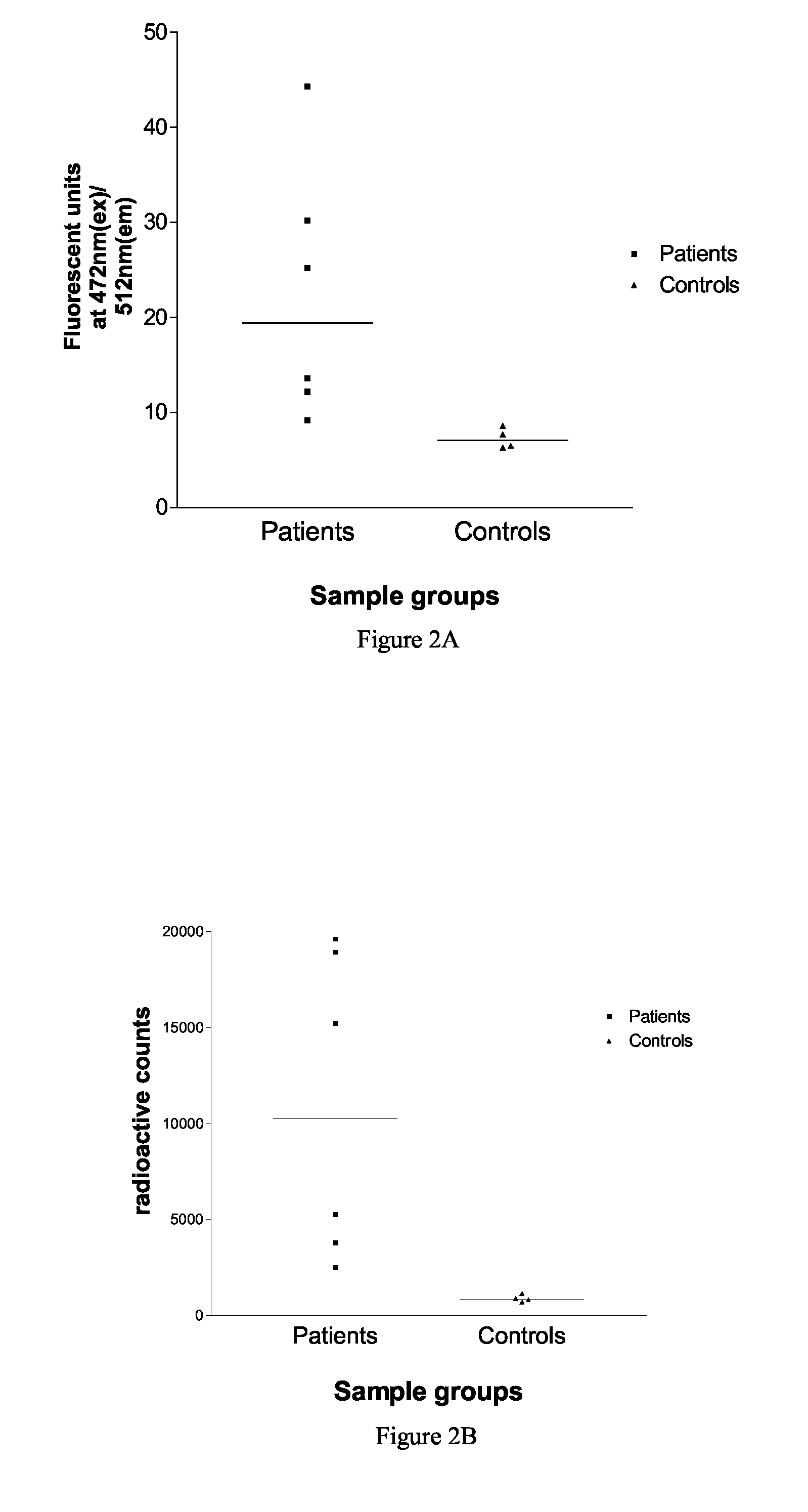
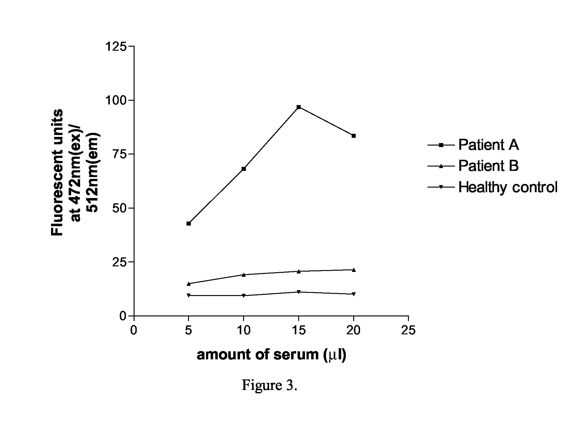
[0009]The use of a fusion protein whereby the fluorescent protein label is incorporated directly in the biological synthesis (i.e. translation) of the target antigen is advantageous because the fluorescent tag can be incorporated at chosen positions within the antigen and because the labelled antigen can maintain immunogenic conformation. Further, when assaying no external label and therefore no purification is required. In particular a radioactive label is not required. The use of such an intrinsically fluorescence-labelled antigen is proving to be a highly efficient method.
[0039]As already noted, the method of the present invention is particularly applicable for the detection of autoantigens and although many autoantigens have been identified, it is believed that a wealth of autoantigenic targets remain to be discovered. The method of the present invention can be used to identify autoantigens by labelling candidate putative antigens with a tag, generally a tag protein, which is fluorescent, and then screening serum samples from particular patient groups to see whether these samples contain antibodies to the putative autoantigen. Thus groups of serum samples can be obtained from patients who have similar clinical phenotypes with characteristics indicative of an autoimmune disorder. This may be a known autoimmune disorder in which case then method can be used to identify autoantigens associated with that disorder. Alternatively, the patients may have characteristics suggestive of an autoimmune disorder but without the disorder yet having been conclusively identified as autoimmune. Thus the patients may respond to immunosuppressive therapy, generally have a fluctuating course of disease, there may be family associations with other autoimmune disorders, and they may share common HLA haplotypes. In this case, as well as identifying autoantigens, the method may assist in confirming disorders as autoimmune in nature.
[0042]Various modifications of the method could be incorporated depending upon the biology of the antigen and the tag. Different tags have different excitation / emission spectra which will require modification of the detection device in accordance with the tag but also allows the possibility of carrying several tests in the same tube as one assay. Other modifications include putting a second (non-fluorescent) tag on the target antigen which can be used for purification prior to adding the patient serum. For example, this can be applied to MuSK using purification via a poly-histidine tag and metal affinity chromatography and this enhances the signal to background ratio of the results.

Problems solved by technology

Working with radioactivity involves inherent dangers and these methods require precautions to be taken against the dangers of using radioactivity.
However, the use of high affinity radio-labelled ligands may mask some autoantibody binding sites on the target antigen.
Similar problems may apply to the use of radioimmunoprecipitation of antibodies generally in samples such as serum, particularly in cases where antibody specificity requires the antigen to be in its native conformation.

Method used


Image

  • Detection of Antibodies
  • Detection of Antibodies
  • Detection of Antibodies

Examples


example 1

An Assay for Serum Antibodies to AChR Using EGFP Tagged AChR α-, γ- and ε-Subunits

Methods

[0055]Construction of Plasmids that Express EGFP-Tagged Human AChR Subunits.

[0056]Oligonucleotides were designed to amplify 615 base pairs (a polypeptide tag of 205 amino acids) encoding the EGFP sequence tag, so that the tag fragment could be ligated “in frame” within the AChR subunit cDNA sequence. The site of insertion within the AChR subunit sequence was chosen to be within the intracellular cytoplasmic domain between transmembrane regions M3 and M4, but not within the MA amphipathic helix region.

[0057]Thus for the human AChR α subunit, oligonucleotides

5′-GCCGATATCATGGTGAGCAAGGGCGAGGAGC-3′and5′-CGCGATATCCTTGTACAGCTCGTCCATGCCGAGAGTGAT-3′

were used to amplify the EGFP sequence. The resulting fragment was cut with EcoR V and ligated into the α-subunit cDNA sequence at the EcoR V restriction site at position 1041. Following transformation, constructs were analysed and colonies isolated in which t...

example 2

An Assay for Serum Antibodies to Muscle-Specific Kinase (MuSK) Using a EGFP-Tagged MuSK

[0072]Serum antibodies to muscle-specific kinase (MuSK) underlie a form of myasthenia gravis in which antibodies to the AChR are absent. Diagnostic tests for anti-MuSK myasthenia gravis at present are performed by radio-labelling purified MuSK with 125Iodine, and using this radiolabelled antigen in standard immunoprecipitation assays. In a method according to the present invention, EGFP-tagged MuSK is synthesised and fluorescence-tagged immunoprecipitation is used as a method for detection of anti-MuSK antibodies in serum samples.

Method

[0073]HEK 293 cells were transiently transfected with the plasmid pSecTagA 1234MuSK-GFP, which is derived from the plasmid pSecTagC (Invitrogen), and which encodes the extracellular region of human MuSK (residues 22-473), a polyhistidine tag and EGFP. The secreted protein was harvested from cell growth medium (Cambrex UltraCHO) after 2 and 5 days. The protein was pu...

example 3

An Assay for Serum Antibodies to Aquaporin-4 (AQP4) Using a EGFP-Tagged AQP4

[0076]Although there are criteria laid down as a guide to differentiate between Multiple Sclerosis (MS) and Neuromyelitis Optica (NMO), they present with similar symptoms. An early difference in the pathogenesis of the two diseases seems to be the presence of detectable levels of autoantibodies to the water channel aquaporin-4 (AQP4) in about 65% of NMO patients. These autoantibodies have not been detected in MS patients. Because of the severity of NMO (Devic's disease) and the different treatment needed for the two diseases, an assay to differentiate them at an early stage would be of immense value. The principle behind this assay, which used immunohistochemical techniques, was published by Lennon et al. J Exp Med (2005) 202 (4): 473-7 and Weinshenker et al. Dis Markers. (2006) 22 (4):197-206.

[0077]Aquaporin 4 is a member of the aquaporin family of membrane water channels. It is abundantly expressed in the ...


PUM

PropertyMeasurementUnit
pHaaaaaaaaaa
excitation at wavelengthaaaaaaaaaa

Abstract

The present invention relates to a method for detecting antibodies against a target antigen in a sample which comprises contacting the sample with labelled target antigen, subjecting the sample to immunoprecipitation to precipitate antibodies in the sample and detecting the presence of antibodies against the target antigen in the sample by means of the presence of labelled target antigen in the immunoprecipitate, wherein the labelled target antigen is a fusion protein comprising the target antigen and a fluorescent protein label and the presence of labelled target antigen in the immunoprecipitate is detected by means of the fluorescence of the fluorescent label. The method is particularly suitable for use where the target antigen is an autoantigen and can also be used to identify autoantigens implicated in a particular autoimmune disorder by screening serum samples from patients with a clinical phenotype indicative or suggestive of an autoimmune disorder and suitable controls. The target protein may be from the cys-loop acetyl choline receptor ion channel gene superfamily, the voltage-gated calcium, sodium or potassium ion channel gene superfamily, the glutamate receptor gene family, a receptor tyrosine kinase, or other membrane associated channels such as aquaporin gene family.

Description

FIELD OF THE INVENTION[0001]This invention relates to the detection of antibodies and in particular to a method which is particularly suitable for detecting and / or identifying autoantibodies.BACKGROUND TO THE INVENTION[0002]Radioimmunoprecipitation of a target antigen is a standard method for the detection of autoantibodies in a sample such as serum and in this detection method the target antigen must be directly or indirectly radio-labelled. For example, the target antigen may be directly labelled by iodination with 125I or by incorporation of 35S-methionine, or the target antigen may be indirectly labelled through binding to a radio-labelled high-affinity ligand, such as 125I-α-bungarotoxin or 125I-α-dendrotoxin. Working with radioactivity involves inherent dangers and these methods require precautions to be taken against the dangers of using radioactivity.[0003]In many cases autoantibody specificity requires that the antigen be in its native conformation and in this case labellin...

Claims


Application Information

Patent Timeline
Patent Type & Authority Applications(United States)
IPC IPC(8): G01N33/53
CPCG01N33/564G01N33/582G01N33/566
Inventor BEESON, DAVID
Owner BEESON DAVID
Learn More