Cytochrome c protein and assay

a cytochrome c protein and assay technology, applied in the field of cytochrome creporter fusion protein constructs, can solve the problems of not being able to meet high throughput live cell screening, using cells in suspension, and not providing homogeneous living cell assays

Inactive Publication Date: 2009-02-26
GE HEALTHCARE LTD
View PDF8 Cites 3 Cited by
  • Summary
  • Abstract
  • Description
  • Claims
  • Application Information

AI Technical Summary

Benefits of technology

[0073]In another embodiment of the method of the invention, the localisation of the fusion construct is determined on non-living, fixed cells. Thus, the living cells over-expressing the fusion construct are fixed at specific time points by conventional means and the location of the fusion protein detected using a detectable moiety, such as a labelled antibody or specific binding chemical. In this way, the effect an agent has upon apoptosis can be determined by comparing localisation of the fusion construct in the presence and the absence of the agent. Alternatively, the effect that the agent has upon apoptosis can be determined by comparing localisation of the construct in the presence of the agent against a known value (for example, one stored on a database) for localisation in the absence of the agent.

Problems solved by technology

However, one problem with flow cytometry is that it can only be used in cells in suspension, such as liquid cell cultures and cells derived from the hemopoietic system.
Furthermore flow cytometry of tissue cells requires physical and enzymatic manipulation to get the cells in suspension, which by itself may trigger apoptosis, necessitating checks by conventional light or fluorescence microscopy.
None of the techniques described above provide a homogeneous living cell assay, based upon the early events in apoptosis, in real time nor discriminate from cellular necrosis.
Furthermore, none of these assays are amenable to high throughput live cell screening which is required to test large numbers of compounds for their ability to modulate apoptosis.
To date, it has not been possible to generate stable cell lines over-expressing this protein which could be reliably used for screening purposes.

Method used


Image

  • Cytochrome c protein and assay
  • Cytochrome c protein and assay
  • Cytochrome c protein and assay

Examples


example 1

Amplification of the Cytochrome c Gene, Fusion to GFP (F64L-S175G-E222G) and Introduction of the K72A (APAF-1 Binding) Mutation

[0092]The fluorescent-cytochrome c mutant fusion proteins of the current invention were produced by joining, in frame, a sequence of the nucleic acid that encodes for the cytochrome c protein to a sequence of the nucleic acid that encodes for a fluorescent protein and then introducing the K72A (APAF-1 binding) mutation (Kluck et al., J Biol Chem., (2000) 275, 16127-16133)) to the nucleic acid of the fusion construct. A preferred sequence of the human cytochrome c gene is described by Zang and Gerstein (Gene, (2003) 312, 61-72); NCBI Accession number NM—018947. (SEQ ID NO: 1) the encoded protein is shown in SEQ ID NO: 2. Alternative human cytochrome c sequences may be used. In addition, alternative sequences around the start and stop codons of the gene may be used to provide useful restriction enzyme sites for protein fusion. Where such alterations change the...

example 2

Influence of Cytochrome c-K72A (APAF-1 Binding) Mutation Upon GFP-Fusion Protein Stable Cell Line Generation in Mammalian Cells

[0100]Plasmid DNA to be used for transfection was prepared for all constructs using the HiSpeed plasmid purification kit (Qiagen, Westberg, NL). In addition to the constructs in example 1, pCORON1000-GFP and pCORON2100-GFP were used as selection controls. DNA was diluted to 100 ng. μl−1 in 18-Megohm water (Sigma, Dorset, UK) and 1 μg used for transfections. For 50-80% confluency on the day of transfection, Hek293 cells were plated at a density of 5×10⁴ / well in 6-well plates and incubated overnight. A 1:3 (1 μg:3 μl) ratio of DNA to FuGene6 reagent (Roche Diagnostics, Basel, Switzerland) was used for each transient transfection reaction; 3 μl FuGene6 was added to 87 μl serum-free DMEM medium (Sigma) (containing penicillin / streptomycin, L-glutamine [Invitrogen, Carlsbad, Calif.]) and gently tapped to mix, then 10 μl (1 μg) construct DNA was added and again gen...


PUM

PropertyMeasurementUnit
temperaturesaaaaaaaaaa
diameteraaaaaaaaaa
fluorescentaaaaaaaaaa

Abstract

The present invention relates to a a cytochrome c-reporter fusion protein construct comprising a modified cytochrome c protein which targets the mitochondria and has a reduced ability to induce apoptosis in a living cell. The invention also relates to nucleic acid constructs encoding such protein fusions and cells stably transfected with such constructs. The stably transfected cells of the invention can be used in assays to detect apoptosis.

Description

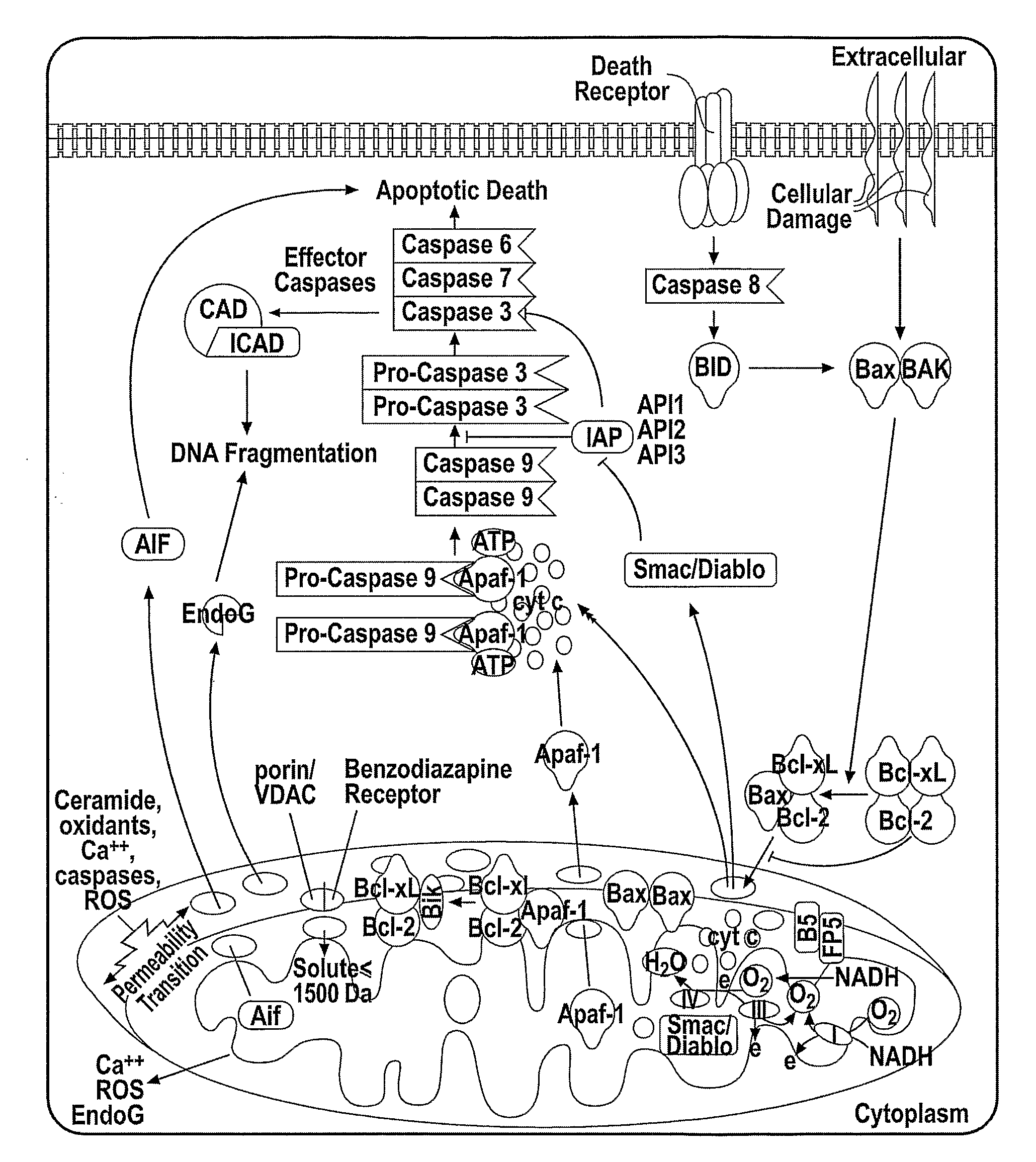
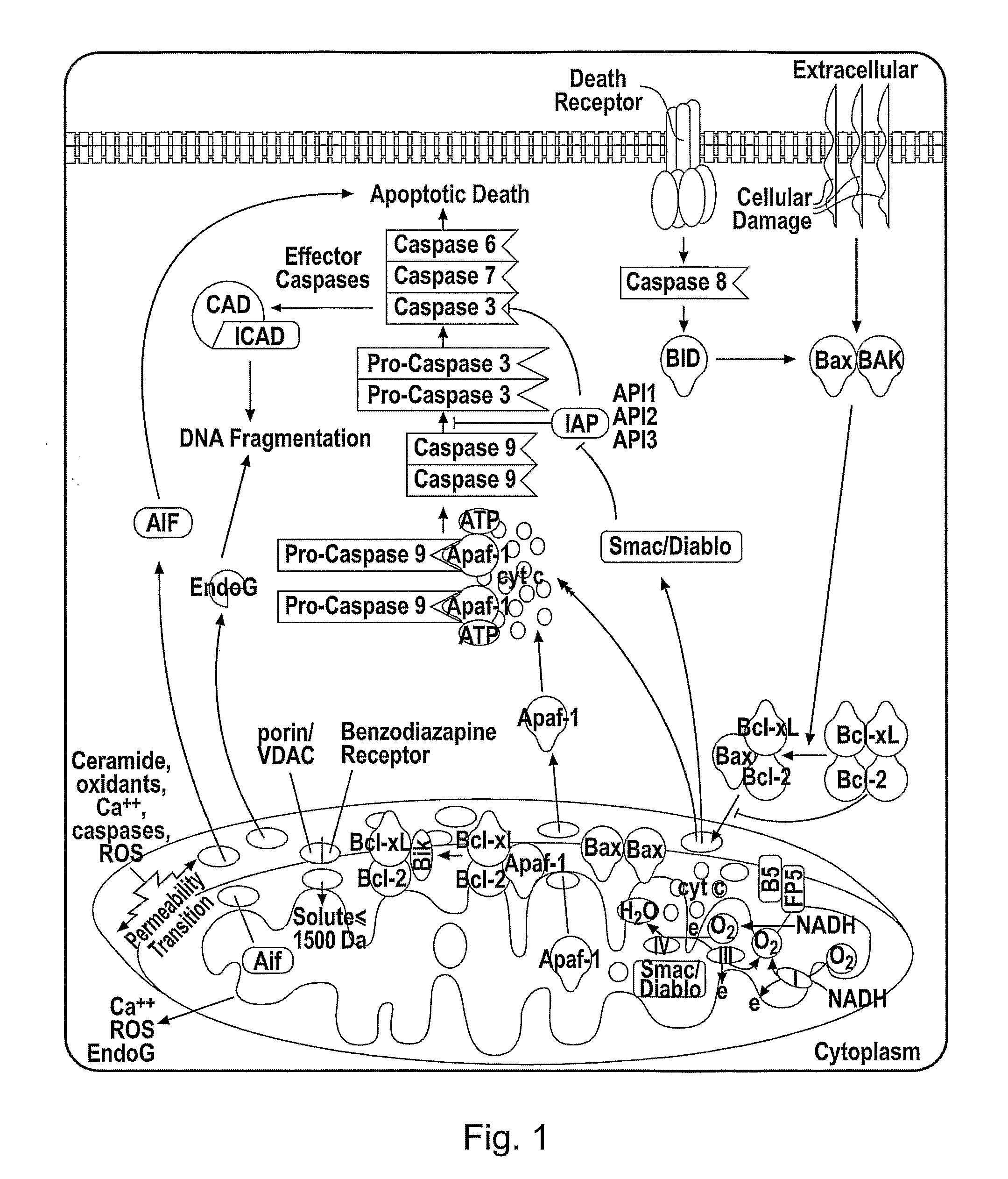
TECHNICAL FIELD[0001]The present invention relates to a cytochrome c-reporter fusion protein construct which targets the mitochondria and has a reduced ability to induce apoptosis in a living cell. The fusion construct of the invention can be used in assays for detecting early events in apoptosis in living cells.BACKGROUND TO THE INVENTION[0002]‘Programmed cell death’ or apoptosis is a key event in multicellular organisms, defining a genetically encoded cell death program which is morphologically, biochemically and molecularly distinct from necrosis (Vermes et al., J Immunol Meth., (2000) 243, 167-190). The characteristic morphological signs of apoptosis (cellular shrinkage, membrane blebbing, nuclear condensation and fragmentation) are the results of a complex biochemical cascade of events which is an integral part of physiological homeostasis.[0003]Apoptosis ensures an equilibrium between cell proliferation and cell death, thus playing a regulatory role in the control of the size ...

Claims


Application Information

Patent Timeline
Patent Type & Authority Applications(United States)
IPC IPC(8): C12Q1/02C07K14/80C07H21/04C07K16/46C12N15/63C12N5/10C12N15/67
CPCC07K14/80G01N2510/00C07K2319/60
Inventor STUBBS, SIMON L.FRANCIS, MICHAEL J.CUSHING, ADRIANISMAIL, RAHMAN A.
Owner GE HEALTHCARE LTD
Why Eureka
Try Eureka