
Detection of nucleic acid differences using endonuclease cleavage/ligase resealing reactions and capillary electrophoresis or microarrays

a nucleic acid and endonuclease technology, applied in microorganism testing/measurement, biochemistry apparatus and processes, nanoinformatics, etc., can solve the problems of patients facing the real possibility, unable to calibrate therapy to the molecular state of the tumor, and difficult to meaningfully predict individual treatment and stratification. , to achieve the effect of extending hairpins and reducing nois

Inactive Publication Date: 2009-05-14
CORNELL RES FOUNDATION INC
View PDF0 Cites 28 Cited by
  • Summary
  • Abstract
  • Description
  • Claims
  • Application Information

AI Technical Summary

Benefits of technology

[0041]The use of different sets of arrays and amplifications and mixing in separate tubes can prevent cross-hybridization of closely related genes to homologue addresses, resulting in a false positive signal.
[0043]By using multiple 50mer sequences to tile across a region, noise is divided by the number of addresses used to tile the targeted region. Proofreading enzymes may also be used to further reduce noise, if needed.
[0045]With the present invention, single stranded DNA on an array cannot fold back on itself and permit polymerase to extend hairpins. This all avoids producing a false positive signal.

Problems solved by technology

It is presently not possible to calibrate therapy to the molecular state of the tumor.
However, there is such large variation from patient to patient, that meaningful individual predictions and treatment stratification are quite difficult.
Thus, patients are confronted with the real possibility that the standard course of adjuvant chemotherapy treatment will be either inadequate or too aggressive for their disease.
Despite the successes of microarrays for expression profiling, with notable exceptions, they have been less than successful in predicting patient outcomes or helping to identify new candidate cancer genes.
For instance, compared to germline analysis, mutation detection in solid tumors is more difficult.
As a result, assays with low specificity are more prone to false positive results.
Although false negative and false positive results are both generally undesirable, a false positive tends to be more deleterious.
This is because a false negative only removes the sample from its appropriate group, whereas a false positive not only removes the sample from its appropriate group, but also places it in the wrong group; a double error.
The total number of genotypes that can be performed is limited by the amount of sample available.
However, the PCR step can generate cross contamination artifacts, affecting the overall specificity and sensitivity, and can yield variable multiplexing results, limiting the throughput of the assay.
Unfortunately, direct sequencing has only limited utility for analysis of solid tumors or pooled samples of DNA due to low sensitivity (Yan, H., et al., Science, 289(5486):1890-2 (2000)).
Although these techniques contain some very desirable characteristics, all fall short in either desired throughput or sensitivity for both SNP association studies and mutational analysis of solid tumors.
The techniques that can identify the position of a polymorphism (ddF and REF) are not sensitive enough to reliably detect low level polymorphisms in pooled or solid tumor samples.
The other techniques are more rapid and can detect low level polymorphisms but do not identify the approximate position of the polymorphism.
This greatly reduces throughput capabilities.
In addition, these techniques lack the ability to distinguish missense from silent polymorphisms, an essential distinction for tumor analysis.
However, in the current formulation, variation detection arrays miss a high percentage of SNPs and mutations when the position is unknown.
Although it is touted as a high-throughput technology, when the position of the mutation is unknown, the need to tile across a large sequence space limits these arrays to just one gene at a time.
These chips are particularly deficient in identifying frameshift mutations (Ahrendt, S., et al., Proc. Natl. Acad. Sci.
Further, since detection of mutations via array hybridization already has too high a rate of false negative signal with germline mutations (Ahrendt, S., et al., Proc. Natl. Acad. Sci.
USA, 96(7382-7387 (1999)), arrays are just too unreliable to accurately score new mutations in tumor samples where stromal contamination becomes an issue.
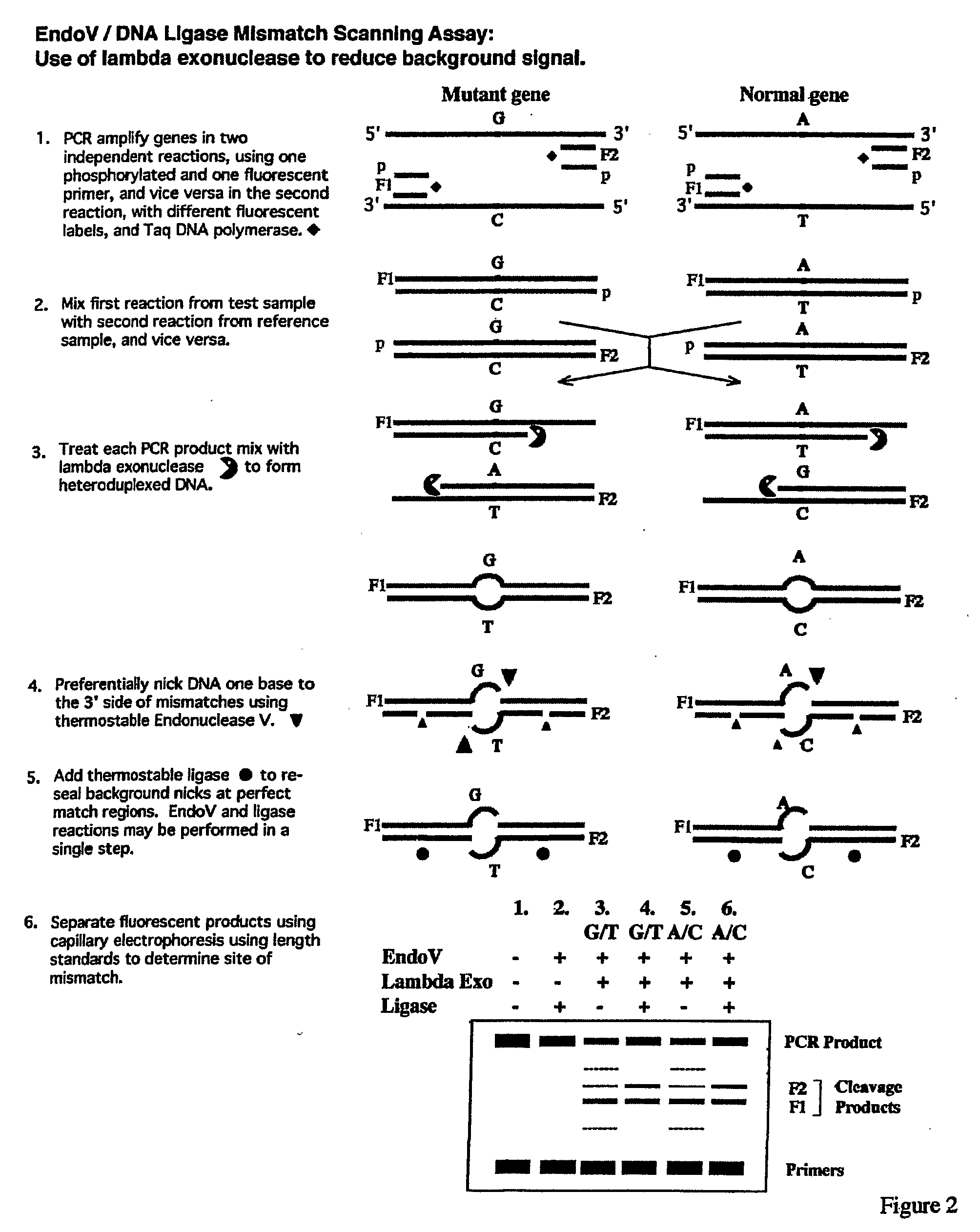
Unfortunately, the cleavage enzymes often nick complementary regions of DNA as well.
This increases background noise, lowers specificity, and reduces the pooling capacity of the assay.
Unfortunately, many enzymes that are commonly used to detect mismatches are incompatible with this solution.
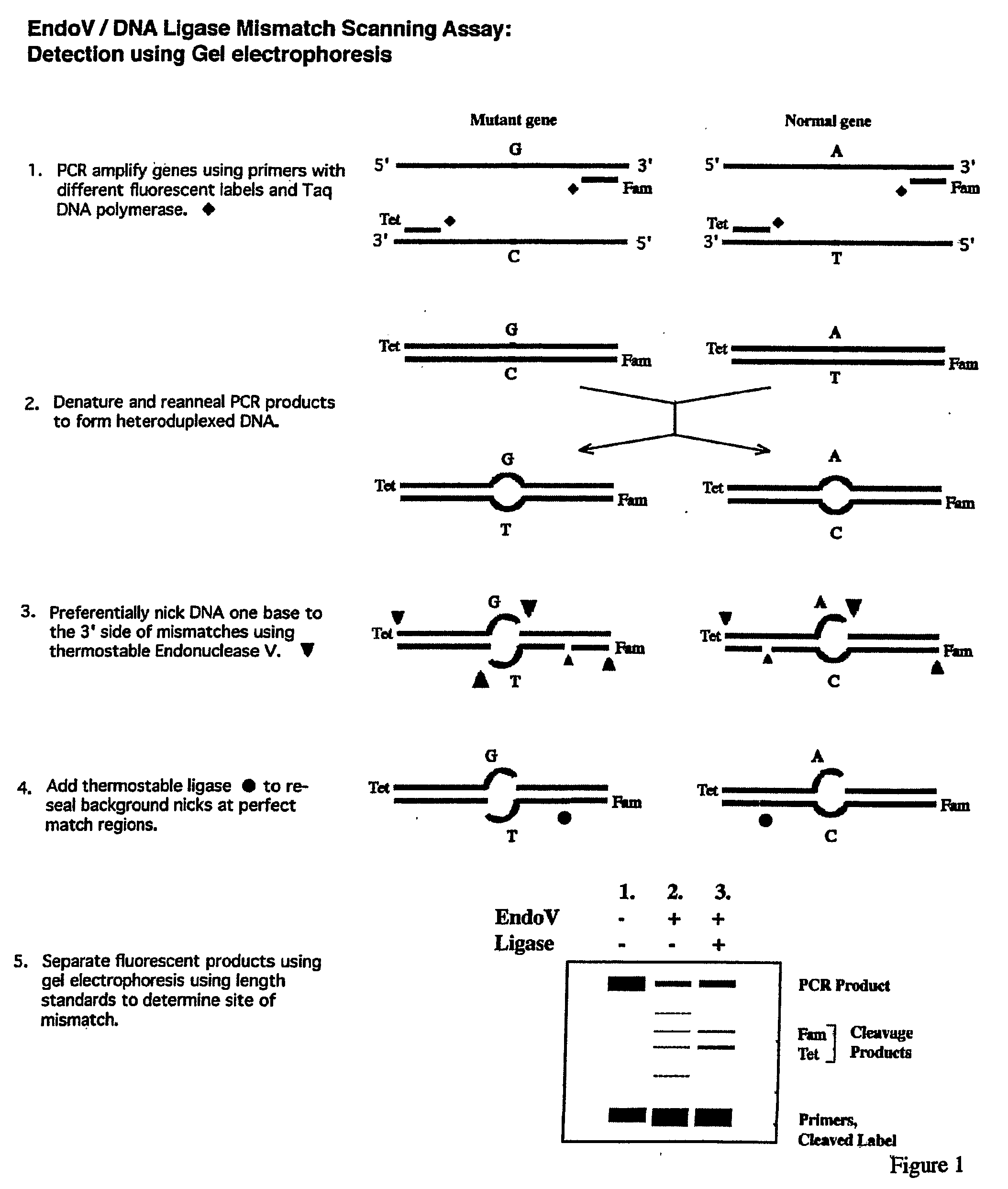
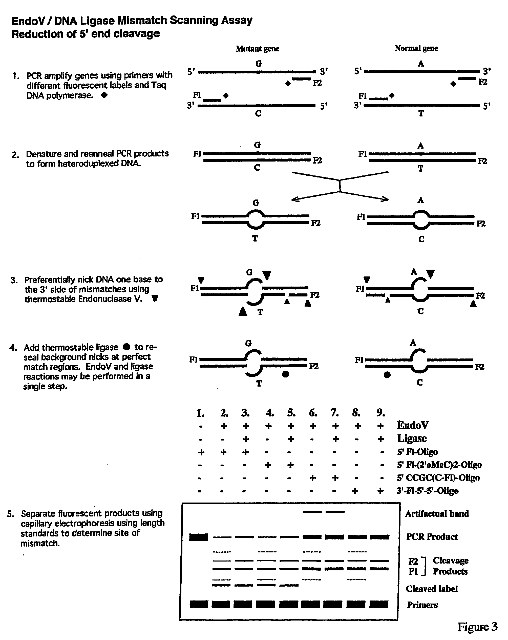
However, the EndoV enzyme cleaves the 5′ fluorescent label off the PCR products, and these run aberrantly in the capillary, providing a potential for false signal.
Few tumor samples will have sufficient material to allow for hybridization in the absence of some form of amplification.
As a result, a false positive signal would be generated.
Another problem with the Endo V / ligase assay is that EndoV will cleave some heteroduplexed DNAs on both strands.
Another problem with the EndoV / ligase assay is that closely related genes like K-ras, N-ras, and H-ras will cross-hybridize to the homologue address.
This will result in a false-positive signal being generated.
Finally, enzyme reactions on solid surfaces (i.e. microarrays) are difficult due to substrate access, causing protein denaturation problems.
In addition, there will be splice site variants which may be difficult to score.
This has the potential of causing false-positives.
Since the position of mutations and the intensity of cleavage will influence fluorescent signal strength, it may be difficult to distinguish true-positives from false-positives in the EndoV / ligase assay.

Method used


Image

  • Detection of nucleic acid differences using endonuclease cleavage/ligase resealing reactions and capillary electrophoresis or microarrays
  • Detection of nucleic acid differences using endonuclease cleavage/ligase resealing reactions and capillary electrophoresis or microarrays
  • Detection of nucleic acid differences using endonuclease cleavage/ligase resealing reactions and capillary electrophoresis or microarrays

Examples


example 1

Standard Procedure

PCR Amplification with Primers Fluorescently Labeled on their 5′-End with Tet and Fam

[0156]All routine chemical reagents were purchased from Sigma Chemicals (St. Louis, Mo., USA) or Fisher Scientific (FairLawn, N.J., USA). GeneScan-500 (TAMRA) size standard, GeneScan-500 LIZ™ size standard, Hi-Di formamide, polymer POP7 and PCR kits were purchased from Applied Biosystems Division of Perkin-Elmer Corporation (Foster City Calif.). Deoxyribonucleoside triphosphate (dNTPs), bovine serum albumin (BSA), ATP, were purchased from Boehringer-Mannheim (Indianapolis, Ind., USA). Proteinase K was purchased from QIAGEN (Valencia, Calif., USA). Deoxyoligonucleotides were ordered from Integrated DNA Technologies Inc. (Coralville, Iowa, USA) and Applied Biosystems Division of Perkin-Elmer Corporation (Foster City, Calif.). Thermotoga maritima Endonuclease V and Thermus species AK16D DNA ligase were purified as described (Huang et al., Biochemistry 40(30):8738-48 (2001); Tong et al...

example 2

Standard Denaturation / Renaturation Procedure

Preparation of Fluorescently Labeled Heteroduplex DNA Substrates

[0159]Aliquots (4 μl) of the p53 exon 8 PCR products were analyzed on a 2% agarose gel, and quantified using the Gel-Doc 2000 imager with Quantity One software (BioRad, Hercules, Calif.). Approximately equal ratios of wild-type PCR amplicons were mixed with mutant (R273H, G->A) PCR amplicons in a 12 μl final volume, with a total of ˜1500 ng DNA. The wild-type control consisted of wild-type DNA PCR products alone in a 12 μl final volume (˜1500 ng total DNA). In order to inactivate Taq DNA polymerase, 1 μl of proteinase K (20 mg / ml, Qiagen, Valencia, Calif.) was added to each PCR mixture, including the wild-type control, and incubated at 65° C. for 30 min. This was followed by a 10 min incubation at 80° C. to inactivate the proteinase K. PCR mixtures were then heated at 95° C. for 2 min, and gradually cooled down to room temperature in a GeneAmp PCR System 960 thermo-cycling mac...

example 3

Performing the EndoV / Ligase Mutation Scanning Assay Under the Standard Conditions

Analysis on the ABI-377 Sequencer

[0160]The EndoV / Ligase assay was performed under the standard two-step reaction conditions, (+EndoV, +Ligase reactions), as described below:[0161]1. Half the volume (˜6.5 μl) of each heteroduplex mixture, including the wild-type homoduplex control, was incubated for 40 min at 65° C. in a 20-μl reaction mixture containing 20 mM Hepes pH 7.5, 5 mM MgCl2, 1 mM DTT, 2% glycerol, 5% DMSO, 1.5 M betaine and 1 μM EndoV.[0162]2. Fifteen μl of each EndoV cleavage reaction mixture were then subjected to the Ligase reaction in a 20-μl final volume during a 30-min incubation at 65° C. This was done by adding 2 μl of 10×supplemental buffer (200 mM Tris pH 8.5, 12.5 mM MgCl2, 500 mM KCl, 10 mM DTT, 200 μg / ml BSA), 1 μl of 20 mM NAD, and 2 μl of 30 nM Ligase.

[0163]In parallel, each heteroduplex mixture, including the wild-type homoduplex control, was subjected to the same protocol, exc...


PUM

PropertyMeasurementUnit
temperatureaaaaaaaaaa
pHaaaaaaaaaa

Abstract

The present invention is directed to various methods for detecting DNA sequence differences, including single nucleotide mutations or polymorphisms, one or more nucleotide insertions, and one or more nucleotide deletions. Labeled heteroduplex PCR fragments containing base mismatches are prepared. Endonuclease cleaves the heteroduplex PCR fragments both at the position containing the variation (one or more mismatched bases) and, to a lesser extent, at non-variant (perfectly matched) positions. Ligation of the cleavage products with a DNA ligase corrects non-variant cleavages and thus substantially reduces background. This is then followed by a detection step in which the reaction products are detected, and the position of the sequence variations are determined.

Description

[0001]This application claims benefit of U.S. Provisional Patent Application Ser. Nos. 60 / 603,937, filed Aug. 24, 2004 and 60 / 603,855, filed Aug. 24, 2004, which are hereby incorporated by reference in their entirety.[0002]The present invention was made with funding from the National Institutes of Health under NCI Grant No. 2 PO1 CA65930-05. The U.S. Government may have certain rights in this invention.FIELD OF THE INVENTION[0003]The present invention is directed to the detection of nucleic acid differences using endonuclease cleavage and ligase resealing reactions in conjunction with electrophoresis or microarrays.BACKGROUND OF THE INVENTIONCancer Detection Generally[0004]Each year in the United States, 130,000 new patients are found to have colorectal cancer and 55,000 patients die with metastatic disease even after attempted surgical resection. Despite extensive information about the morphology and the molecular biology of this disease, only a small number of patients actually be...

Claims


Application Information

Patent Timeline
Patent Type & Authority Applications(United States)
IPC IPC(8): C12Q1/68
CPCB82Y5/00B82Y10/00C12Q1/683C12Q2537/143C12Q2521/501C12Q2521/301C12Q2549/119C12Q2531/113C12Q2521/514
Inventor BARANY, FRANCISPINCAS, HANNAHUANG, JIANMIN
Owner CORNELL RES FOUNDATION INC
Learn More