Cell labeling with perfluorocarbon nanoparticles for magnetic resonance imaging and spectroscopy

a technology of perfluorocarbon nanoparticles and magnetic resonance imaging, which is applied in the direction of biocide, diagnostics, diagnostic recording/measuring, etc., can solve the problem of subject exposed to a magnetic field of reduced strength, and achieve the effect of minimizing the loss of input cells and minimal effects on cell viability

Inactive Publication Date: 2009-10-22
WASHINGTON UNIV IN SAINT LOUIS
View PDF3 Cites 13 Cited by
  • Summary
  • Abstract
  • Description
  • Claims
  • Application Information

AI Technical Summary

Benefits of technology

[0009]It is in view of the above problems that the present invention was developed. The invention is first drawn to a method of obtaining an endothelial precursor cell suitable for magnetic resonance imaging or spectroscopy comprising the steps of providing an endothelial precursor cell; incubating said endothelial precursor cell in a cell culture media containing a plurality of perfluorocarbon nanoparticles for a period of time and at a perfluorocarbon nanoparticle concentration sufficient to result in internalization of a detectable level of perfluorocarbon nanoparticles; and separating said endothelial precursor cell from said culture media containing perfluorocarbon nanoparticles. The perfluorocarbon nanoparticles comprise a perfluorooctylbromide core component or a perfluoro-15-crown-5-ether core component. When the perfluorocarbon nanoparticles comprise a perfluorooctylbromide core component, a detectable level of internalized perfluorocarbon nanoparticles is an intracellular perfluorocarbon nanoparticle concentration of at least 2.8 pmol per cell. When the perfluorocarbon nanoparticles comprise a perfluoro-15-crown-5-ether core component, a detectable level of internalized perfluorocarbon nanoparticles is an intracellular perfluorocarbon nanoparticle concentration of at least 0.5 pmol per cell. The endothelial precursor cell may be provided by isolating mononuclear cells from human umbilical cord blood and growing the cells in a modified endothelial cell culture media. This modified endothelial cell culture media may comprise the growth factors hEGF, VEGF, hFGF-B, and R3-IGF-1. The endothelial precursor cell may be any one of a CD34+ cell, CD133+ cell, CD31+ cell, a Tie-2+ cell, a CD31+ / CD34+ cell, CD34+ / CD133+ / CD31+ cell, a CD34+ / Tie-2+ cell, a CD34+CD133+Tie-2+CD45+ cell, and a CD34+ / CD133+ cell. Alternatively, the endothelial precursor cell may be characterized by an ability to internalize acetylated-Low Density Lipoprotein (LDL) and / or by the presence of fucose at its surface. The endothelial precursor cell suitable for magnetic resonance imaging that is obtained by this method can typically internalize acetylated-Low Density Lipoprotein (LDL) and has fucose present at its surface. By using this method, one skilled in the art can obtain an endothelial precursor cell suitable for magnetic resonance imaging without using methods such as electroporation or transfection to introduce the perfluorocarbon nanoparticles into the cells. The advantages of using this technique are that introduction of the perfluorocarbon nanoparticles has minimal effects on cell viability and minimizes loss of input cells.

Problems solved by technology

Advantages of this imaging method are that the subject is exposed to a magnetic field of reduced strength and that the image is acquired in a shorter period of time.

Method used


Image

  • Cell labeling with perfluorocarbon nanoparticles for magnetic resonance imaging and spectroscopy
  • Cell labeling with perfluorocarbon nanoparticles for magnetic resonance imaging and spectroscopy
  • Cell labeling with perfluorocarbon nanoparticles for magnetic resonance imaging and spectroscopy

Examples


example 1

Labeling Stem / Progenitor Cells with PFC Nanoparticles

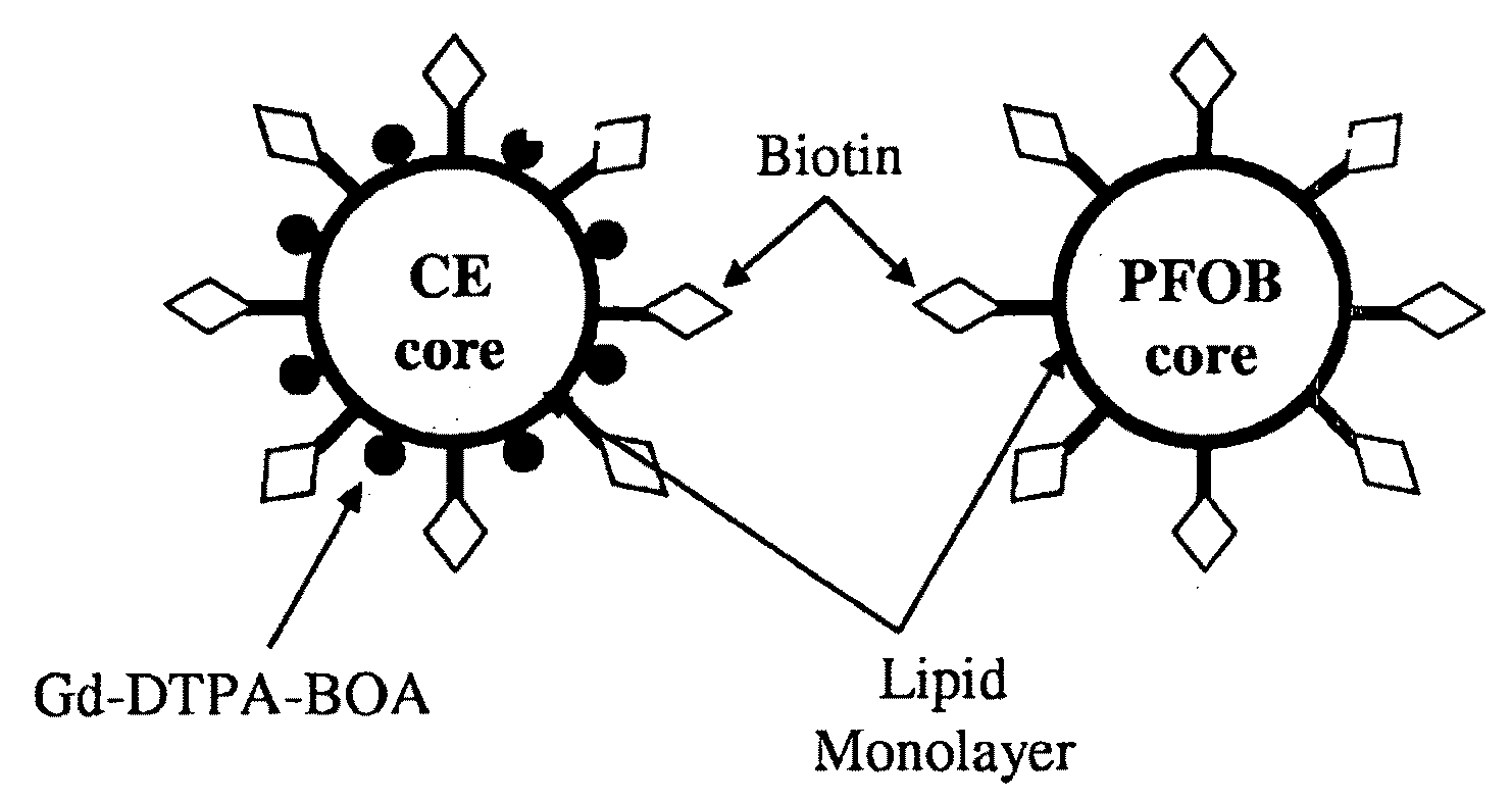
[0070]Liquid PFC nanoparticles were formulated using methods previously developed in our laboratories (Lanza, G. M. et al. Circulation 94: 3334-3340, 1996). Briefly, the emulsions comprised 20% (v / v) perfluorocarbon (PFC) such as either perfluorooctylbromide (PFOB) or 15-crown-5 ether (CE), 1.5% (w / v) of a surfactant / lipid co-mixture, and 1.7% (w / v) glycerin in distilled, deionized water. Fluorescent nanoparticles contained fluorescent-conjugated phospholipids of either 2.05 mole % of NBD (1,2-dioleoyl-sn-glycero-3-phosphoethanolamine-N-(7-nitro-2-1,3-benzoxadiazol-4-yl)) or 0.135 mole % of rhodamine (1,2-dioleoyl-sn-glycero-3-phosphoethanolamine-N-(lissamine rhodamine B sulfonyl)) (Avanti Polar Lipids, Inc., Alabaster, Ala.) in the surfactant layer. The mixture of surfactant components, PFC and water was blended and then emulsified at 20,000 PSI for four minutes in an ice bath with an estimated temperature range of about 0° C. to...

example 2

Confocal Microscopic Imaging and Immuno-Characterization of PFC-Labeled Cells

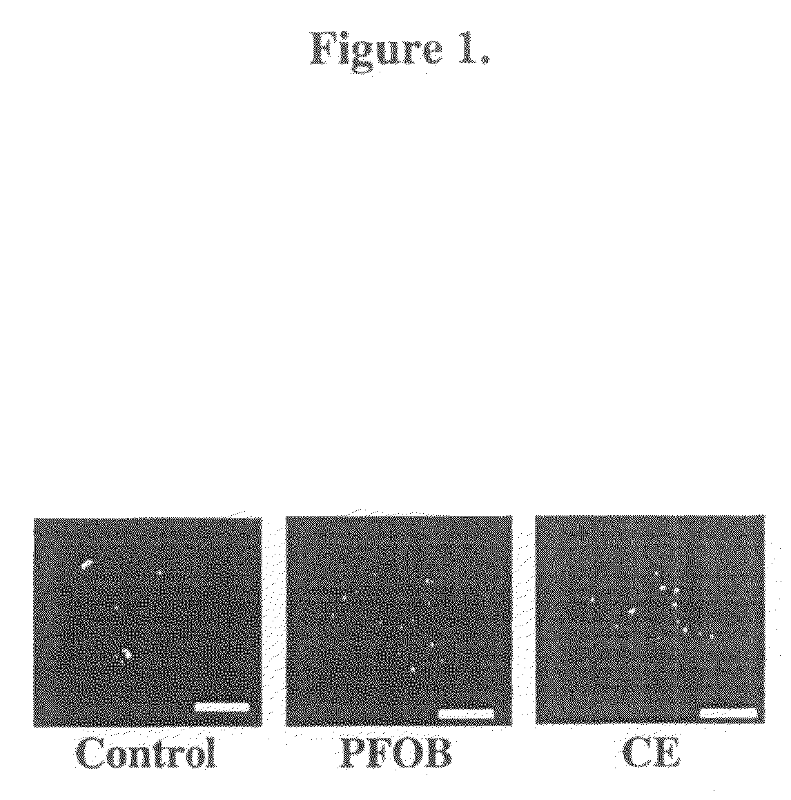
[0072]To image PFC-labeled cells with confocal microscopy, labeled cells treated as described in Example 1 were first subjected to 2% paraformaldehyde fixation for 30 minutes. For confocal imaging fixed cells were placed in 1.5 glass bottom culture dishes (Bioptechs Inc., Butler, Pa.). Fluorescence imaging of cell sections was conducted with a confocal microscope (Zeiss Meta 510, Thornwood, N.Y.), using standard filter sets. The location of nanoparticles with respect to the cell was determined with simultaneous differential interference contrast (DIC) imaging.

[0073]Internalization of nanoparticles occurred without aid of any additional transfection agents or methods, characterized by abundant uptake and distribution throughout the cytosol for both PFOB and CE nanoparticles. Qualitatively for individual cells that internalized nanoparticles, a greater uptake of nanoparticles appeared for PFOB-loaded as compa...

example 3

Effects of PFC Nanoparticle Labeling on Cell Viability and Function

[0077]To determine if PFC nanoparticle labeling affected cell viability, the percent (%) cell survivability after labeling of cells as described in Example 1 was determined by trypan blue exclusion. Cells labeled with either CE or PFOB nanoparticles were removed with trypsin, resuspended in PBS, and diluted 1:1 with 0.4% trypan blue (Sigma, St. Louis, Mo.). For a positive control, cells were heated at 45° C. for 15 min. The number of viable and nonviable cells was counted using a hemocytometer with the percentage of trypan blue positive cells was used to calculate cell survival. We found high cell survivability (˜90%) of cells subjected to labeling procedure of Example 1 with no significant difference from non-labeled control cells (FIG. 3a). Positive control cells exposed to high temperature manifested a substantial loss of cell viability (˜60% cell survivability). Accordingly, neither PFOB nor CE nanoparticles exer...


PUM

PropertyMeasurementUnit
diameteraaaaaaaaaa
magnetic fieldaaaaaaaaaa
slice thicknessaaaaaaaaaa

Abstract

Methods of obtaining cells internally labeled with perfluorocarbon nanoparticles suitable for magnetic resonance imaging and spectroscopy are disclosed. Also disclosed are methods for obtaining magnetic resonance imaging data from labeled under clinically relevant scan times and field strengths. Finally, the application further discloses methods of specifically detecting and distinguishing magnetic resonance imaging and spectroscopy data from two distinct sets of cells labeled with distinct types of perfluorocarbon nanoparticles.

Description

CROSS REFERENCE TO RELATED APPLICATIONS[0001]This patent application claims priority to the Feb. 24, 2006 filing date of U.S. Provisional Patent Application No. 60 / 776,743, which is incorporated by reference herein in its entirety.STATEMENT REGARDING FEDERALLY SPONSORED RESEARCH OR DEVELOPMENT[0002]National Institutes of Health (U54-CA-119342 and HL-073646 to S. A. Wickline, CO-37007 to G. M. Lanza).FIELD OF THE INVENTION[0003]This invention relates generally to methods of obtaining labeled cells suitable for magnetic resonance imaging or magnetic resonance spectroscopy. The invention further relates to methods of magnetic resonance imaging or magnetic resonance spectroscopy that permit data acquisition from labeled cells under clinically relevant conditions (i.e., magnetic field strengths of 1.5 T with imaging times of less than about 12 minutes). Finally, this invention further provides for methods of obtaining two distinct magnetic resonance imaging or spectroscopy data sets deri...

Claims


Application Information

Patent Timeline
Patent Type & Authority Applications(United States)
IPC IPC(8): A61K49/00C12Q1/02
CPCB82Y5/00A61K49/1896
Inventor WICKLINE, SAMUEL A.LANZA, GREGORY M.
Owner WASHINGTON UNIV IN SAINT LOUIS
Why Eureka
Try Eureka