Compounds and Kits for the Detection and the Quantification of Cell Apoptosis
- Summary
- Abstract
- Description
- Claims
- Application Information
AI Technical Summary
Benefits of technology
Problems solved by technology
Method used
Image
Examples
example 1
Development of a New Probe with High Selectivity to Cell Plasma Membranes and Drastic Sensitivity to Surface Potential
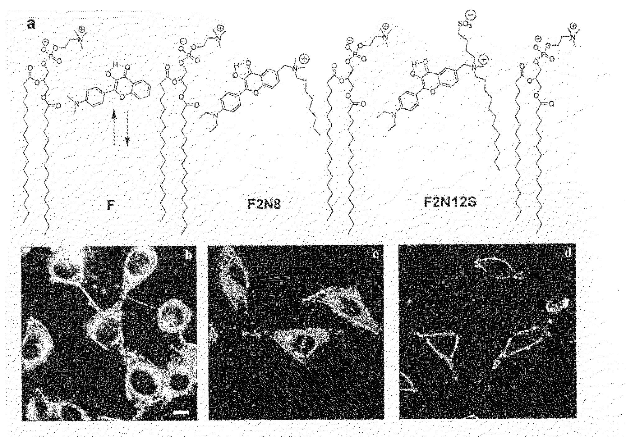
[0116]Confocal laser microscopy have been performed on adherent L 929 cells stained with these first generation molecules (FIG. 1.b, c). The obtained fluorescence images reveal a rapid penetration of these dyes inside the cells.
[0117]Images of adherent L 929 cells stained with probes F2N12S (FIG. 1.d) demonstrate emission exclusively from the plasma membranes. The probes stay in the membrane during the whole observation time under the microscope, which is limited by the lifetime of the cells in HBSS buffer (approx. 1 hour). Moreover, F2N12S probe was found to be of sufficient brightness for fluorescence microscopy of individual cells. Some inhomogeneity in the distribution of the fluorescence intensity at the plasma membrane has been observed. This is probably connected with the heterogeneous lipid distribution in the bilayer that affects the probe distribution.
[0118...
example 2
Effect of Surface Potential on F2N12S Fluorescence
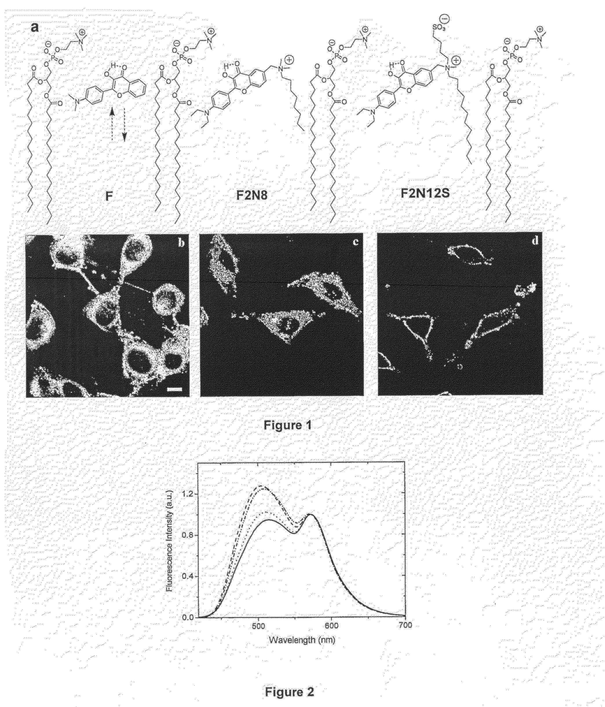
[0119]To model the increase in the negative surface charge at the outer leaflet of the plasma membrane during apoptosis, large unilamellar vesicles (LUV) composed of neutral (phosphatidylcholine PC and phosphatidylethanolamine PE) and negatively charged (phosphatidylglycerol PG) and phosphatidylserine PS) phospholipids have been used. The probe of the invention F2N12S demonstrates strong two-band response to the surface charge in large unilamellar vesicles (LUV). Indeed, in anionic vesicles (PG and PS) this probe shows a higher relative intensity of the short-wavelength band as compared to neutral PC vesicles (FIG. 2). A significantly smaller increase in the short-wavelength band relative intensity is observed also for neutral PE vesicles.
example 3
Response of F2N12S to Apoptosis in Cell Suspensions and Ca2+-Dependence of this Response
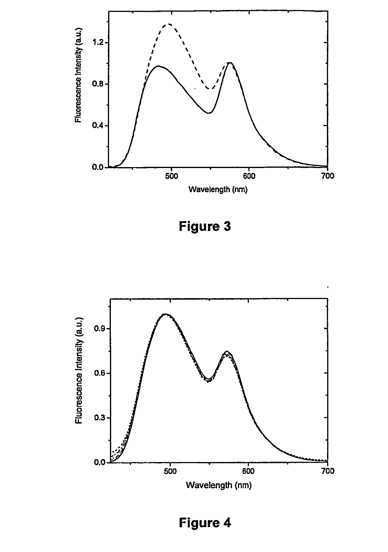
[0120]CEM T cells apoptosis was induced by actinomycin D, an inhibitor of DNA-primed RNA polymerase. Then, apoptotic cells were separated from living cells by cytometry using the FITC-annexin marker. Next, the living and apoptotic cells were labeled with F2N12S and their emission spectra were recorded at an excitation wavelength of 400 nm. At this wavelength, the excitation of FITC is minimal and its residual emission was subtracted from the emission spectrum of F2N12S. The results are presented on FIG. 3. Apoptosis results in dramatic changes of the fluorescence spectrum of probe F2N12S since the relative intensity of the short-wavelength band is nearly 2-fold higher than that in living cells. This result is in line with the model experiments on the surface charge effect in lipid vesicles of example 2 (FIG. 2). In living cells, the separation of the two bands of F2N12S is significantly larger th...
PUM
 Abstract
Description
Claims
Application Information
 - R&D
- Intellectual Property
- Life Sciences
- Materials
- Tech Scout
- Unparalleled Data Quality
- Higher Quality Content
- 60% Fewer Hallucinations
Browse by: Latest US Patents, China's latest patents, Technical Efficacy Thesaurus, Application Domain, Technology Topic, Popular Technical Reports.
© 2025 PatSnap. All rights reserved.Legal|Privacy policy|Modern Slavery Act Transparency Statement|Sitemap|About US| Contact US: help@patsnap.com


