
Implantable devices for promoting reendothelialization and methods of use thereof

Inactive Publication Date: 2009-11-05
ESTRACURE
View PDF4 Cites 0 Cited by
  • Summary
  • Abstract
  • Description
  • Claims
  • Application Information

AI Technical Summary

Benefits of technology

[0012]The present invention is concerned with the surprising finding that administration of a dosage of estrogen receptor agonist of at least 16.7 μg / mm of implantable device at the injured site of a vessel accelerates and optimizes vessel repair.
[0036]As used herein the terms “effective amount of biodegradable polymer” refers to an amount of polymer that enables the loading of as much estrogen receptor agonist as possible in accordance with the present invention. The precise amount of polymer thus depends on its nature and on the nature of the estrogen receptor agonist. Polymers such as PEA from Medivas enables the loading of therapeutic agent in an amount about equal to its own weight (e.g. for 500 μg of polymer, up to 500 μg of estrogen receptor agonist can be loaded). A top coat of polymer can also be applied in addition to this amount to decrease release speed. The present invention also encompasses the chemical coupling of the estradiol receptor agonist to the polymer to slow down its release from this polymer.
[0044]One embodiment of the invention provides a method for biologically stenting a procedurally traumatized mammalian blood vessel. The method comprises administering to the blood vessel an amount of an estrogen receptor agonist in a vehicle effective to biologically stent the vessel. As used herein, “biological stenting” means the fixation of the vascular lumen in a dilated state near its maximal systolic diameter, e.g., the diameter achieved following balloon dilation and maintained by systolic pressure. The method comprises the administration of an effective amount of an estrogen receptor agonist to the blood vessel. Preferably, the estrogen receptor agonist is dispersed in a pharmaceutically acceptable liquid carrier. Preferably, a portion of the amount administered penetrates to at least about 6 to 9 cell layers of the inner tunica media of the vessel (or much deeper than that in the case of where 17 beta estradiol is used as estrogen receptor agonist) and is thus effective to biologically stent the vessel.
[0047]The implantable device of the present invention possesses at least one of the following advantageous properties: accelerates reendothelialization, reduces macrophage infiltration, reduces leukocyte and / or platelet adhesion and reduction of type I collagen. This last effect allows estradiol's deep penetration and its consequent reduction of migration of smooth muscle cells from the media to the injured region exposed to blood circulation as detected in the adventitia and peri-coronary muscular and white fat tissues (FIG. 10). The adventitia includes the vasa vasorum, small vessels which provide nutriment to the vascular wall but which are also an entry door for inflammatory cells.

Problems solved by technology

These experiments did not suggest optimal dosage for controlled delivery administration of estrogen receptor agonist by implantable device.
Nevertheless, neointimal proliferation was not completely abolished by these estradiol-eluting stents.
These unsatisfying results were attributed to the suboptimal estradiol elution of their delivery system which provided an elution that stopped within 24 hours after administration.
Although this trial again confirmed that there were no safety concerns with estradiol administration (i.e. death, or stent thrombosis), the results were again disappointing.
It has been reported that delayed or incomplete reendothelialization is likely a cause of late susceptibility to stent thrombosis.

Method used


Image

  • Implantable devices for promoting reendothelialization and methods of use thereof
  • Implantable devices for promoting reendothelialization and methods of use thereof
  • Implantable devices for promoting reendothelialization and methods of use thereof

Examples


example 1

Material and Methods for Examples 2 to 4

[0063]Pre-Operative Procedures Animals were monitored and observed at least 5 days prior to experimental use. Within 24 hours prior to the procedure, the animals were started on a dosage of 650 mg aspirin, 75 mg Plavix™ and 30 mg nifedipine orally.

[0064]Stents and hormone used for the procedures The polymer poly (ester-amide) (PEA) / estradiol (E2) stents used in Examples presented herein were prepared by MicroPort (MicroPort Medical Co, Ltd. Shanghai). Stainless steel bare metal stents were of 18 mm and 23 mm length and were spray coated by MicroPort with 300 μg or 500 μg of E2 matrixed with MediVas™ poly (ester-amide) (MVPEA.I.(Ac)) tempo polymer. In addition to the polymer-steroid matrixed base coat, a 200 ug MVPEA.I.(Ac).tempo protective topcoat was applied to the stents to decrease drug elution speed. The stents were sterilized by ethylene oxide at the MicroPort Medical Co facilities under the recommendation of Medivas. The sizes of Cypher™...

example 2

Effect of 16.7 μg / mm (300 μg on 18 mm stent) and 27.8 μg / mm (500 μg on 18 mm of Stent) 17 Beta-Estradiol on Neointima Formation

[0072]Animal groups Vascular healing (efficacy study) was evaluated in six animals for each type of stent (bare metal stent (BM), polymer poly (ester-amide) (PEA) only, PEA with 300 μg E2, PEA with 500 μg E2, at 24 hours, 14 days, 1 and 3 months after the stent implantation and 2 animals for Cypher™ and Taxus™ at 1 month post-implantation.

[0073]Stents Three stents were implanted in each animal. All animal groups were successfully completed and survived until the pre-determined time point.

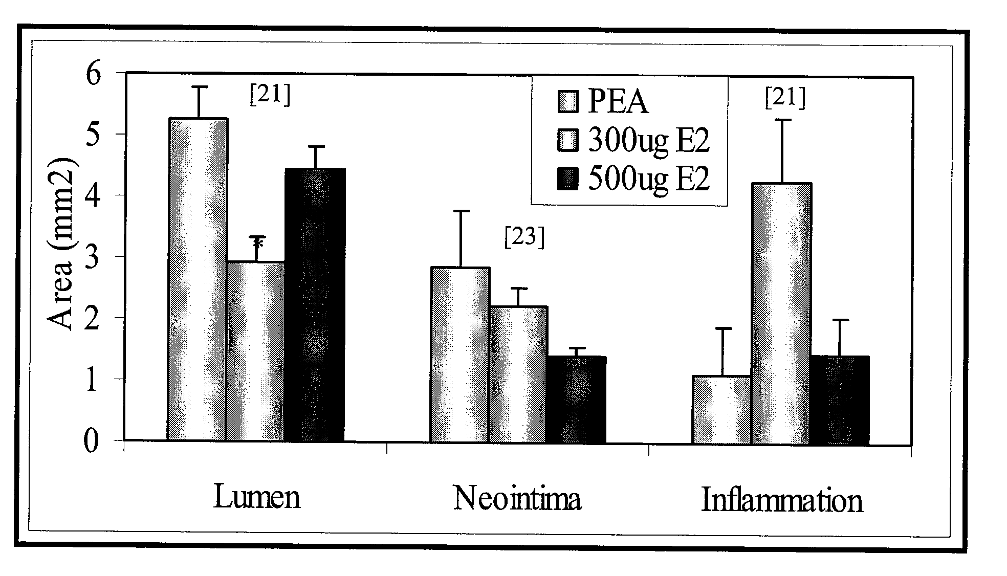
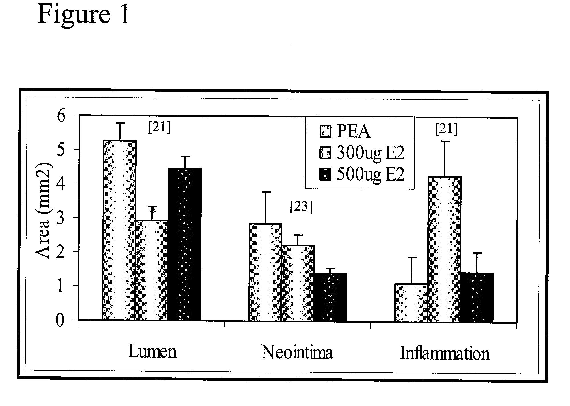
[0074]Morphometric analysis was performed for each stented artery. Measurements of the neointima area (Neointima), the lumen area (Lumen), media area (Media) (i.e. thickness of intima and adventitia together), and the percentage of stenosis are summarized for control bare metal stent (BM), PEA polymer only (PEA), coated with PEA+300 μg of E2 (300 μg E2) or PEA+500 μg of E2 (...

example 3

Effect of 16.7 μg / mm (300 μg on 18 mm stent) and 27.8 μg / mm (500 μg on 18 mm of Stent) 17 Beta-Estradiol on Macrophage Infiltration

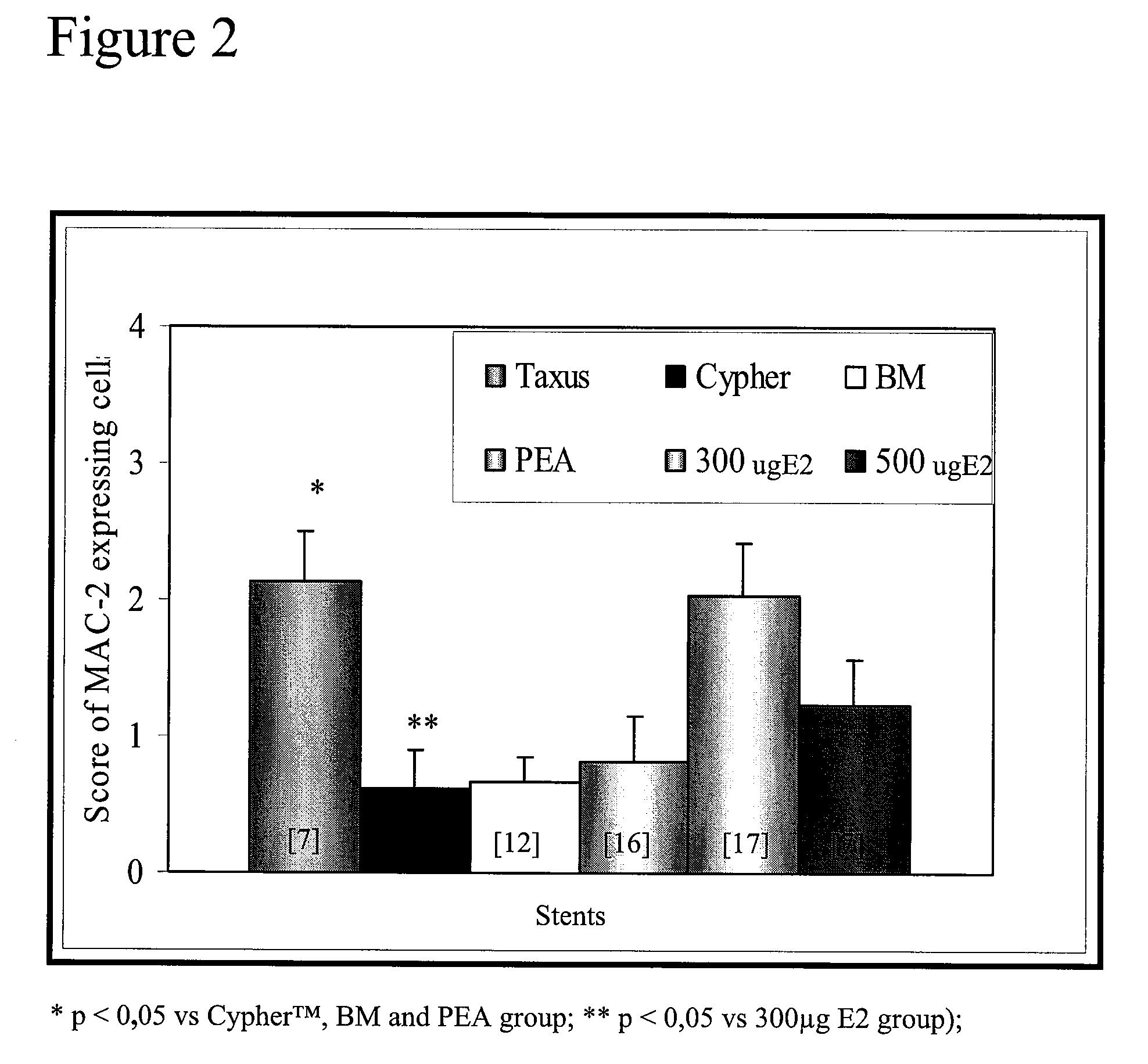
[0076]Immunohistology analysis was performed to determine the degree of macrophage infiltration in the wall of the stented arteries. Macrophage infiltration is used as an inflammation marker and as an indication of risk of thrombosis. Macrophage infiltration was evaluated using an anti-MAC-2 specific antibody (MAC-2), a cell surface marker for macrophages. Macrophage infiltration was scored for each strut as follow: 0: absence of macrophages, 1: very rare number of macrophage, 2: limited number macrophages, 3: high number of MAC-2 positive cells, 4: very high number of macrophages around the strut and between the struts. The mean of scores of the struts corresponds to the score for the artery. As may be seen in FIG. 2, after 1 month, the 300 μg E2 group had more macrophages (mean grade of 2) than three other groups but it is comparable to infiltration le...


PUM

PropertyMeasurementUnit
Timeaaaaaaaaaa
Timeaaaaaaaaaa
Timeaaaaaaaaaa

Abstract

An implantable device for the controlled delivery of an estrogen receptor agonist to an injured site in the lumen of a mammalian blood vessel, wherein the estrogen receptor agonist is present in an amount of at least about 16.7 μg / mm implantable device length. Methods of use thereof.

Description

FIELD OF THE INVENTION[0001]The present invention relates to implantable devices for promoting reendothelialization and methods of use thereof. More specifically, the present invention is concerned with implantable devices for the controlled delivery of an estrogen receptor agonist at an injured site of a injured vessel.BACKGROUND OF THE INVENTION[0002]17-β-Estradiol is known to inhibit proliferation of smooth muscle cells (neointima formation) and promote reendothelialization in vitro and in vivo. Co-pending application Ser. No. 10 / 088,405 has shown that estradiol inhibits restenosis and promotes reendothelialization in a pig model for restenosis.[0003]Results of administering a bolus of either 600 μg (co-pending application Ser. No. 10 / 088,405 and Chandrasekar B et al. J Am Coll Cardio 2001; 38(5):1570-6) or 100 ug / kg and 200 ug / kg (total of 2 to 4 mg) (Chandrasekar B et al. Thromb Haemost 2005; 94:1042-7) by catheter at the site of the injury are known. With such a type of admini...

Claims


Application Information

Patent Timeline
IPC IPC(8): A61F2/00
CPCA61L27/34A61L27/507A61L27/54A61L2300/43A61L31/16A61L2300/412A61L31/10
Inventor TANGUAY, JEAN-FRANCOIS
Owner ESTRACURE
Learn More