
Physiologically Active Polypeptide- or Protein-Encapsulating Polymer Micelles, and Method for Production of the Same

Active Publication Date: 2009-11-26
NANOCARRIER
View PDF6 Cites 11 Cited by
  • Summary
  • Abstract
  • Description
  • Claims
  • Application Information

AI Technical Summary

Benefits of technology

[0016]As a result of much diligent research conducted in light of the current circumstances explained above, the present inventors have discovered that physiologically active polypeptides and proteins can be efficiently encapsulated in polymer micelles by using a block copolymer comprising hydrophilic segments composed of polyethylene glycol, and hydrophobic segments composed of a polyamino acid selected the group consisting of acidic amino acids, hydrophobic derivatives thereof and mixtures of said acidic amino acids and said hydrophobic derivatives. Furthermore, by adjusting the pH used for preparation of the polymer micelles in consideration of the isoelectric points of the physiologically active polypeptides and proteins, we successfully accomplished more efficient encapsulation. The present inventors have completed this invention upon finding that this method can be applied for numerous physiologically active polypeptides and proteins regardless of their acidity or basicity, that modifying the block copolymer can contribute to hydrophobic interaction between the hydrophobic segments and the polypeptide or protein, thus allowing the encapsulation efficiency and release rate of the drug to be improved, and in particular that the structure of the hydrophobic side-chains in the block copolymer contributes significantly to the drug release rate.
[0020]The invention affords the advantage of allowing efficient encapsulation of high-molecular-weight drugs such as physiologically active polypeptides and proteins in polymer micelles, while also permitting control of their release rate.

Problems solved by technology

Such polypeptides, however, generally have a short half-life in vivo due to their extremely rapid enzymolysis, metabolism and the like, and in most cases it has not been possible to obtain satisfactory effects when they are administered as drugs.
This has resulted in less frequent administration and thus reduced burden on patients, but such polymer-modified proteins generally exhibit lower activity due to the modification, and it has been difficult to control the modification sites and modification rates in a reproducible manner.
However, the particle size is usually in the micrometer range and is not suitable for intravenous administration.
However, the particle sizes obtained by such methods are at minimum a few hundred nanometers (Int. J. Pharm. 149, 43-49 (1997)), while the surface modification is laborious and it has also been difficult to control the organ distribution in a reproducible manner.
However, such liposomes do not necessarily have adequate drug retention properties.
Specifically, the amounts of drugs that can be encapsulated per unit liposome formulation are currently inadequate and more efficient methods are desired.
In addition, the problems such as insufficient stability in vivo and difficulty of industrial production have still not been satisfactorily overcome.
However, these cannot be applied for water-soluble drugs with large molecular weights, and especially proteins and polypeptides.
However, the micelles themselves are poorly stable and, when actually administered to the body, are believed to undergo an immediate breakdown, because they have no hydrophobic portions and form only under electrical charge.
These interactions lead to inactivation of the proteins and destabilization of the is micelles.
Therefore, this method cannot be applied for most proteins or polypeptides.
The following techniques have also been proposed to date in an attempt to fulfill the specifications mentioned above, in order to increase the therapeutic effects of physiologically active polypeptides and proteins, but not all of them have been successful.
Specifically, it is believed that attack by digestive enzymes in the body causes rather rapid decomposition of the protein or other substance on the nanoparticle surfaces, resulting in its inactivation.
In addition, since the isoelectric points of the proteins and polypeptides that are to be encapsulated are not considered in forming the nanoparticles, the release may be relatively rapid, making it impossible to obtain a long-lasting effect.
The particle sizes of the fine particles obtained by this technique are a few hundred nanometers at the smallest, and since they rapidly accumulate in the reticuloendothelial system after intravenous administration, they cannot easily produce long-lasting effects.
This strategy is characterized by using biodegradable polyamino acid as the hydrophilic segments, but compared to polyethylene glycol, it is expected to have higher immunogenicity and increased interaction with serum proteins after intravenous administration, leading to shorter retention in blood circulation, making it impossible to obtain a long-lasting effect.
Because this method includes a heating step at about 70° C., it is considered poorly suitable for heat labile proteins.

Method used


Image

  • Physiologically Active Polypeptide- or Protein-Encapsulating Polymer Micelles, and Method for Production of the Same
  • Physiologically Active Polypeptide- or Protein-Encapsulating Polymer Micelles, and Method for Production of the Same
  • Physiologically Active Polypeptide- or Protein-Encapsulating Polymer Micelles, and Method for Production of the Same

Examples


example 1

Human IgG-Encapsulating Micelle Preparation 1

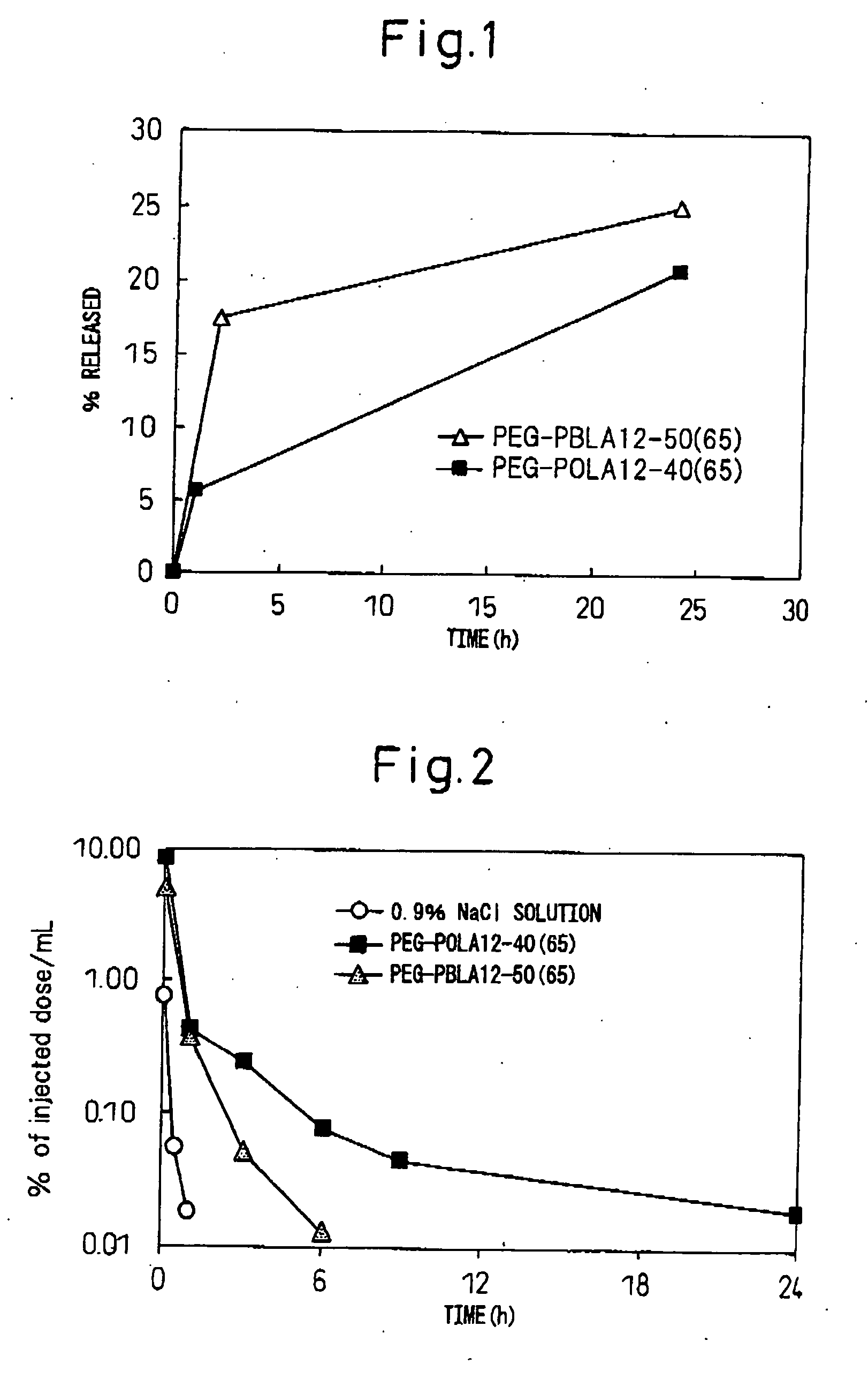
[0063]The block copolymer used was polyethylene glycol-co-polyaspartic acid benzyl ester (hereinafter, PEG-PBLA. In this polymer, the aspartic acid residues without benzyl esters are of general formula (I) wherein R5 is —O— and R6 is hydrogen (same hereunder). Also, all of the following block copolymers are of general formula (I) wherein R1 is CH3, L1 is —O—(CH2)3NH and R2 is COCH3. After precisely weighing out 10 mg of PEG-PBLA 12-50(65) into a glass vial, 1 ml of dichloromethane was added for dissolution. The solution was dried into a film under a nitrogen gas stream, and then further dried for about 1 hour under reduced pressure. To this there was added 62 μL of a 20 mM phosphate buffer (pH 6, 16.5 mg / mL) solution containing purified human IgG (MP Biomedicals Co.) (pI: approximately 8), and then 1.938 mL of 20 mM phosphate buffer (pH 6) or 20 mM TAPS buffer (pH 8) was slowly added while gently stirring at 4° C. After stirring overnight...

example 2

Human IgG-Encapsulating Micelle Preparation 2

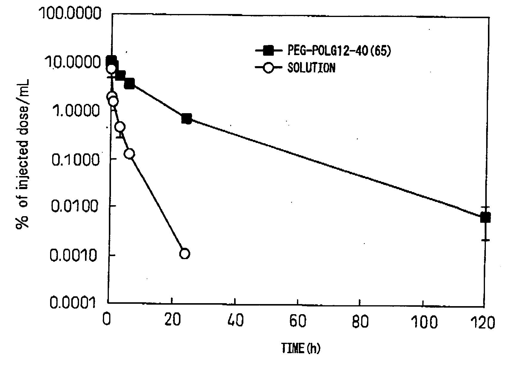
[0065]The block copolymer used was polyethylene glycol-co-polyglutamic acid benzyl ester (hereinafter, PEG-PBLG. In this polymer, the glutamic acid residues without benzyl esters are of general formula (I) wherein R5 is —O— and R6 is hydrogen (same hereunder)). After precisely weighing out 10 mg of PEG-PBLG 12-40(65) into a glass vial, 1 mL of dichloromethane was added for dissolution. The solution was dried into a film under a nitrogen gas stream, and then further dried for about 1 hour under reduced pressure. To this there was added 62 μL of a phosphate buffer solution (pH 6, 16.5 mg / mL) containing purified human IgG (MP Biomedicals Co.) (pI: approximately 8), and then 1.938 mL of 20 mM phosphate buffer (pH 6) or 20 mM TAPS buffer (pH 8) was slowly added while gently stirring at 4° C. After stirring overnight at 4° C., a Biodisruptor (High Power Unit, product of Nissei Corp.) was used for sonication for about 10 seconds (1 second interm...

example 3

Human IgG-Encapsulating Micelle Preparation 3

[0067]The block copolymer used was polyethylene glycol-co-polyaspartic acid octyl ester (hereinafter, PEG-POLA. In this polymer, the aspartic acid residues without octyl esters are of general formula (I) wherein R5 is —O— and R6 is hydrogen (same hereunder)). After precisely weighing out 10 mg of PEG-POLA 12-40(65) into a glass vial, 1 mL of dichloromethane was added for dissolution. The solution was dried into a film under a nitrogen gas stream, and then further dried for about 1 hour under reduced pressure. To this there was added 62 μL of a 20 mM phosphate buffer (pH 6, 16.5 mg / mL) solution containing purified human IgG (MP Biomedicals Co.) (pI: approximately 8), and then 1.938 mL of 20 mM phosphate buffer (pH 6) or 20 mM TAPS buffer (pH 8) was slowly added while gently stirring at 4° C. After stirring overnight at 4° C., a Biodisruptor (High Power Unit, product of Nissei Corp.) was used for sonication for about 10 seconds (1 second in...


PUM

PropertyMeasurementUnit
Fractionaaaaaaaaaa
Acidityaaaaaaaaaa
Hydrophilicityaaaaaaaaaa

Abstract

The invention provides a physiologically active polypeptide- or protein-encapsulating polymer micelle composition derived from a block copolymer comprising hydrophilic segments and hydrophobic segments.

Description

TECHNICAL FIELD[0001]The present invention relates to polymer micelles containing physiologically active polypeptides or proteins at a high content, and which can be biologically administered and are stable in vivo.BACKGROUND ART[0002]Advances in genetic engineering techniques have allowed numerous physiologically active polypeptides and proteins to be provided in a stable manner by cell culturing methods, for application in the treatment or prevention of diseases. Such polypeptides, however, generally have a short half-life in vivo due to their extremely rapid enzymolysis, metabolism and the like, and in most cases it has not been possible to obtain satisfactory effects when they are administered as drugs. A great deal of research has been conducted to date toward solving this issue, with focus on modification of the polypeptides and proteins with polymers or their sustained-release formulations.[0003]For example, polyethylene glycolation is a polymer modification technique current...

Claims


Application Information

Patent Timeline
IPC IPC(8): A61K9/127A61K38/02
CPCA61K9/0019A61K47/34A61K38/00A61K9/1075A61P1/04A61P1/16A61P1/18A61P13/02A61P13/12A61P15/00A61P35/00A61K9/127A61K38/16A61K38/21
Inventor OHUCHI, MIHOHARADA, MITSUNORIAMANO, YUKOKATO, YASUKI
Owner NANOCARRIER
Learn More