Methods and Apparatus for Optical Spectroscopic Detection of Cell and Tissue Death

a cell and tissue death and optical spectroscopic technology, applied in the field of tissue diagnosis, can solve the problems of inability to perform real-time monitoring of the reaction, the complexity of histological procedures, and the inability to detect cell death

Inactive Publication Date: 2009-11-26
VANDERBILT UNIV +1
View PDF69 Cites 21 Cited by
  • Summary
  • Abstract
  • Description
  • Claims
  • Application Information

AI Technical Summary

Problems solved by technology

The lack of ATP and / or cell membrane integrity leads to imbalance in ion, e.g., Na+, influx and efflux, and hence cell death occurs.
In addition to requiring cell or tissue samples from the host, these histological procedures usually are complex and very time-consuming.
Therefore, real time monitoring of the reaction cannot be performed, as the cell or tissue must be killed during the assay.
All of these techniques are costly, time consuming and labor intensive, and require the sacrifice of large numbers of animals in order to achieve meaningful results.
There are no current techniques available for real time monitoring of the cellular processes leading to cell death in the petri dish (in vitro) or in animal and human therapeutics trials (in vivo).
However, relatively few studies have addressed the diagnostic potential of optical spectroscopy in tumors, such as brain tumor and liver tumor.
Particularly, optical spectroscopic characteristics of liver tissues have not yet been developed.
Cancers, such as liver cancers, pose a significant problem to public health worldwide.
Unfortunately, about 70% of patients cannot have this surgery due to the size or location of the tumors or other health factors.
Currently, all thermotherapy procedures suffer from the lack of an adequate feedback control system, making it difficult to know precisely when to cease coagulation.
The accuracy of this approach, unfortunately, is hindered by the limitations of tumor margins detection using tumor echogenicity and stiffness.
This practice often yields unsatisfying therapeutic outcomes due to the fact that tissue characteristics vary drastically from patient to patient.
In addition, the dynamics of tissue characteristics, such as temperature-dependent tissue properties, further complicate the process of thermal coagulation of liver tissues.
Since the coagulation zones are often not visible during thermotherapy, it is difficult, if not impossible, to precisely determine the endpoint of therapy.
In addition, knowledge of A and Ea of tissues is largely unavailable.
These limitations make temperature-based feedback control of thermotherapies of liver tumors less than optimal.
Therefore, a heretofore unaddressed need exists in the art to address the aforementioned deficiencies and inadequacies.

Method used


Image

  • Methods and Apparatus for Optical Spectroscopic Detection of Cell and Tissue Death
  • Methods and Apparatus for Optical Spectroscopic Detection of Cell and Tissue Death
  • Methods and Apparatus for Optical Spectroscopic Detection of Cell and Tissue Death

Examples


example 1

Cell and / or Tissue Death Process Detection

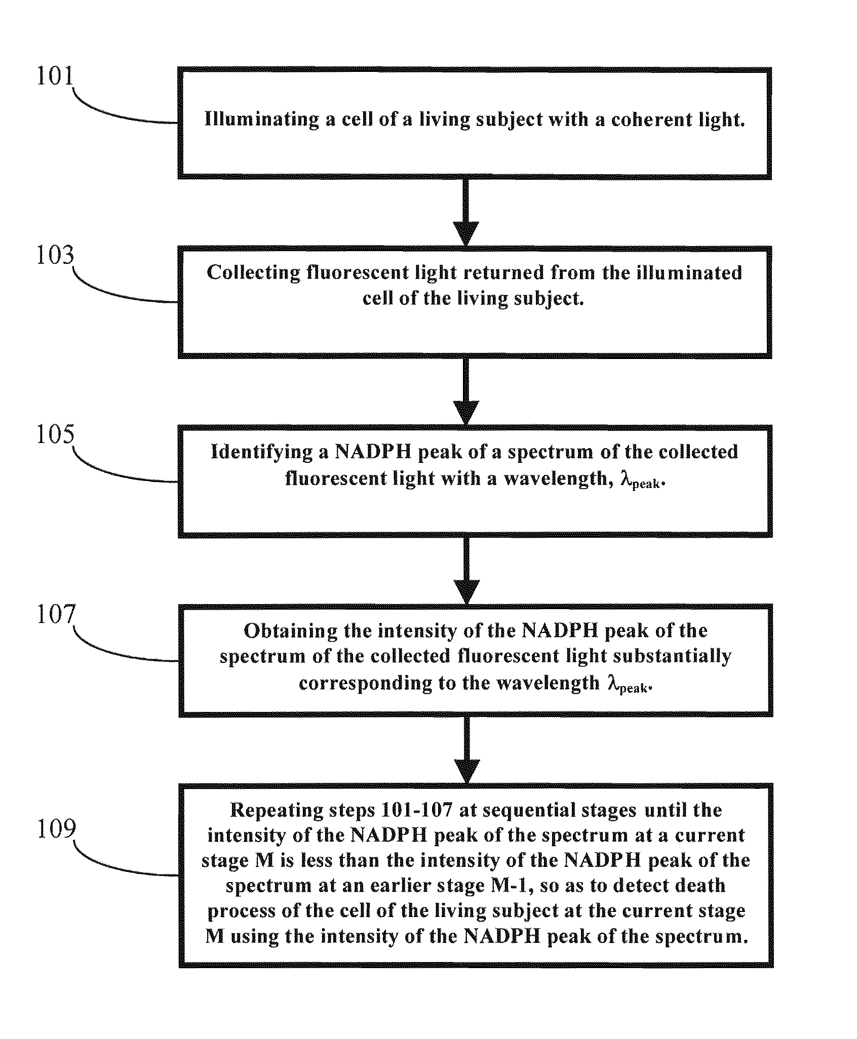
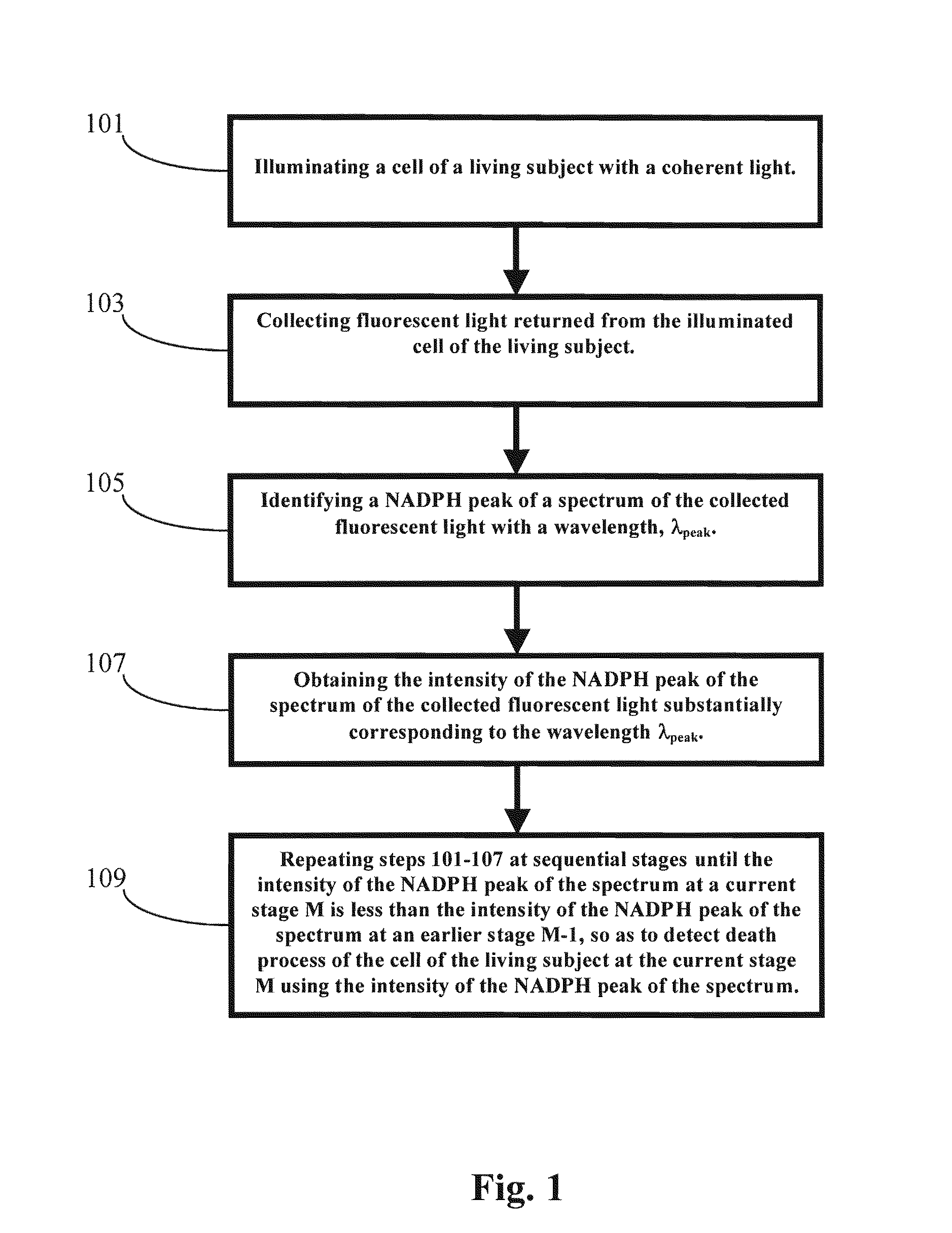
[0075]In one embodiment of the present invention, a cell or tissue of a living subject was native at a first stage and was subject to hyperthermia and / or cisplatin for various periods of time at the sequential stages. The cells or tissues of the living subject to be diagnosed were human colon adenocarcinoma cells (SW480). The NAD(P)H peak of the spectrum of the returned fluorescent light from the cell or tissue of the living subject, which was illuminated by the coherent light with an incident wavelength around 337 nm, was substantially at a wavelength around 460 nm. The NAD(P)H peak wavelength λpeak did not shifted from 460 nm for the cell or tissue of the living subject at different stages. However, the corresponding intensity of the NAD(P)H peak of the returned fluorescent spectrum dropped dramatically when the cell or tissue of the living subject were subject to hyperthermia and / or cisplatin for various periods of time at the sequential ...

example 2

In Vitro Liver Tissue Discrimination

[0083]In one embodiment, liver samples, including normal and malignant liver tissues, were obtained from patients receiving a hepatic tumor resection and liver transplant. The liver samples were stored in a freezer at about − / 80° C. if not used immediately. The frozen liver samples were passively thawed at room temperature for at least 30 minutes prior to the optical spectral data collections.

[0084]Fluorescence emission characteristics of the normal and malignant liver tissues were characterized with excitation of a coherent light having a wavelength from about 250 nm to about 550 nm in about 10 nm increments using a spectrofluorometer, such as a LS-50 (Perkin Elmer Inc., Shelton, Conn.): The fluorescence spectra collected from a single sample were then used to form an excitation-emission matrix (hereinafter “EEM”). The liver samples of the normal and malignant liver tissues were dissected into about 5 mm thick slabs with a surface area larger tha...

example 3

In Vivo Liver Tumor Differentiation

[0091]In vitro studies, small-scale human clinical trials were conducted at Vanderbilt University Medical Center. The experimental protocol for the clinical trials was approved by Vanderbilt Institution Review Board. Hepatic tumor patients receiving hepatic tumor resection were selectively recruited by a surgeon, according to the tumor sites and patient's medical condition. Prior to the clinical trials, the experimental procedures were carefully explained to the participants, and written consent was obtained from each of them.

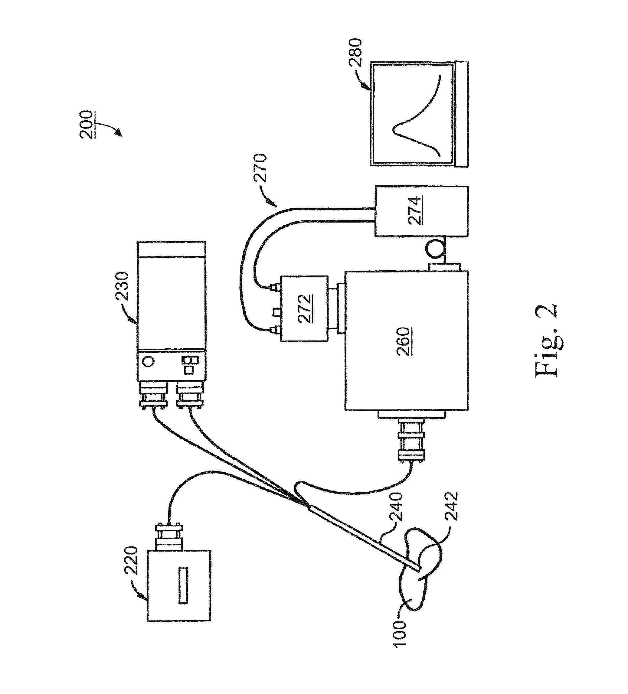
[0092]In the clinical studies, fluorescence and diffused reflectance spectra were acquired from perfused (i.e., prior resection) and non-perfused (i.e., post resection) liver tissue using the fiberoptic spectroscopic system described above. The fiber optical probe was sterilized, so it could be used in the operating field. Once the liver of the studied patient was exposed, perfused fluorescence and diffused reflectance spectra...


PUM


Abstract

A method for detecting death process of a cell or tissue of a living subject. In one embodiment, the method includes the steps of illuminating the cell or tissue of the living subject with a coherent light, collecting fluorescent light returned from the illuminated cell or tissue of the living subject, identifying a NAD(P)H peak of a spectrum of the collected fluorescent light with a wavelength, λpeak, and obtaining the intensity of the NAD(P)H peak of the spectrum of the collected fluorescent light substantially corresponding to the wavelength λpeak. These steps are repeated at sequential stages until the intensity of the NAD(P)H peak of the spectrum at a current stage is less than the intensity of the NAD(P)H peak of the spectrum at an earlier stage immediately prior to the current stage so as to detect death process of the cell of the living subject at the current stage using the intensity of the NAD(P)H peak of the spectrum.

Description

CROSS-REFERENCE TO RELATED PATENT APPLICATION[0001]This application is a continuation-in-part of U.S. patent application Ser. No. 10 / 613,225, filed Jul. 3, 2003, entitled “APPARATUS AND METHODS OF DETECTION OF RADIATION INJURY USING OPTICAL SPECTROSCOPY,” by Wei-Chiang Lin, Steven A. Toms, Anita Mahadevan-Jansen, Paul J. Phillips, Mahlon Johnson, and Robert J. Weil, the disclosure for which is hereby incorporated herein by reference in its entirety, which itself claims the benefit, pursuant to 35 U.S.C. § 119(e), of provisional U.S. patent application Ser. No. 60 / 394,217, which was filed on Jul. 5, 2002, entitled “APPARATUS AND METHODS OF DETECTION OF RADIATION INJURY USING OPTICAL SPECTROSCOPY,” by Wei-Chiang Lin, Steven A. Toms, Anita Mahadevan-Jansen, Paul J. Phillips, Mahlon Johnson, and Robert J. Weil, which is incorporated herein by reference in its entirety. This application also is a continuation-in-part of PCT patent application Serial No. PCT / US2003 / 031163, filed Sep. 30, ...

Claims


Application Information

Patent Timeline
Patent Type & Authority Applications(United States)
IPC IPC(8): A61B6/00A61BA61B5/00
CPCA61B5/0071A61B5/4244A61B5/0084A61B5/0075
Inventor LIN, WEI-CHIANGTOMS, STEVEN A.MAHADEVAN-JANSEN, ANITACHARI, RAVI S.
Owner VANDERBILT UNIV
Why Eureka
Try Eureka