Restoration of visual responses by in vivo delivery of rhodopsin nucleic acids

a rhodopsin and nucleic acid technology, applied in the field of molecular biology and medicine, can solve the problems of severe visual impairment in the centrally, no effective treatment or cure, complete blindness, etc., and achieve the effects of restoring visual evoked responses, restoring light sensitivity to the retina, and producing robust membrane depolarization or action potential firing

Active Publication Date: 2010-01-21
WAYNE STATE UNIV +1
View PDF9 Cites 89 Cited by
  • Summary
  • Abstract
  • Description
  • Claims
  • Application Information

AI Technical Summary

Benefits of technology

[0019]The present invention is directed to the genetic conversion of surviving light-insensitive inner retinal neurons in a retina in which photoreceptors are degenerating or have already died, into directly photosensitive neuronal cells, thereby imparting light sensitivity to such retinas and restoring one or more aspects of visual responses and functional vision to a subject suffering from such degeneration. By restoring light sensitivity to a retina lacking this capacity, due to disease, the invention provides a mechanism for the most basic light-responses that are required for vision. Said another way, the present invention introduces a “light sensors” into retinal neurons that normally do not have them, to compensate for loss of retinal photoreceptor cells.

Problems solved by technology

335-466), and can result in complete blindness.
Age-related macular degeneration (AMD) is also a result of the degeneration and death of photoreceptor cells, which can cause severe visual impairment within the centrally located best visual area of the visual field.
There are currently no effective treatments or cures for these conditions.
Unfortunately, for patients in advanced stages of retinal degeneration, this approach is not applicable, and the photoreceptor cells must be replaced.
However, both methods face many fundamental obstacles.
The electrica

Method used


Image

  • Restoration of visual responses by in vivo delivery of rhodopsin nucleic acids
  • Restoration of visual responses by in vivo delivery of rhodopsin nucleic acids
  • Restoration of visual responses by in vivo delivery of rhodopsin nucleic acids

Examples


example i

Materials and Methods

DNA and Viral Vector Constructions

[0172]The DNA fragment encoding the N-terminal fragment (Met1-Lys315) of Chop2 (Nagel et al., 2003) was cloned into pBluescript vector (Stratagene) containing the last exon of a mouse protamine 1 gene containing polyadenylation signal (mP1) and GFP cDNA inserted in frame at the 3′ end of the Chop2 coding fragment to produce a Chop2-GFP fusion protein. The function of Chop2-GFP chimera was verified in transfected HEK293 cells.

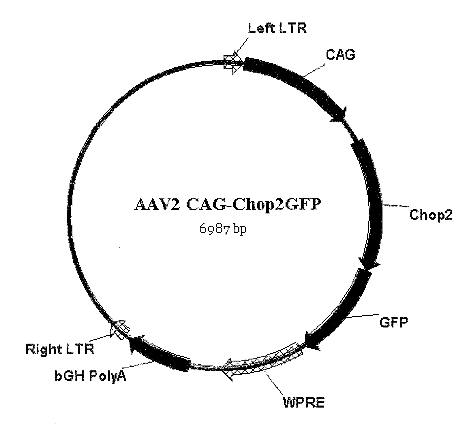
[0173]The viral expression construct rAAV2-C AG-Chop2-GFP-WPR E was made by subcloning the Chop2-GFP fragment into an adeno-associated (serotype-2) viral expression cassette. The viral cassette comprised a hybrid CMV enhancer / chicken β-actin promoter (CAG), a woodchuck posttranscriptional regulatory element (WPRE), and a bovine growth hormone (BGH) polyadenylation sequence. Viral vectors were packaged and affinity purified (GeneDetect).

[0174]The vector map is shown in FIG. 7.

[0175]The nucleic acid sequence o...

example 2

Expression of Chop2 in Retinal Neurons In Vivo

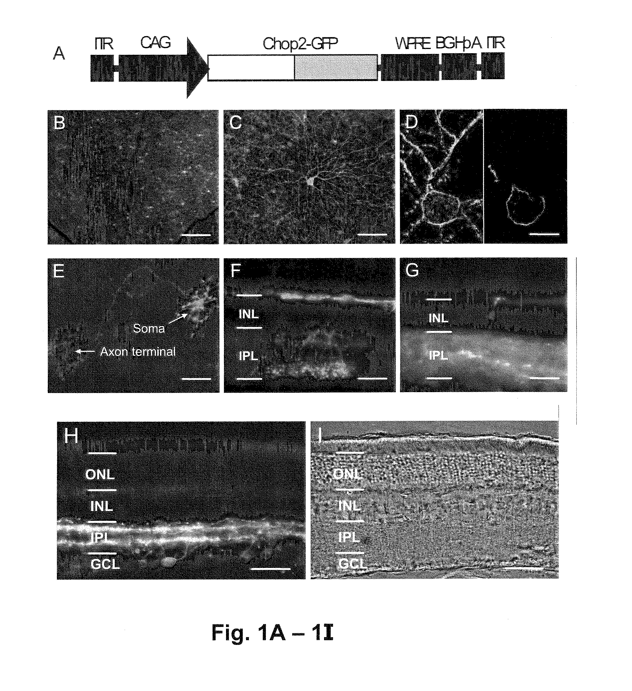
[0198]To directly visualize the expression and localization of Chop2 proteins, the C-terminal portion of the Chop2 channel was replaced with GFP, to make a Chop2-GFP chimera. The adeno-associated virus (AAV) vectors was selected to target the expression of Chop2-GFP fusion protein into retinal neurons because the capability of AAV vectors to deliver transgenes into nondividing cells, including inner retinal neurons (Harvey et al., 2002 and Martin et al., 2003), and to integrate the transgenes into the host genome (Flotte, 2004).

[0199]A viral expression cassette, rAAV2-C AG-Chop2-GFP-WPRE, was made by subcloning the Chop2-GFP chimera into an AAV serotype-2 expression cassette containing a hybrid CMV enhancer / chicken β-actin (CAG) promoter (FIG. 1A). To establish the expression and function of Chop2 channels in retinal neurons in general, we first examined the expression of Chop2 in nondystrophic retinas. The viral vector was injected into...

example 3

Properties of Light-Evoked Currents of ChR2-Expressing Inner Retinal Neurons

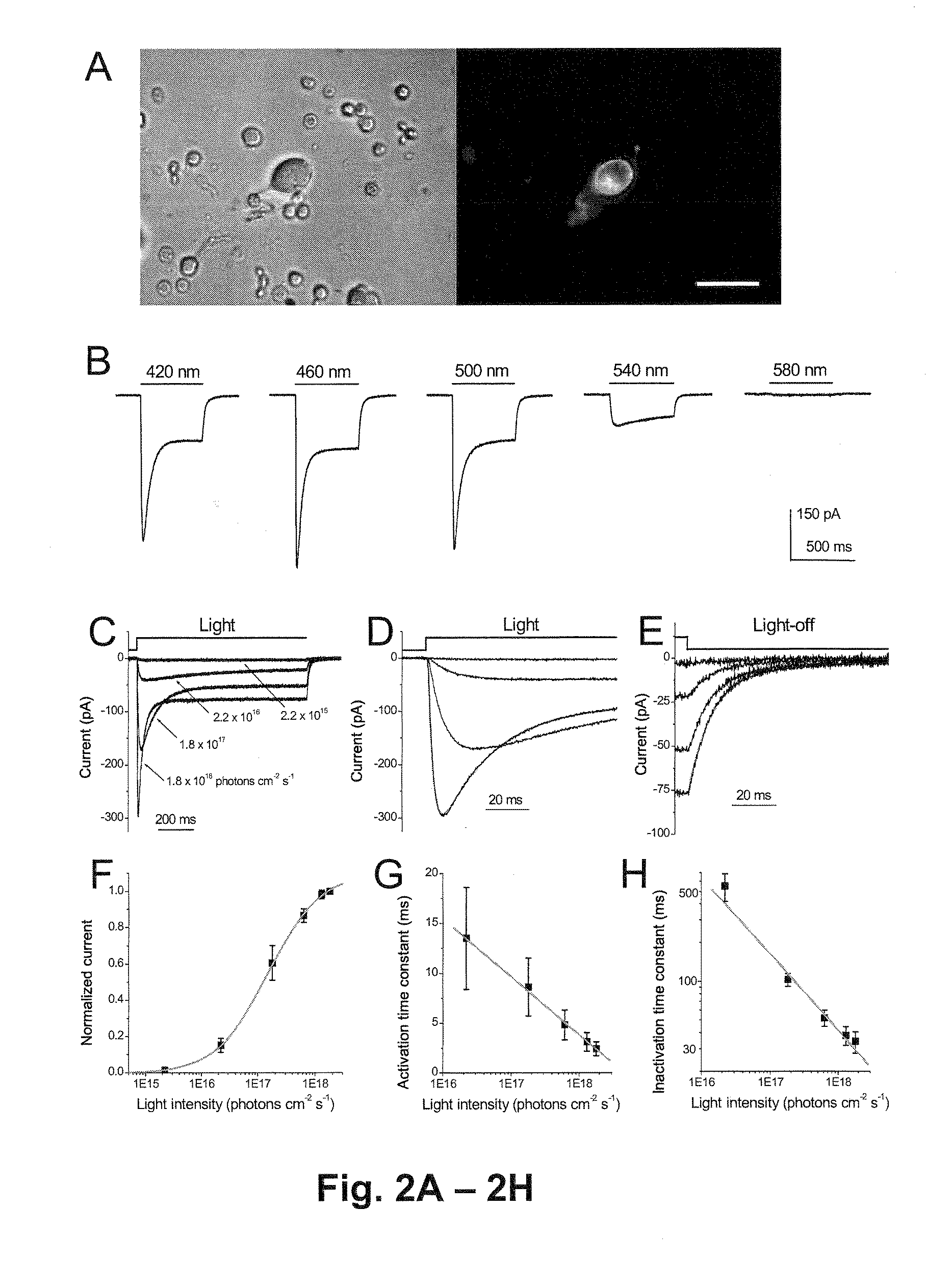
[0202]Functional properties of the Chop2 channels were examined in inner retinal neurons by using whole-cell patch-clamp recordings. The recordings were performed in acutely dissociated cells so that photoreceptor-mediated light responses were confidently excluded. Chop2-GFP-positive cells were identified by their GFP fluorescence (FIG. 2A). The precursor for the Chop2 chromophore group, all-trans retinal, was not added because it might be ubiquitously present in cells (Kim et al, 1992 and Thompson and Gal, 2003). Light-evoked responses were observed in all recorded GFP fluorescent cells (n=34), indicating that functional ChR2 (Chop2 with the chromophore attached) can be formed in retinal neurons with the retinal chromophore groups already present in the cells. Consistently, the expression of functional ChR2 channels has also been recently reported in cultured hippocampal neurons without the supply of exogen...


PUM

PropertyMeasurementUnit
Timeaaaaaaaaaa
Biological propertiesaaaaaaaaaa
Sensitivityaaaaaaaaaa

Abstract

Nucleic acid vectors encoding light-gated cation-selective membrane channels, in particular channelrhodopsin-2 (Chop2), converted inner retinal neurons to photosensitive cells in photoreceptor-degenerated retina in an animal model. Such treatment restored visual perception and various aspects of vision. A method of restoring light sensitivity to a retina of a subject suffering from vision loss due to photoreceptor degeneration, as in retinitis pigmentosa or macular degeneration, is provided. The method comprises delivering to the subject by intravitreal or subretinal injection, the above nucleic acid vector which comprises an open reading frame encoding a rhodopsin, to which is operatively linked a promoter and transcriptional regulatory sequences, so that the nucleic acid is expressed in inner retinal neurons. These cells, normally light-insensitive, are converted to a light-sensitive state and transmit visual information to the brain, compensating for the loss, and leading to restoration of various visual capabilities.

Description

STATEMENT OF RIGHTS TO INVENTIONS MADE UNDER FEDERALLY SPONSORED RESEARCH[0001]This invention was funded in part by grants from the National Institutes of Health grants (EY12180, EY-04068, EY16087, EY17130 and EY11522) which provides to the United States government certain rights in this invention.BACKGROUND OF THE INVENTION[0002]1. Field of the Invention[0003]The present invention in the field of molecular biology and medicine relates to the use of microbial-type rhodopsins, such as the light-gated cation-selective membrane channel, channelrhodopsin-2 (Chop2) to convert inner retinal neurons to photosensitive cells in photoreceptor-degenerated retina, thereby restoring visual perception and various aspects of vision.[0004]2. Description of the Background Art[0005]Vision normally begins when rods and cones, also called photoreceptors, convert light signals to electrical signals that are then relayed through second- and third-order retinal neurons and the optic nerve to the lateral g...

Claims


Application Information

Patent Timeline
IPC IPC(8): A61K35/76A61K31/7088
CPCA61K48/005C07K14/705C12N15/86A61K38/177C12N2830/002C12N2830/008A61K38/00C12N2750/14143A61P27/02
Inventor PAN, ZHUO-HUADIZHOOR, ALEXANDER M.
Owner WAYNE STATE UNIV
Why Eureka
Try Eureka