Eureka AIR delivers breakthrough ideas for toughest innovation challenges, trusted by R&D personnel around the world.

MRI trackable drug delivery particles, uses and methods thereof

a technology of magnetic resonance imaging and drug delivery particles, applied in the field of drug delivery particles, can solve the problems of limited traditional medical treatment, lack of specificity, and imposing limitations on therapy

Inactive Publication Date: 2010-03-11
EPITARGET
View PDF9 Cites 7 Cited by
  • Summary
  • Abstract
  • Description
  • Claims
  • Application Information

AI Technical Summary

Benefits of technology

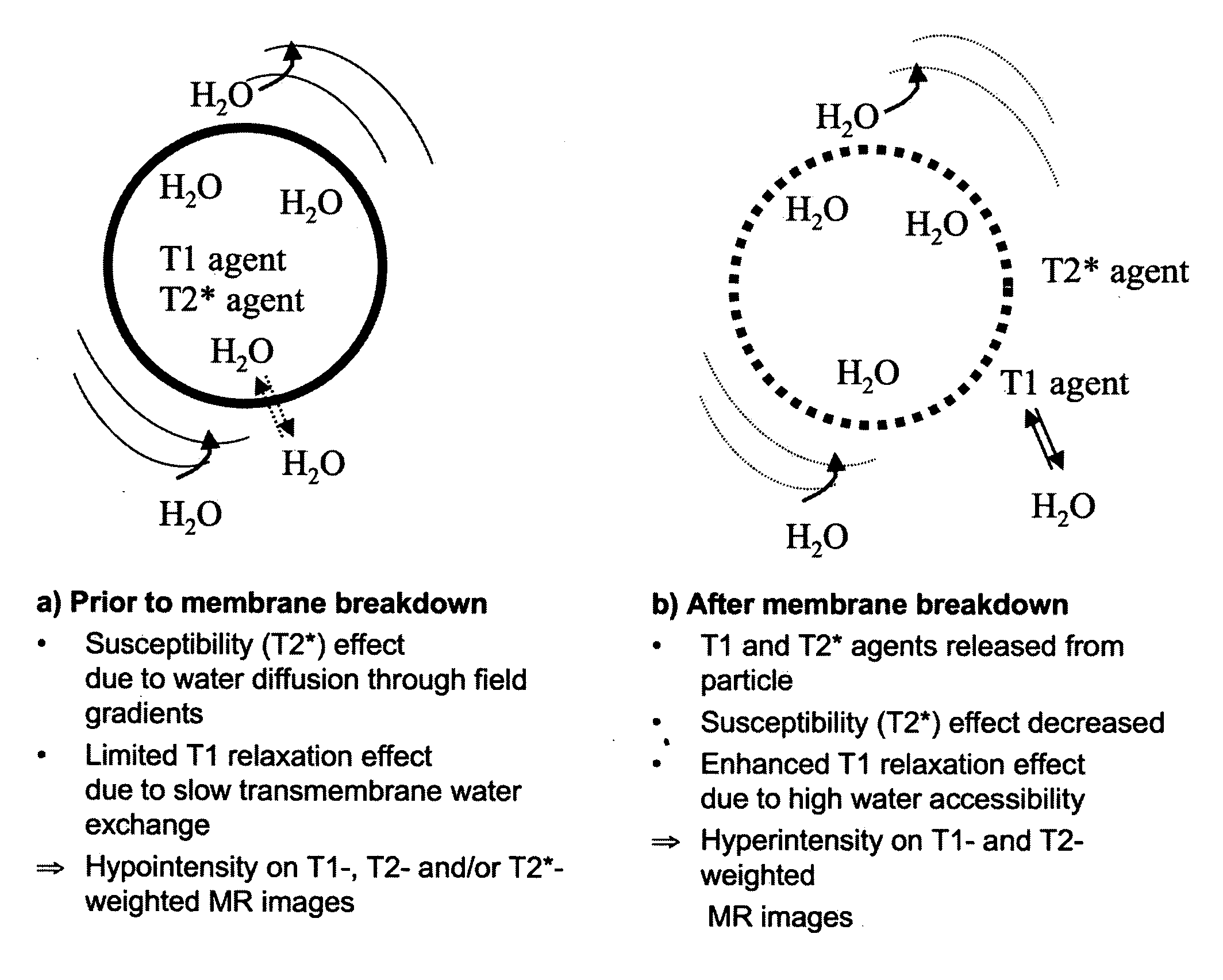
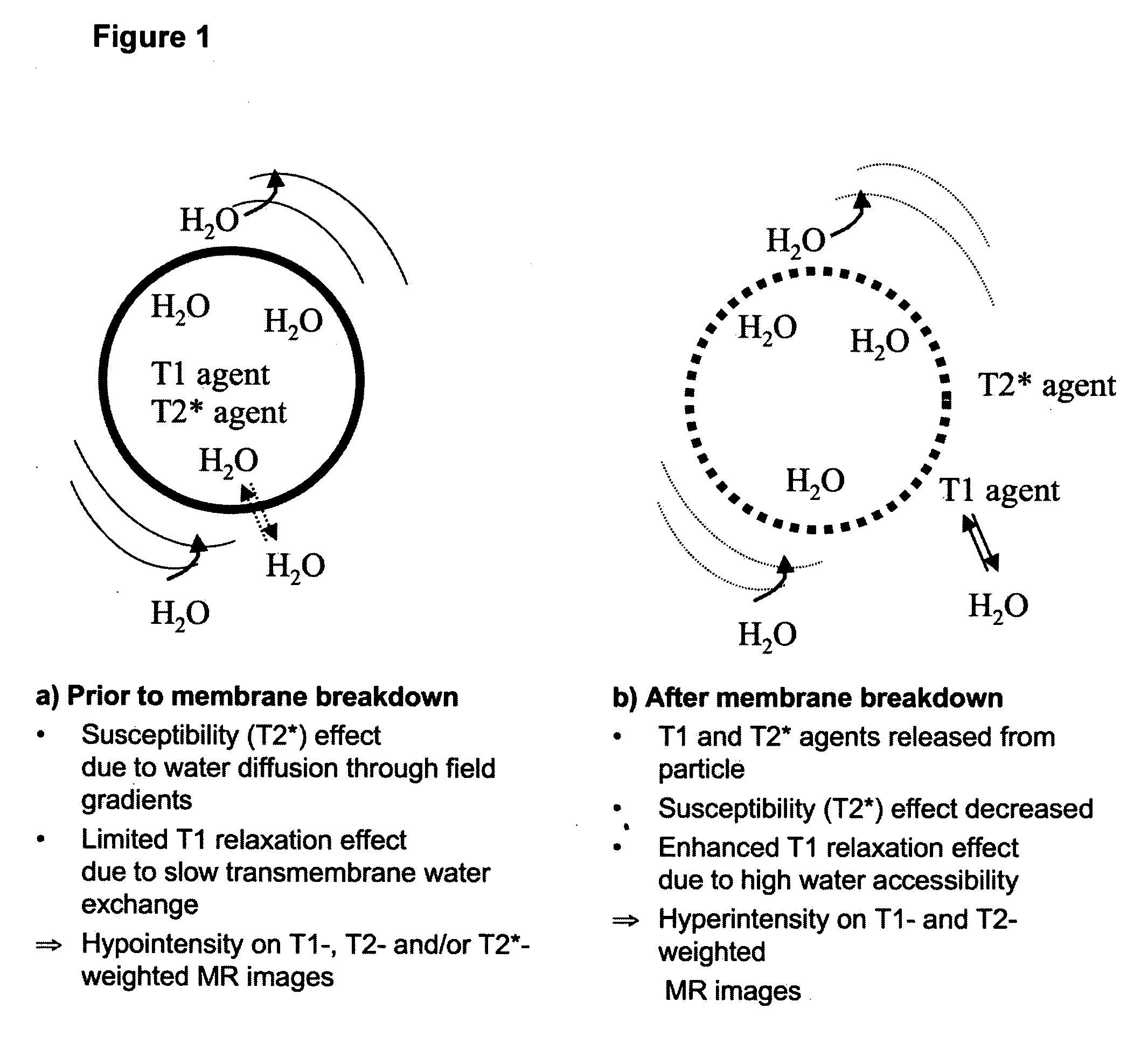
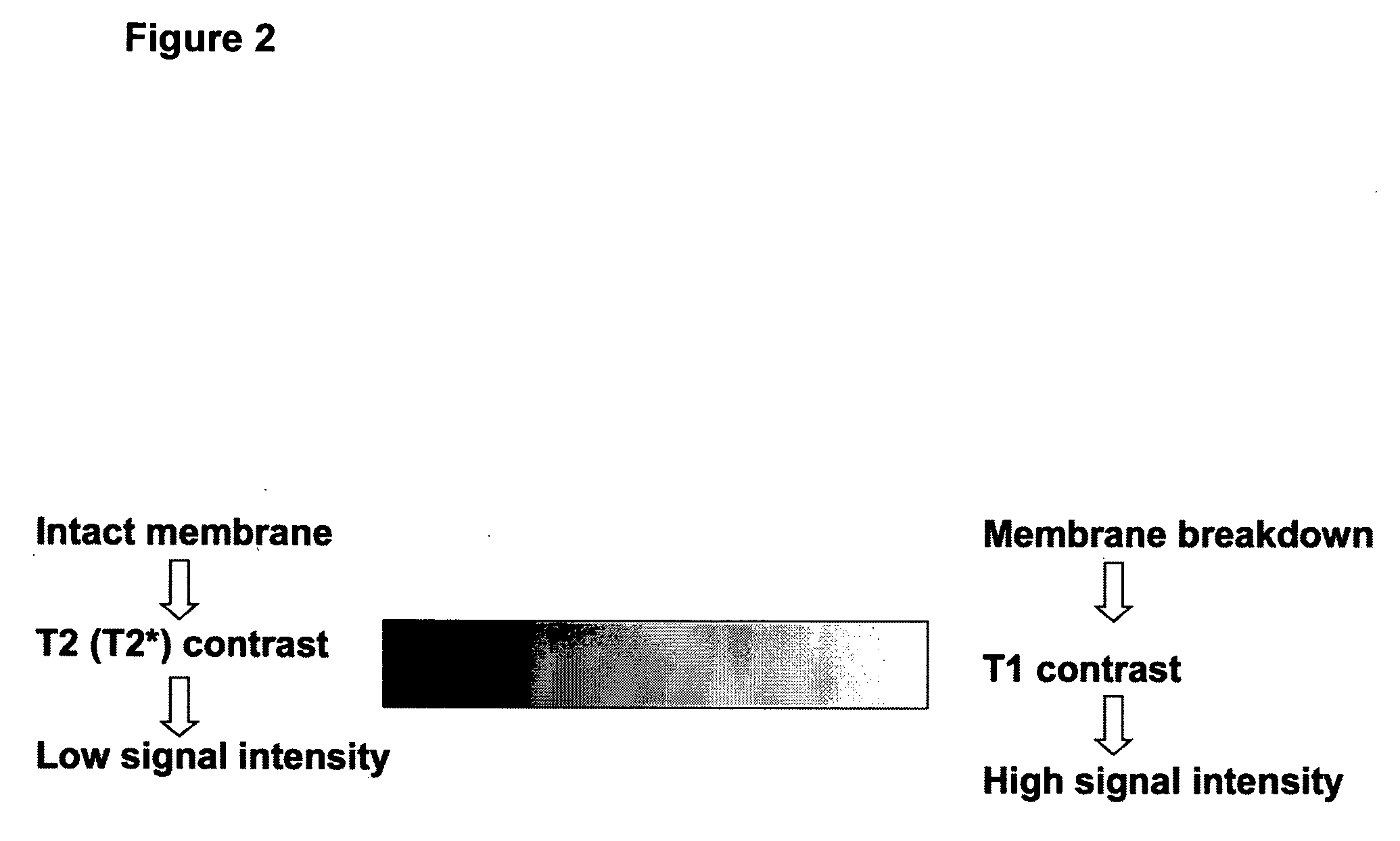
[0029]Even more specifically, the current invention comprises a trackable particulate material for drug delivery comprising a matrix or membrane material, a drug, and a T1 and a susceptibility (T2*) magnetic resonance contrast agent (or contrast agent per se), said matrix or membrane material being responsive only to non-physiological parameters and the response is chemical or physical breakdown of the matrix or membrane material, to cause the relaxation efficiency of only the T1 agent to increase.
[0033]A liposomal product for parenteral administration demands high chemical and colloidal stability both during storage and use. Additionally, it must be non-toxic and biologically compatible, e.g. isotonic and isohydric. The composition and design of the liposome depend upon the properties and applications of the liposomal product. Charge stabilization of liposomes is achieved by imparting a surface charge to the liposome surface, which is accomplished by employing negatively or positively charged phospholipids. Polymeric coating materials, such as polyethylene glycol (PEG), are also used to prevent particle fusion or aggregation by steric hindrance, thus increasing colloidal stability. Liposomes of high chemical stability are normally obtained by using saturated phospholipids with a gel-to-liquid crystal phase transition temperature (Tc) above 42° C., in practice phospholipids having saturated fatty acid portions with an acyl chain length of 14 carbon atoms or more are used. This is a crucial feature for liposome encapsulated material as the risk of leakage during storage and also in vivo is minimized. For membrane incorporated material, the use of saturated phospholipids is not so critical for minimizing leakage; however the use of saturated phospholipids is preferred to achieve acceptable chemical stability.
[0040]The diameter of the particulate material should not exceed 1000 nm. Preferably the diameter is below 1000 nm, more preferably below 250 nm, more preferably below 150 nm, and even more preferably around 100 nm. The current inventors prefer that the size of the material is within the range 50 to 150 nm. Small size is preferred to is maximize the probability of passive accumulation in target tissue due to the Enhanced Permeability and Retention Effect (EPRE) (Maeda et al, 1989).
[0042]As mentioned above, the T1 relaxation efficiency of the second magnetic resonance contrast generating species varies in response to drug release, more specifically, the second contrast species is only MR visible with respect to T1 effect during and / or after drug release. This is possible because drug release, particularly ultrasound induced drug release, will always coincide with relief of the relaxation exchange limitations. Hence, said second species is a T1 contrast agent of any type known to a skilled person, see e.g. EP 1069 88 B1 incorporated herein by reference. Typically, Gd chelates and Mn compounds are used. One or several T1 agent species may be comprised in the drug delivery particle, however, one species is preferred. In a preferred embodiment the T1 contrast agent is a Gd chelate either encapsulated in the aqueous phase of the particulate carrier and / or attached to the inner surface of the particulate carrier membrane. The T1 contrast agent, more particularly the Gd compound, renders qualitative and quantitative monitoring of the drug release process possible.
[0047]A further aspect of the present invention is use of the particulate material of the invention for monitoring spatial position of said material before drug release and efficiency of drug release.

Problems solved by technology

A serious limitation of traditional medical treatment is lack of specificity, that is, drugs do not target the diseased area specifically, but affect essentially all tissues.
This limitation is particularly evident in chemotherapy where all dividing cells are affected imposing limitations on therapy.
One challenge in this regard is to monitor both accumulation of the drug delivery entity in the diseased area and the extent of drug release.
Due to toxicity, these paramagnetic metal ions need to be administered in the form of stable chelates or other stabilizing entities.
Liposomes have also been extensively investigated as carriers for paramagnetic and superparamagnetic materials, but so far no liposomal MRI contrast agents are commercially available.
Hence, the need to concomitantly monitor position, particle concentration and drug release is neither realized nor solved.
None of the disclosed inventions are suitable for both monitoring particle position and drug release efficiency.

Method used


Image

  • MRI trackable drug delivery particles, uses and methods thereof
  • MRI trackable drug delivery particles, uses and methods thereof
  • MRI trackable drug delivery particles, uses and methods thereof

Examples


example 1

Preparation and Characterization of Liposome Containing a Gd Chelate and a Dy Chelate (“Paramagnetic Liposomes”)

[0058]DSPC and DSPE-PEG 2000 were purchased from Genzyme Pharmaceuticals (Liestal, Switzerland). Chloroform, methanol, calcein, HEPES, sodium azide and sucrose were all obtained from Sigma Aldrich. GdDTPA-BMA (Omniscan®) and DyDTPA-BMA (Sprodiamide) were kindly supplied by GE Healthcare and Rikshospitalet-Radiumhospitalet, respectively, both Oslo, Norway.

[0059]DSPC / DSPE-PEG 2000 (mole %; 92:8) liposomes were prepared by the thin film hydration method. The phospholipids were dissolved in a chloroform / methanol mixture (volume ratio; 10:1) and the organic solution was evaporated to dryness under reduced pressure. Liposomes were formed by hydrating the lipid film with a pre-heated (65 deg C) 10 mL aqueous solution containing GdDTPA-BMA chelate (150 mM), DyDTPA-BMA chelate (150 mM) and 4 mM HEPES (pH 7.4). The resulting liposome dispersion at a nominal phospholipid concentratio...

example 2

Preparation and Characterization of Liposome Containing a Gd Chelate, a Dy Chelate and the Drug Marker Calcein (“Paramagnetic Liposomes Containing Calcein”)

[0063]DSPC / DSPE-PEG 2000 (mole %; 92:8) liposomes were prepared analogously to Example 1, except that the aqueous solution used for lipid film hydration also contained 20 mM of the fluorescent dye calcein. The dialysed liposome dispersion was characterised with respect to key physicochemical properties as described in Example 1.

[0064]The intensity-weighted liposome size was 88 nm. The osmolality of the dialysed liposome dispersion was 640 mosmol / kg water. The effective concentration of Gd and Dy in the dialysed liposome dispersion was 5.2 mM.

example 3

Ultrasound Treatment of Liposome Samples

[0065]For the ultrasound experiments, the dialysed liposome dispersions (from Examples 1 and 2) were further diluted with isosmotic sucrose / 10 mM HEPES (pH 7.4) / 5 mM EDTA solution containing 0.02% w / v sodium azide. EDTA was supplied as the disodium and diydrate salt from Sigma Aldrich.

[0066]The ultrasound experiments were performed with a ‘Vibra-Cell’ 40 kHz ultrasonic processor, VC754, with a 1.9 cm diameter transducer, purchased from Sonics and to Materials, Inc. (CT, US). Ultrasound with a 20% amplitude was applied for 4 minutes to the diluted liposome dispersions (dispensed in plastic flask). The plastic flasks containing liposomes were during ultrasound exposure placed in a water / ice bath to minimise any ultrasound mediated heating effect. The sample temperature rose from 10 deg C. prior to ultrasound treatment to 20 deg C. after completed treatment.


PUM

PropertyMeasurementUnit
temperatureaaaaaaaaaa
physiological temperaturesaaaaaaaaaa
Tcaaaaaaaaaa

Abstract

The current invention discloses a drug delivery system allowing monitoring of spatial position and drug release, as well as methods and uses thereof. More particularly, the drug delivery system comprises drug carrying particles comprising novel combinations of magnetic resonance imaging contrast agents.

Description

FIELD OF THE INVENTION[0001]The present invention relates to a drug delivery particle allowing monitoring of spatial position and drug release. More particularly, the invention relates to drug carrying particles comprising magnetic resonance imaging contrast agents, as well as methods and uses thereof.BACKGROUND OF THE INVENTION[0002]A serious limitation of traditional medical treatment is lack of specificity, that is, drugs do not target the diseased area specifically, but affect essentially all tissues. This limitation is particularly evident in chemotherapy where all dividing cells are affected imposing limitations on therapy. One strategy to achieve improved drug specificity is incorporation or encapsulation of drugs for example in liposomes, plurogels and polymer particles. To further improve efficiency, ultrasound (US) mediated drug release from such particles has been disclosed in several publications, for a review see Pitt et al. 2004. Other approaches are heat mediated rele...

Claims


Application Information

Patent Timeline
Patent Type & Authority Applications(United States)
IPC IPC(8): A61K49/18
CPCA61K9/1271A61K49/1812A61K41/0028
Inventor FOSSHEIM, SIGRID L.NILSSEN, ESBEN A.
Owner EPITARGET
Why Eureka
Eureka Blog
Learn More