
Cell-Scaffold Constructs

a cell-scaffold and construct technology, applied in the field of cell-scaffold constructs, can solve the problems of abnormal bladder development and surgical augmentation, small bladder capacity that generates high pressure, and inconformity with bladder regulations

Inactive Publication Date: 2010-05-27
ORGAGEN INC
View PDF40 Cites 65 Cited by
  • Summary
  • Abstract
  • Description
  • Claims
  • Application Information

AI Technical Summary

Benefits of technology

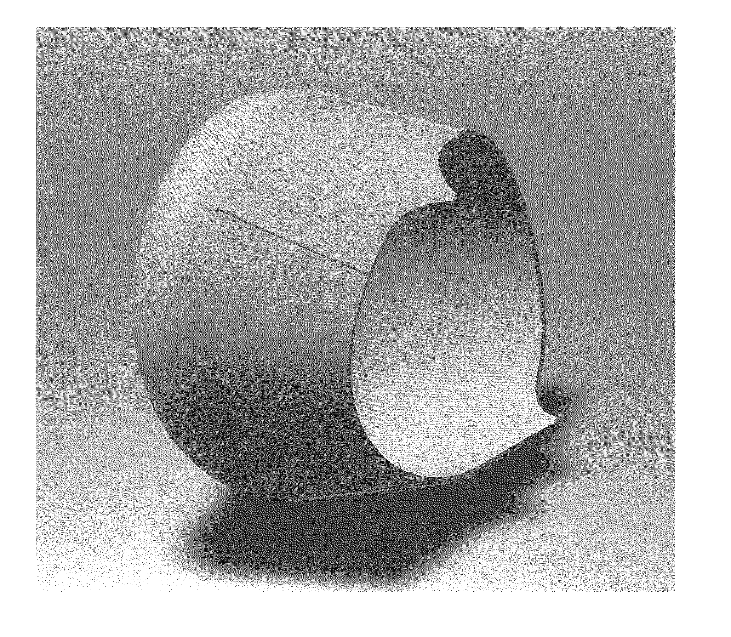
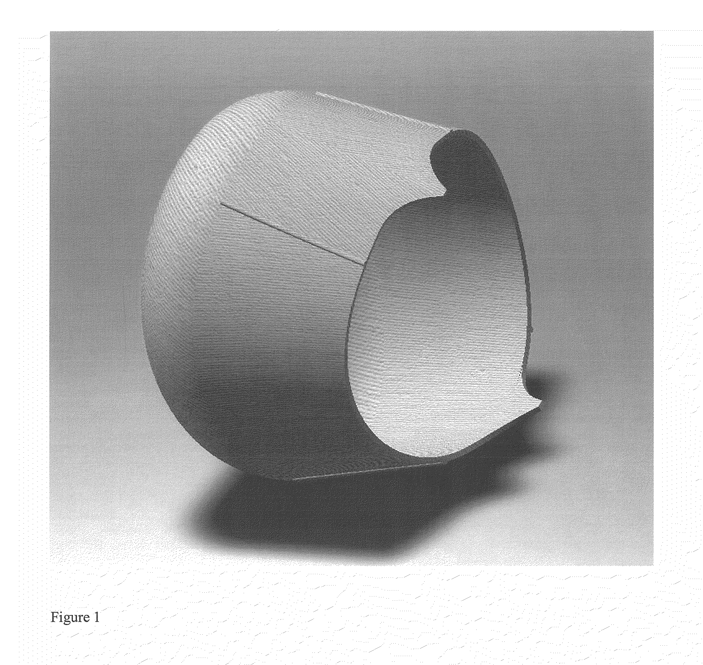
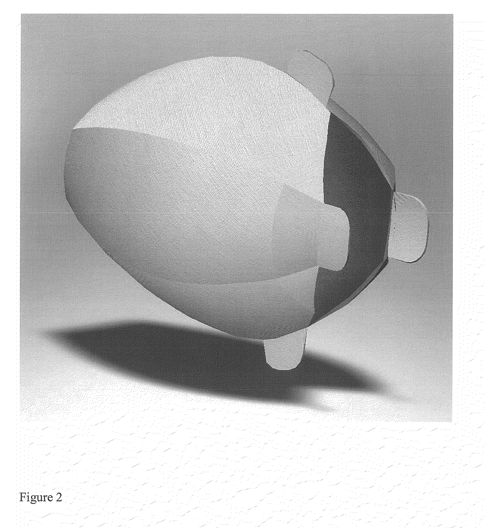
[0057]In another aspect, the instant invention provides methods for increasing bladder volumetric capacity of a bladder in a subject in need of such treatment by providing a biocompatible synthetic or natural polymeric matrix shaped to conform to at least a part of the bladder in need of such treatment and of a sufficient size to be laparoscopically implanted, depositing an autologous cell population that is not derived from the subject's bladder on or in a first area of the polymeric matrix, and laparoscopically implanting the shaped polymeric matrix construct laparoscopically into the subject at the site of the treatment such that bladder volume capacity is increased. In one embodiment, the bladder volume capacity is increased about 50 mL. In another embodiment, the bladder volume capacity is increased about 100 mL.
[0059]In still another aspect, the present invention provides methods for the treatment of urinary incontinence in a subject in need of such treatment by providing a biocompatible synthetic or natural polymeric matrix shaped to conform to at least a part of the subject's bladder and of a sufficient size to be laparoscopically implanted, depositing an autologous cell population that is not derived from the bladder on or in a first area of the polymeric matrix, laparoscopically implanting the shaped polymeric matrix construct laparoscopically into the subject at the site of the treatment such that bladder volume capacity is increased.

Problems solved by technology

Several anomalies can cause the bladder to develop abnormally and require surgical augmentation.
Conditions such as posterior urethral valves, bilateral ectopic ureters, bladder extrophy, cloacal extrophy, and spina bifida (ie, myelomeningocele) may cause the bladder to be noncompliant, resulting in a small capacity bladder that generates high pressures.
Clinically this causes patients to suffer from incontinence while increasing their risk for renal failure due to the high pressures in the genitourinary system.
The surgeries are relatively complex and expensive.
Even in patients with a good technical result, the procedure is associated with numerous immediate risks and chronic complications.
The invasiveness, cost, and complications of these surgeries limit their use to only the most severe bladder deficiencies.
The urinary bladder is subject to numerous maladies and injuries which cause deterioration of the urinary bladder in patients.
Further, bladder deterioration may also occur as a result of trauma such as, for example, car accidents and sports injury.
Other problems have been reported with the use of certain gastrointestinal segments for bladder surgery including stone formation, increased mucus production, neoplasia, infection, metabolic disturbances, long term contracture and resorption.
The use of natural materials for urinary diversion has shown that bladder tissue, with its specific muscular elastic properties and urothelial impermeability functions, cannot be easily replaced.
The requirement of multiple surgeries increases the overall cost of the procedures, the risk to the patient, and patient's overall comfort.
Urinary incontinence is a prevalent problem that affects people of all ages and levels of physical health, both in the community at large and in healthcare settings.
Economically, the costs are astounding; in the United States alone, over ten billion dollars per year is spent managing incontinence.
Despite all of the knowledge associated with incontinence, the majority of cases of urge incontinence are idiopathic, which means a specific cause cannot be identified.
Consequences of detrusor malfunction such as hyperreflexia include poor bladder compliance, high intravesical pressure, and reduction in bladder capacity, all of which may result in deterioration of the upper urinary tract.
The large size of the botulinum toxin molecule can limit its ability to diffuse, and thus prohibits it from reaching both afferent and efferent nerve fibers.
As a result, current methods of administration for overactive bladder (OAB), for example, require many injections (typically 20 to 50) of botulinum toxin into the bladder muscle wall, thus increasing the number of doctor visits and associated cost of treatment.
Moreover, the safety of chronic long-term impact of inhibition of sensory neurotransmitter release from bladder has not yet been determined.
Often, however, such drug therapies do not achieve complete success with all classes of incontinent patients, and often results in the patient experiencing significant side effects.
While endoscopically directed injections of collagen around the bladder neck has a quite high success rate in sphincter deficiency with no significant morbidity, the use of collagen can result in failures that occur after an average of two years and considerations need to be given to its cost effectiveness (Khullar V. et al., British J. Obstetrics & Gynecology, 104:96-99 (1996)).
Despite of the limited success rate, transurethral collagen injection therapy remains an acceptable treatment for intrinsic sphincter deficiency, due to the lack other suitable alternatives.
However, if these non-invasive pharmaceutical products fail, physicians offer a more invasive solution.
However, there are challenges associated with a dependence upon the development and maintenance of cell culture systems from the primary organ site as the basic unit for developing new and healthy engineered tissues.
For example, the treatment of a defective bladder poses a particular challenge regarding cell sourcing because it stands to reason that culturing bladder cells from a defective bladder will result in the cultured cells also being defective.
Such cells are not a wise choice for populating an implantable neo-bladder scaffold or matrix.

Method used


Image

  • Cell-Scaffold Constructs
  • Cell-Scaffold Constructs
  • Cell-Scaffold Constructs

Examples


example 1

Peripheral Blood and Adipose Tissue as a Source of SMCs

Blood-Derived Cells

[0372]Smooth muscle cells have been successfully isolated from canine, porcine, and human peripheral blood. Briefly, a dilution of 50 ml of peripheral blood 1:1 with phosphate buffered saline (PBS; 100 mL final volume) was prepared and layered onto Histopaque, a density gradient material, and centrifuged at 1,354×g for 20 minutes at room temperature. After centrifugation, four layers will be clearly defined in the density gradient (from top to bottom): serum, buffy coat, Histopaque, red blood cells. The mononuclear cells are located in the buffy coat, which appears as an opaque white / gray band. The buffy coat was withdrawn and transfered into a separate 50 ml conical tube. Dilute to 50 mL with PBS. Centrifuge the samples at 711×g for 10 minutes at room temperature to pellet cells. Resuspend pellet and culture the cells. When appropriate cell numbers are reached by subsequent cell passaging, an aliquote is fixe...

example 2

MCP-1 Production and Cell Density

[0413]Conditioned medium from cultures of bladder smooth muscle cells were analyzed using commercially available kits for the detection and quantitation of MCP-1. Conditioned media samples from 9 constructs (3 from each of 3 seeding levels) and the paired SMC cells used for seeding the constructs were tested for MCP-1 levels. The results are shown in Table 2.1.

TABLE 2.1Sam-TestplecIL2cIL6cIL10cMCP-1cIFNgcTNFacTGFbIDIDpg / mlpg / mlpg / mlpg / mlpg / mlpg / mlpg / ml1TT11.02TT28.839.6

[0414]In order to quantitate MCP-1 present in the construct medium, an ELISA based assay system specific for Canine MCP-1 from R&D Systems was employed. Samples were assayed in duplicate and compared to a standard curve to provide estimated MCP-1 levels in construct medium. As shown in FIG. 25, the results from this analysis show a positive correlation between MCP-1 production and the density of cells seeded. Table 2.2 shows MCP-1 quantitation of construct medium as determined by R&D S...

example 3

Implantation of Neo-Urinary Conduit Constructs in Swine

[0417]The objective of this study was to investigate the surgical implantation of the neo-urinary conduit and evaluation of the post surgical care, as well as to assess the regeneration of urinary-like tissue following implantation of the Neo-Urinary Conduit (NUC) test articles and the ability of swine peritoneum to provide a vascular supply and water-tightness to the implant.

[0418]Neo-Urinary Conduit (NUC) construct test articles were comprised of a scaffold formed from nonwoven polygycolic acid (PGA) felts and poly(lactic-co-glycolic acid) polymers (PLGA) with or without autologous smooth muscle cells (SMC). Cells previously removed from an animal were used to produce the construct that was implanted in the same animal. A construct refers to the sterile tube-shaped biomaterial comprised of a scaffold formed from nonwoven polygycolic acid (PGA) felts and poly(lactic-co-glycolic acid) polymers (PLGA) combined with autologous SMC...


PUM

PropertyMeasurementUnit
biocompatibleaaaaaaaaaa

Abstract

The present invention relates to the regeneration, reconstruction, repair, augmentation or replacement of organs or tissue structures using scaffolds and autologous cells that are not derived from such organs or tissues.

Description

RELATED APPLICATIONS[0001]This application claims priority to under Section 119(e) and the benefit of U.S. Provisional Application Ser. Nos. 61 / 111,242 filed on Nov. 4, 2008; 61 / 113,542 filed on Nov. 11, 2008; 61 / 114,021 filed on Nov. 12, 2008; 61 / 114,382 filed on Nov. 13, 2008; 61 / 114,388 filed on Nov. 13, 2008; 61 / 201,555 filed on Dec. 10, 2008; 61 / 201,550 filed on Dec. 10, 2008; and 61 / 201,554 filed on Dec. 10, 2008, the disclosures of which are incorporated by reference herein in their entirety.FIELD OF THE INVENTION[0002]The present invention relates to the regeneration, reconstruction, repair, augmentation or replacement of laminarly organized luminal organs or tissue structures using scaffolds seeded with cells obtained from autologous sources.BACKGROUND OF THE INVENTION[0003]Several anomalies can cause the bladder to develop abnormally and require surgical augmentation. Conditions such as posterior urethral valves, bilateral ectopic ureters, bladder extrophy, cloacal extroph...

Claims


Application Information

Patent Timeline
Patent Type & Authority Applications(United States)
IPC IPC(8): A61F2/04B05D5/00
CPCA61L27/18A61L27/3804A61L27/3826A61F2/042A61L2430/22A61L27/3882C08L67/04A61F2/02A61L27/40
Inventor LUDLOW, JOHN W.JAYO, MANUEL J.BASU, JOYDEEPBERTRAM, TIMOTHY A.GENHEIMER, CHRISTOPHER W.GUTHRIE, KELLY I.ILAGAN, ROGER M.JAIN, DEEPAKKNIGHT, OLUWATOYIN A.PAYNE, RICHARDQUINLAN, SARAH F.RAPOPORT, H. SCOTTSANGHA, NAMRATA D.
Owner ORGAGEN INC
Learn More