

Second ion mass spectrometry method and imaging method
a mass spectrometry and second ion technology, applied in the field of secondary ion mass spectrometry method and imaging method, can solve the problems of insufficient measurement, poor ionization efficiency, low secondary ions derived from organic molecules, etc., and achieve excellent ionization efficiency, high sensitivity, and suppress the destruction of organism-related materials
- Summary
- Abstract
- Description
- Claims
- Application Information
AI Technical Summary
Benefits of technology
Problems solved by technology
Method used
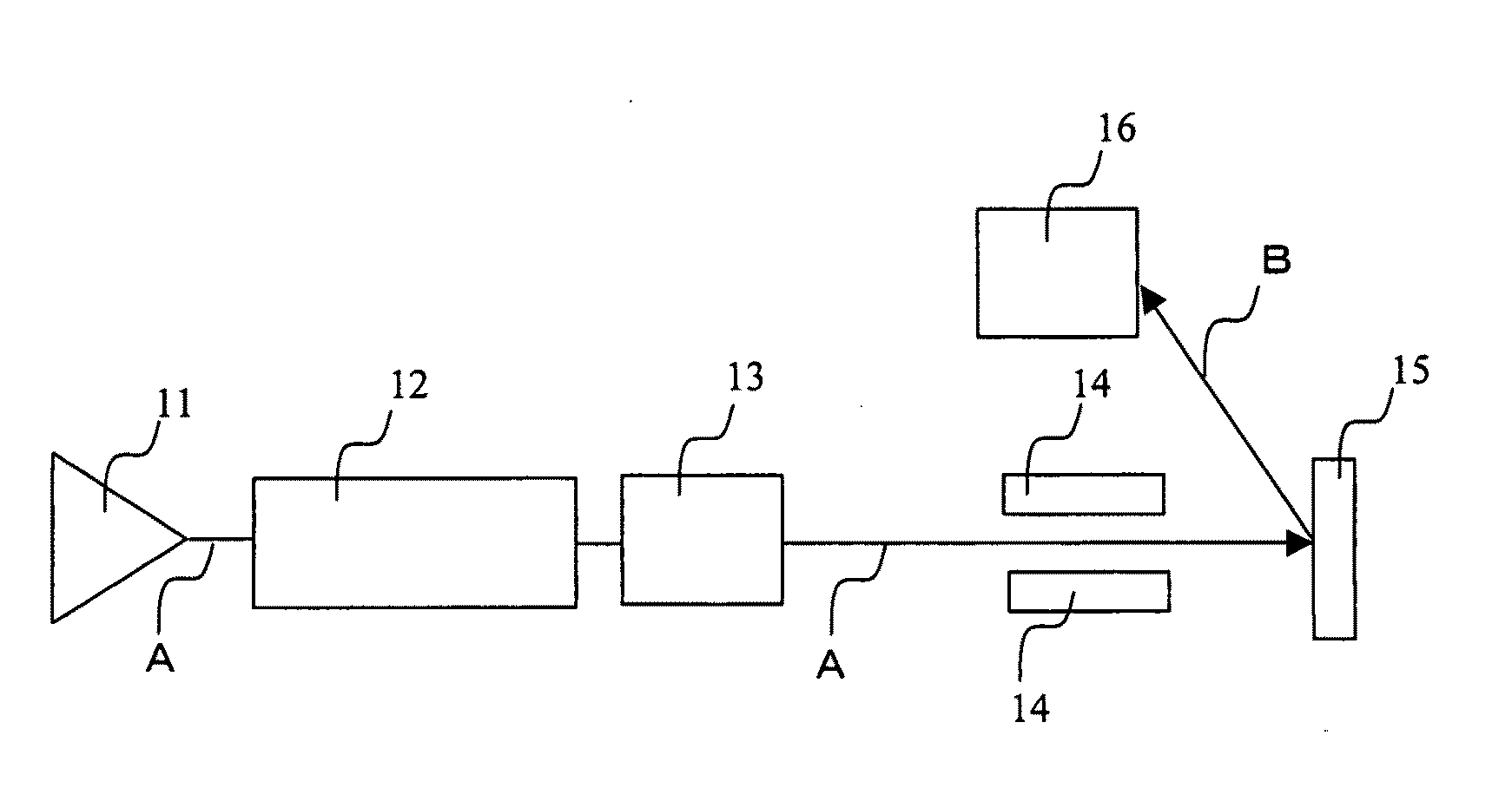
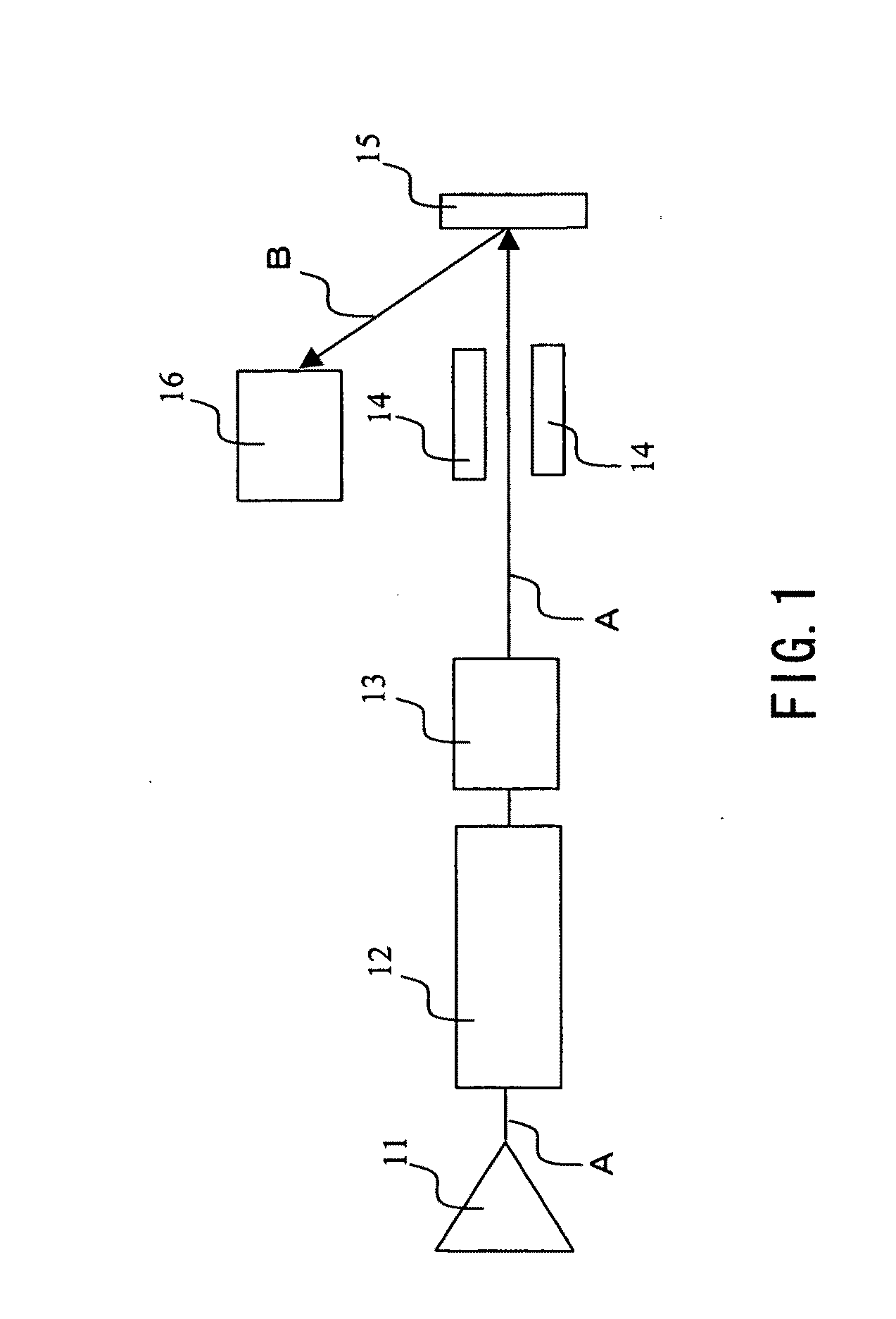
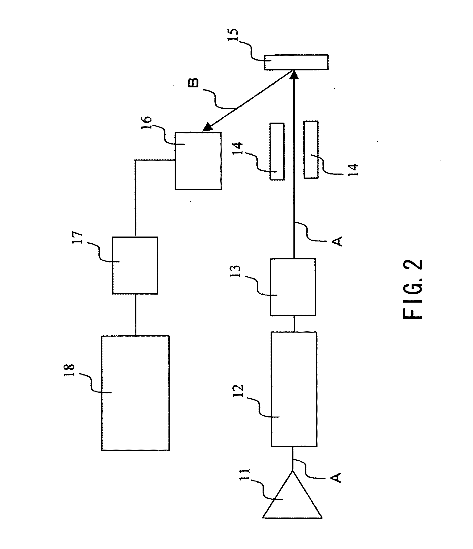
Image

Examples
example 1
[0069]A fast heavy ion beam of MeV was irradiated, and secondary ions thus generated were detected, whereby trehalose was analyzed.
[0070]A trehalose solution was spincoated on a single crystal Si substrate so as to form a trehalose thin film having a thickness of 100 nm. Then, the trehalose thin film was irradiated with a fast heavy ion beam under the following conditions, and secondary ions (negative ions) thus generated were detected. FIG. 4 shows a resultant mass spectrum obtained when an ion beam of 9 MeV (Au5+) was irradiated.
[0071](Condition)
[0072]Incident ion: 3 MeV (15 keV / amu)[0073]6 MeV (30 keV / amu)[0074]9 MeV (45 keV / amu)
[0075]Sample: trehalose thin film (molecular weight: 342.30)
[0076]Beam amount: −10 pA (F.C. measurement with a suppressor)
[0077]Beam diameter: 2 mm
[0078]Pulse: 50 nanoseconds, repetition: 10 kHz
[0079]Measuring time: 500 seconds
[0080]Irradiation amount per measurement: −106 ions[0081](−108 ions / cm2)
[0082]Incident angle: 30°
[0083]As shown in FIG. 4, by the ...
example 2
[0088]A fast heavy ion beam (Au5+) of 9 MeV was irradiated, and secondary ions thus generated were detected, whereby arginine was analyzed.
[0089]An arginine solution was spincoated on a single crystal Si substrate so as to form an arginine thin film (molecular weight: 174.2) having a thickness of 100 nm. Then, the arginine thin film was irradiated with an ion beam of MeV under the same conditions as in Example 1, and secondary ions (positive ions) were detected. FIG. 7 shows a resultant mass spectrum.
[0090]As shown in FIG. 7, a peak of arginine was detected. In particular, a large peak of parent ions (Arg+H)+ was observed, which proved that amino acid was less likely to be decomposed even by the irradiation of an ion beam of MeV.
example 3
[0091](1) A trehalose thin film and an arginine thin film were formed on respective surfaces of Si substrates in the same manners as in Examples 1 and 2, and the relationship between the yield of secondary ions generated and an electronic stopping power was confirmed. The yield of secondary ions was obtained as a ratio between secondary ions and primary ions (secondary ion / primary ion). The ion species, the energy, and the normalized energy (square of the speed) of an ion beam to be irradiated are as follows.
TABLE 1EnergyNormalized energyIon species10 keV 0.25 keV / amu Ar+0.5 MeV 2.5 keV / amu Au+1 MeV 5 keV / amuAu2+1.5 MeV 7.5 keV / amu Ar+3 MeV15 keV / amuAr3+6 MeV30 keV / amuAu4+9 MeV45 keV / amuAu5+
[0092]The results are shown in FIG. 8. In the figure, a number represents the energy (unit: MeV) of an ion beam, and symbols (▪), (□), (▴), and (Δ) represent results of positive ions of arginine, negative ions of arginine, positive ions of trehalose, and negative ions of trehalose, respectively. ...
PUM
 Abstract
Description
Claims
Application Information