Xenograft Bone Matrix for Orthopedic Applications

a bone matrix and orthopedic technology, applied in the field of orthopedic treatment of defective bones, can solve the problems of limited graft material volume and morbidity at the donor site, and achieve the effect of diminishing the immunological response of human to pigs and increasing immunocompatibility

Inactive Publication Date: 2010-08-05
APERION BIOLOGICS
View PDF2 Cites 0 Cited by
  • Summary
  • Abstract
  • Description
  • Claims
  • Application Information

AI Technical Summary

Benefits of technology

[0008]This invention provides an effective xenograft demineralized and deantigenated bone matrix, with osteoconductive and osteoinductive potential following treatment with ∝-galactosidase to eliminate ∝-Gal epitopes. This bone matrix has an increased immunocompatibility. The invention also provides a treatment strategy for xenograft bone particulate and shows the osteoconductive and osteoinductive properties while diminishing the hum

Problems solved by technology

Problems with autogenous bone harvest from the iliac crest site are donor site morbidity and limita

Method used


Image

  • Xenograft Bone Matrix for Orthopedic Applications
  • Xenograft Bone Matrix for Orthopedic Applications

Examples


example i

Evaluation of Xenograft Materials in Primates—Cancellous and Cortical Bone Models

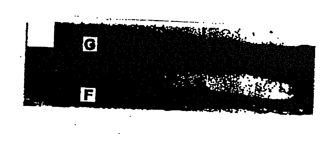
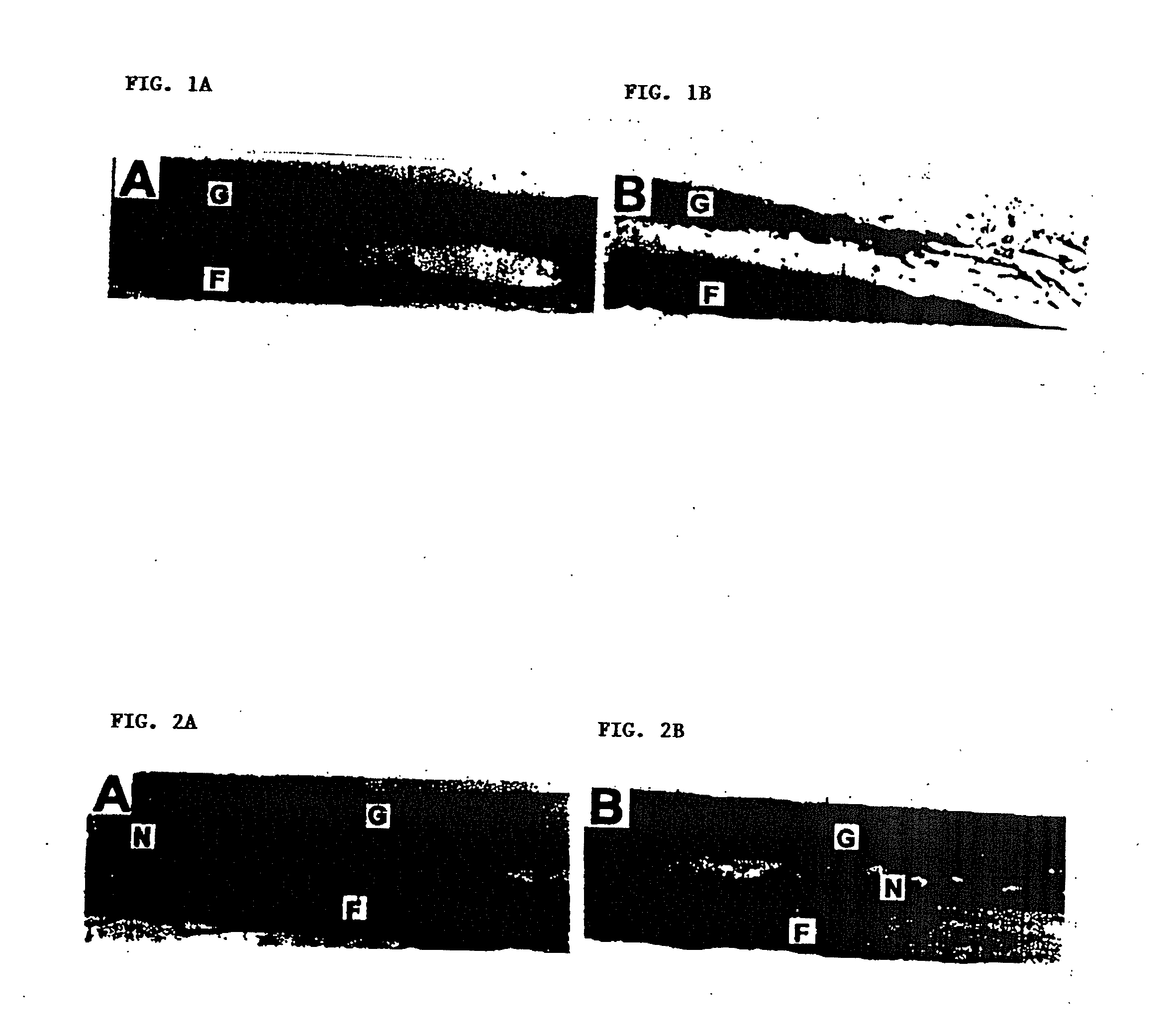
[0016]In this EXAMPLE, methods have been developed for decreasing the immune response against porcine tissue implanted in monkeys, by eliminating the α-Gal epitopes (Galα1-3Galβ1-4GlcNAc-R) with recombinant α-galactosidase, and mild glutaraldehyde fixation. Results using non-decalcified bone struts provide supporting evidence for the use of bone particulates for bone repair.

[0017]Background. Previously, CrossCart Inc. (San Francisco, Calif., USA) has extensively characterized a porcine bone patellar tendon bone anterior cruciate ligament (ACL) reconstruction device. This composite device consists of a sterile and biocompatible collagen tendon with cortical / cancellous bone plugs on each end. An irradiation-processing step significantly reduces viral agents from spiked samples. In previous primate bone testing, CrossCart used porcine bone grafts in the femur of primates to evaluate solid bony fusion to sc...

example ii

Xenograft Bone Matrix for Orthopedic Applications

[0031]This EXAMPLE refines the treatment regimen of EXAMPLE I to obtain maximum benefit in removal of α-Gal epitopes from xeno-active tissues and promote accelerated osseus union.

[0032]Process Development. Diaphyseal bone is harvested from 6 to 12 month old swine from a medical grade abattoir that also supplies porcine aortic heart valves for human implantation. After dissection of soft tissue, manual periosteal stripping and marrow removal, bone pieces are subjected to consecutive hypertonic, hypotonic and alcohol rinses. The bone is then milled to sieve standardized 150 to 500 μM particle size (Zhang et al., J. Periodontal. 68(11): 1085-92 (1997)). After sizing, the particles are subjected to consecutive hydrogen peroxide and alcohol washes. Downstream processing includes separate hydrochloric acid decalcification and enzymatic treatment. Protocols have been established to characterize the α-galactosidase enzyme, as described below:...

example iii

Z-Bone Process

[0048]TABLE VII below provides steps for one embodiment of the Z-bone process:

TABLE VII1.Scrub frozen porcine thighs with disinfectant2.Allow tissues to thaw3.Remove soft tissue with boning knife4.Scape remaining soft tissue with periosteal elevator5.Remove proximal and distal metaphysis with oscillating saw6.Cut bone shaft into manageable segments with oscillating saw7.Ream out marrow with rotary reamer8.Cut into small pieces and pool into basin with isopropanol9.Transfer segments to vessel with hexane / methanol for 12-18 hours with constantagitation at 4° C.10.Wash with WFI for 10-12 hours with constant agitation at 4° C., repeat 2 times.11.Wash with WFI w / 1.5M NaCl for 10-12 hours with agitation with lighting mixer (a310impeller) at 4° C.12.Inspect segments, remove any remaining soft tissue and transfer for new WFI bath forholding13.Remove segments and reduce to appx. 2 cm pieces14.Mill cold to 15.Suspend resulting slurry in 70% IPA 0.1% Tween 20 and pour through st...


PUM

PropertyMeasurementUnit
Timeaaaaaaaaaa
Accelerationaaaaaaaaaa

Abstract

The invention provides for the use of an improved xenograft bone particulate with respect to osteo-integration and bone remodeling, while diminishing the primate-to-pig immunological response using established bone-processing technique. Work was carried out using undecalcified bone to determine immunocompatibilty and bone remodeling potential of processed porcine bone struts following onlay graft implantation. New bone formation was evident, including the infiltration of cellular materials responsible for fusion and bone reconstruction.

Description

CLAIM OF PRIORITY[0001]This application is a continuation-in-part of U.S. patent application Ser. No. 09 / 585,509, filed Jun. 1, 2000, now allowed, which claims priority to U.S. patent application Ser. No. 09 / 248,476, filed Feb. 11, 1999, now U.S. Pat. No. 6,231,608, which issued on May 15, 2001. This patent application is also a continuation-in-part of U.S. patent application Ser. No. 09 / 646,376, filed Sep. 14, 2000, which is the U.S. National Stage of Int'l Patent Application No. PCT / US99 / 05661, filed Mar. 15, 1999, published as WO 99 / 47080 on Sep. 23, 1999, and which claims priority to U.S. Provisional Patent Application Ser. No. 60 / 078,139, filed Mar. 16, 1998, and Ser. No. 60 / 100,755, filed Sep. 17, 1998. This patent application is also a continuation-in-part of U.S. patent application Ser. No. 09 / 647,726, filed Dec. 4, 2000, which is the U.S. National Stage of Int'l Patent Application No. PCT / US99 / 05646, filed Mar. 15, 1999, published as WO 99 / 51170 on Oct. 14, 1999, and which ...

Claims


Application Information

Patent Timeline
IPC IPC(8): A61K35/32A01N1/00A61L27/00A61F2/24A61F2/28A61F2/30A61F2/46A61K35/34A61L27/36A61L27/54C12N5/08C12N5/28
CPCA01N1/00A61F2/2412A61F2/28A61F2/3094A61F2002/2835A61L2300/254A61K35/34A61L27/3608A61L27/365A61L27/3695A61L27/54A61F2002/4649
Inventor STONE, KEVIN R.TUREK, THOMAS J.
Owner APERION BIOLOGICS
Why Eureka
Try Eureka