
Non-imaging, weakly focused fluorescence emission apparatus and method

a fluorescence emission and weak focus technology, applied in the field of non-imaging, can solve the problems of limited in the capacity to generate high-resolution tissue images, limited in the scattering of incident laser pulses, and limited in the useful detection and imaging of multi-photon excited fluorescence deep within strongly scattering media, so as to improve background discrimination and focus precision, the effect of reducing light intensity and maximizing the generated signal

Inactive Publication Date: 2010-10-28
CORNELL UNIVERSITY
View PDF50 Cites 7 Cited by
  • Summary
  • Abstract
  • Description
  • Claims
  • Application Information

AI Technical Summary

Benefits of technology

[0015]Another embodiment of the invention is directed to a non-imaging, multiphoton fluorescence emission optical system as outlined above that includes a temporal focus controller. The temporal focus controller is disposed in the excitation beam path to spatially segregate spectral components of the short duration, multichromatic excitation pulse. The spectral components are then recombined and the beam is weakly focused on the target as described above. The temporal focus controller provides focal geometry decoupling of the lateral and axial dimensions of the beam and allows shaping and, particularly confinement, of the focal volume in contrast to a conventional spatial focus in which the lateral spot size essentially determines the axial focal dimension. The interested reader is directed to Zhu, G. H. et al., Optics Express, 13 (6) p. 2153 (2005). The benefits of temporal focusing-induced decoupling include the ability to place an axially loosely confined excitation only at the target region of interest and reduced excitation outside the target volume. According to an exemplary aspect, the temporal focus controller is a dispersive device, such as a grating, a hologram, a prism, or other known component that provides optical dispersion.
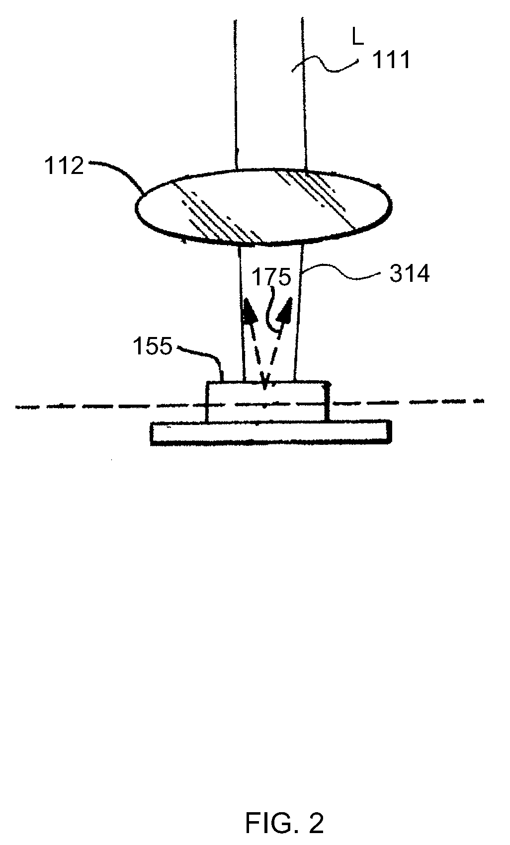
[0020]Thus according to an aspect of the present invention, non-imaging, two-photon fluorescence is generated from a target medium by illumination with a very high instantaneous intensity, ultra-short, weakly focused or unfocused, excitation field, in sharp contrast to conventional MPM imaging employing a, diffraction-limited excitation beam focus having a beam waist of less than one micron in diameter. The picosecond or shorter duration, high intensity pulses provide high instantaneous power making it probable that a fluorophore (e.g., a fluorescent dye) in the target material will absorb two long wavelength photons.
[0021]The total fluorescence excited by conventional, ‘strongly focused’ multiphoton excitation of a locally uniform distribution of fluorophores is roughly independent of the focal volume because the number of illuminated molecules increases for a larger focal volume at about the same rate that the square of the excitation intensity decreases, thus compensating for a decrease in light intensity. Therefore, the degree of illumination focus does not affect the total fluorescence emission. However, two-photon fluorescence excitation by scattered laser light is limited to a characteristic length based on the distances that the excitation photons travel after scattering during the duration of the laser pulse. Thus two-photon fluorescence excitation by scattered laser photons occurs within limited scattering lengths that are tissue and wavelength dependent, and on the order of 30 to 100μ. With reduced scattering, the background is not excited thereby improving background discrimination and focus precision. According to the embodiments of the present invention, the generated signal is maximized in strongly scattering tissues by avoiding strong focusing with weakly focused, high-energy pulses.

Problems solved by technology

As is known, single-photon fluorescence emission increases with the excitation intensity, however the exciting light may typically photo-bleach the fluorophores during fluorescence excitation with the consequent disadvantages known in the art.
The absorption of two or more of these longer wavelength photons provides excitation energy equivalent to the absorption of a single photon of a shorter wavelength, which usually results in excitation confined to the focal volume due to its non-linear nature.
MPM imaging of intrinsic fluorescence is thus a reliable diagnostic tool used, for example, in the practice of human and animal medicine, but it is limited in its capacity to generate high-resolution tissue images at depths greater than 500μ, which are known to be of interest.
Furthermore, useful detection and imaging of multiphoton excited fluorescence from deep within strongly scattering media, such as, e.g., biological tissues, may be limited by scattering of the incident laser pulses.
The high-intensity, short (femtosecond (fs) to picosecond (ps)), focused infrared laser illumination pulses required for multiphoton excitation of fluorescent markers deep in tissues may be scattered strongly enough therein that the quality of the sought-after high spatial resolution multiphoton excited fluorescence images is poor, rendering the images unusable.
Moreover, the scattered illumination can be sufficiently bright to excite excessive fluorescence background, thereby further degrading the image quality.

Method used


Image

  • Non-imaging, weakly focused fluorescence emission apparatus and method
  • Non-imaging, weakly focused fluorescence emission apparatus and method
  • Non-imaging, weakly focused fluorescence emission apparatus and method

Examples


Embodiment Construction

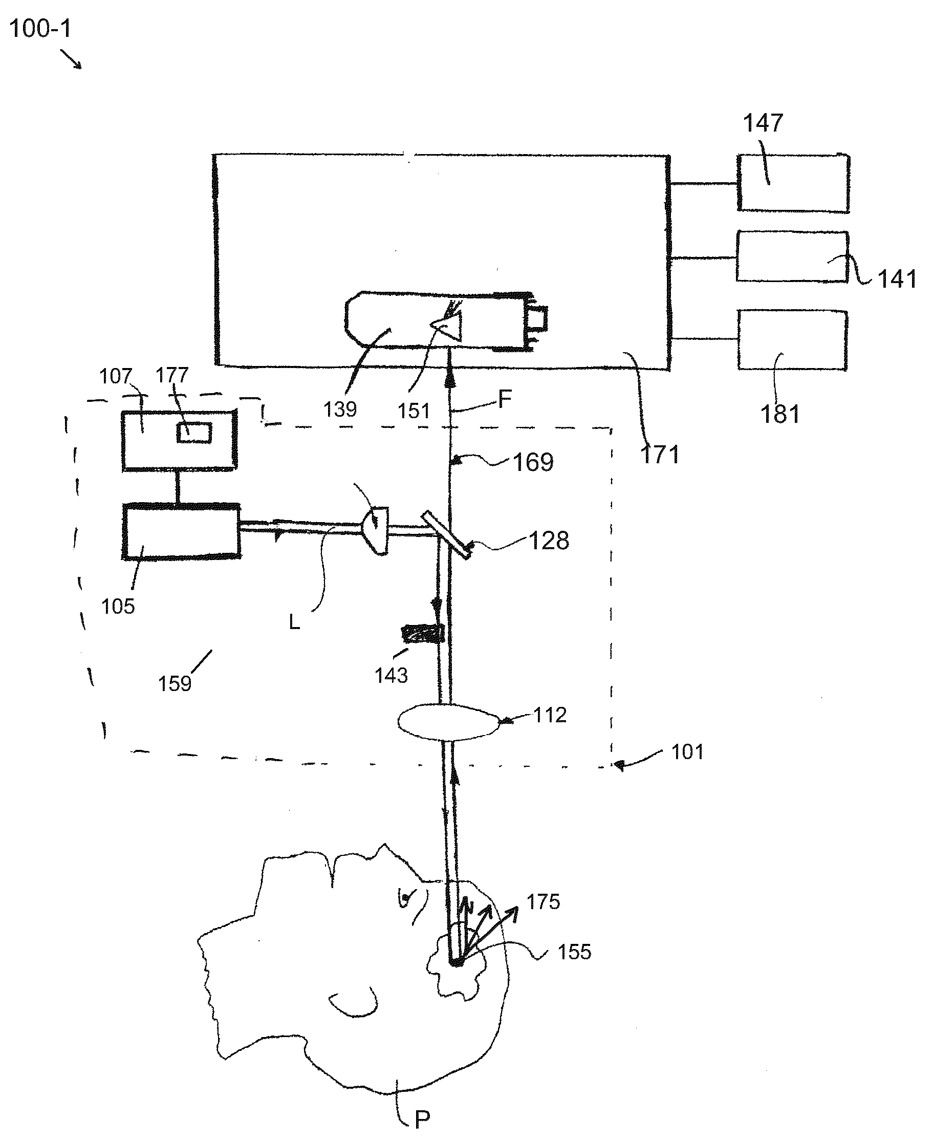
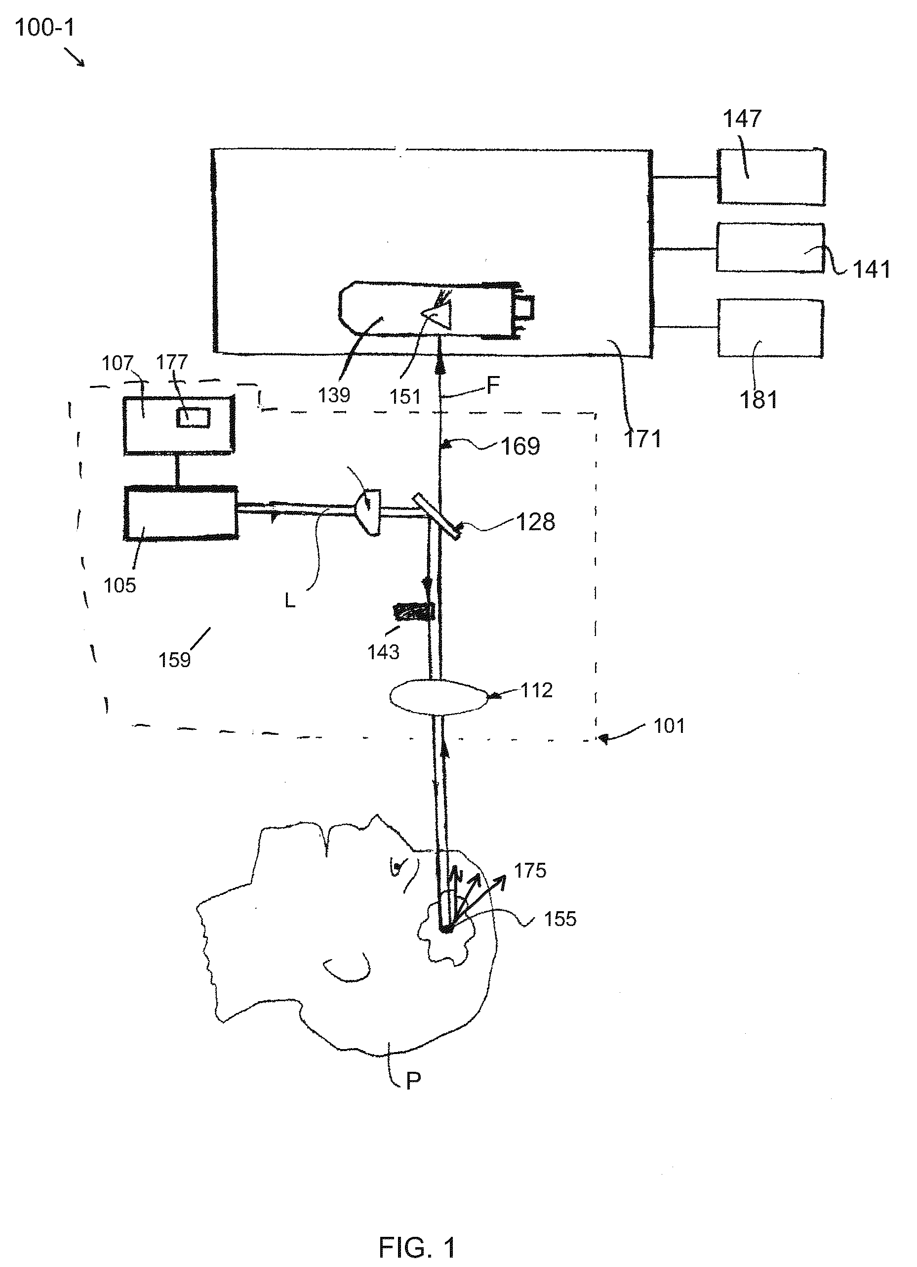
[0012]Embodiments of the invention are generally directed to apparatus and methods for determining, detecting, and analyzing an amount of fluorescence (or fluorophore concentration) that is excited by non-imaging fluorescence emission from a target medium. The disclosed apparatus and methods are less susceptible to collection and measurement errors due to scattering and other disadvantageous attributes associated with conventional MPM and SHG imaging techniques, referred to above and as known in the art. As is known, in a conventional multiphoton imaging system, the excitation beam is focused in the target medium and the fluorescence signal is assigned to a microscopic spatial location referred to in the art as the focal volume. The focal volume is typically on the order of one cubic micrometer (1 μm3). In the non-imaging case consistent with the embodiments of the present invention, there is no assignment of the fluorescence signal to any specific target area; rather, the weakly fo...


PUM


Abstract

Apparatus and methods relating to non-imaging, multiphoton fluorescence and optical second harmonic generation (SHG) (and higher harmonic generation) emission and detection. A weakly focused excitation beam is used to generate fluorescence emission in a volume of between about 0.1 cm3 to one cubic centimeter (1 cm3), which is significantly larger than the conventional MPM focal volume. A method for shaping and / or controlling (confining) the focal volume of a non-imaging, fluorescence emission excitation field in a target medium involves decoupling the axial dimension dependence of the focal volume from the lateral spot size of the excitation field. The method involves the step of spatially separating at least some of the spectral components of a short duration, multichromatic excitation field outside of the focal volume and spatially recombining the spectral components in a short duration, high intensity, weakly focused field incident on the target medium. The apparatus and methods described herein are particularly suitable for, but not limited to, non-invasive, in-vivo biological assay and disease state indication in target tissue and, more particularly, to potential early detection of Alzheimer's and other diseases.

Description

CROSS-REFERENCES TO RELATED APPLICATIONS[0001]This application claims priority to U.S. Provisional application Ser. No. 60 / 987,270 filed on Nov. 12, 2007, the subject matter of which is incorporated by reference herein in its entirety.STATEMENT REGARDING FEDERALLY SPONSORED RESEARCH OR DEVELOPMENT[0002]This invention was made with government support under Grant No. 1R21-AG026650 sponsored by the National Institute on Aging, and Grant No. 5-P41EB001976 sponsored by the National Institute of Biological Imaging and Bioengineering, at the National Institutes of Health. The government has certain rights in the invention.BACKGROUND OF THE INVENTION[0003]1) Field of the Invention[0004]Embodiments of the invention are most generally related to the field of non-linear optics. More particularly, embodiments of the invention are directed to non-imaging, weakly focused, multiphoton-excited-fluorescence emission and detection, and optical (second) harmonic generation (SHG) apparatus and methods....

Claims


Application Information

Patent Timeline
IPC IPC(8): G01N21/64A61B5/374
CPCG01N21/6428A61B5/0059A61B5/4088
Inventor WEBB, WATT W.XU, CHRIS
Owner CORNELL UNIVERSITY
Learn More