Radical therapeutic agent for keloid and hypertrophic scar

a keloid scar and radical technology, applied in the direction of lyase, peptide/protein ingredient, drug composition, etc., can solve the problems of restricting the elasticity of the skin, excessive accumulation of extracellular matrix and cell proliferation, and functional impediments, and achieve the strongest inhibitory effect and more inhibitory

Inactive Publication Date: 2011-01-13
SUZUKI SHIGEHIKO +1
View PDF2 Cites 3 Cited by
  • Summary
  • Abstract
  • Description
  • Claims
  • Application Information

AI Technical Summary

Benefits of technology

[0105]The results of Reference Example 4 suggested that the excessive accumulation of CS causes the deficiency in the elastic fiber formation (inhibition of elastin deposition to fibrillin) in the keloid tissue. In view of this, in an in vitro culture system (elastogenesis assay), keloid cells derived from a patient were artificially treated with only one of CS-A, CS-B and CS-C, or with CSs in combination, and it was examined which combination most prominently causes the deficiency in the elastic fiber formation (inhibition of elastin deposition in the extracellular matrix).
[0106]The culture plate which is 13 mm in diameter is inserted into a 24-well cell-culture plate (manufactured by Iwaki), and keloid cells collected from a human keloid tissue by an explant method were inoculated into the culture plate at a concentration of 1×10⁴ cells / well and cultured in DMEM medium (manufactured by Gibco, Inc.). The CS-added groups were set up with addition of only one of CS-A, CS-B and CS-C, or two or three of CS-A, CS-B and CS-C in combination. One ml of medium containing one of the above 400 μg / ml CS-A, CS-B and CS-C, or two or three of 400 μg / ml CS-A, CS-B and CS-C in combination was added to the keloid cells at an interval of 2 days. Further, as a control, the keloid cells were cultured without an addition of CS(s) (CS-free group). On day 9 of culture, the culture plate which has the cells and the extracellular matrix around the cells were taken out from the 24-well cell-culture plate and fixed with 100% methanol and then with 2% BSA. Thereafter, the thus fixed cells and the matrix were allowed to react with the primary antibodies, anti-elastin antibody (1:100; manufactured by Elastin Products Company, Inc., PR533) and anti-fibrillin-1 antibody (1:200; manufactured by Elastin Products Company, Inc., PR217). Alexa Fluor 546 goat anti-rabbit IgG antibody (1:200; manufactured by Molecular Probes, Inc.) was used as the secondary antibody, and nuclear staining was performed on the cells using Hoechst (manufactured by Sigma). Subsequently, the thus stained cells and the matrix were observed under a confocal laser scanning microscope system (manufactured by Nikon Corporation, Digital Eclipse C1si), and images of the stained cells and the matrix were obtained. Then, the thus obtained images of elastin staining were analyzed using an image analysis software (Image pro). Gray-scaling and image inversion were performed on the images. The image range was selected while comparing with the images of nuclear staining, so that the stained areas not containing the regions of stained nuclei could be extracted. The areas of the elastin-stained parts in the matrix were measured. The thus obtained values were indicated in relative %, taking the value for the CS-free group as 100%.
[0107]The photographs of elastin staining and fibrillin-1 staining of the CS-free group were shown in FIG. 4. The stained elliptical parts indicate the parts of cell nuclei stained by Hoechst staining, that is, the localization of cells, while the spaces between the cells indicate the parts of extracellular matrix. For the CS-free group, elastin deposition in the extracellular matrix (photograph on the right: indicated by white arrows) and the fiber structures of fibrillin-1 (photograph on the left: indicated by white arrows) were confirmed. The images of all the CS-added groups were analyzed using the image analysis software, and the results thereof were shown in a graph and table (FIG. 5 and Table 2). In those groups of which the cells were added with only one of CS-A, CS-B and CS-C, or with two of CS-A, CS-B and CS-C in combination, a slightly more inhibitory effect on the deposition of fibrous elastins was found in the extracellular matrix compared to the CS-free group, and the, strongest inhibitory effect on the deposition of fibrous elastins was found in the group of which the cells were added with three types of CSs, CS-A, CS-B and CS-C, simultaneously. That is, it indicated that the most prominent inhibitory action of the elastic fiber formation is induced when these three types of CSs were allowed to coexist.

Problems solved by technology

However, hypertrophic scars and keloids are common in that both of their lesion portions are a red-colored elevated lesion, which is primarily characterized by an excessive accumulation of extracellular matrix and cell proliferation.
The lesion portions are extremely hard, thereby markedly restricting the elasticity of the skin.
Because of this, these affected areas not only accompany pain, but also cause a functional impediment if located over a joint, such as restriction of the range of the joint motion.
However, there is no appropriate model for animal experimentation for hypertrophic scars and keloids, thus the clarification of the etiology and pathology has not seen much progress up to present.
Such pressuring flattens the lesion portion and relieves pain and itchiness; however, the termination of such pressuring results in re-swelling of the lesion portion and reemergence of pain and itchiness.
Further, the surgical therapy is not applicable to a wide lesion portion or hypertrophic scar extending over a large area, such as a thermal injury.
In order to prevent this recurrence, radiotherapy is necessary after the lesion removal; however, radiotherapy does nothing more than lowering the recurrence rate; therefore, this therapy is not a radical treatment (non-Patent Document 16).
Yet, this therapy does nothing more than improving the symptoms and the effects thereof are not stable.
In addition, there have been many cases where even an improvement of the symptom was not observed; therefore, this therapy is not a radical treatment.
However, depending on the dosage, systemic side effects, such as atrophoderma, hypopigmentation, excessive pigmentation and telangiectasia, may occur; therefore, these steroid drugs cannot be administered over a prolonged period.
Further, the injection is not applicable to lesions extending over a large area as well (non-Patent Document 12 and non-Patent Document 16).
Furthermore, women may also experience a side effect such as menstrual irregularity, even at a small dosage.
In addition, since recurrence is observed in many patients upon the termination of steroid drug injection, steroid drugs are not a radical treatment, and patients are forced to attend a hospital for a prolonged period.
There is also a method in which a tape agent containing a steroid such as betamethazone or fludroxycortide is patched other than the topical injection; however, the effects thereof do not go beyond the improvement of the symptoms and are unstable.
Further, some patients may develop skin rushes by such tape.
This also does nothing more than improving the symptoms such as itchiness in some cases, and in many cases, it has to be taken for not less than 3 months.
In this manner, these conventional therapies for hypertrophic scars and keloids, although they are called “treatment”, are nothing more than supportive measures.
However, even if this is to be applied to keloids, it would not provide something beyond the conventional concept of flattening the lesion portion by inhibiting the proliferation of fibroblasts.
In view that steroid drugs, which have an inhibitory effect on cell proliferation, cannot be used for a radical treatment of hypertrophic scars and keloids and that keloids and the like reoccur upon the termination of steroid drug administration, normalization of the lesion portion cannot be expected by simply inhibiting the proliferation of fibroblasts.
That is, it has been considered that such a radical treatment is impossible.

Method used


Image

  • Radical therapeutic agent for keloid and hypertrophic scar
  • Radical therapeutic agent for keloid and hypertrophic scar
  • Radical therapeutic agent for keloid and hypertrophic scar

Examples


reference example 1

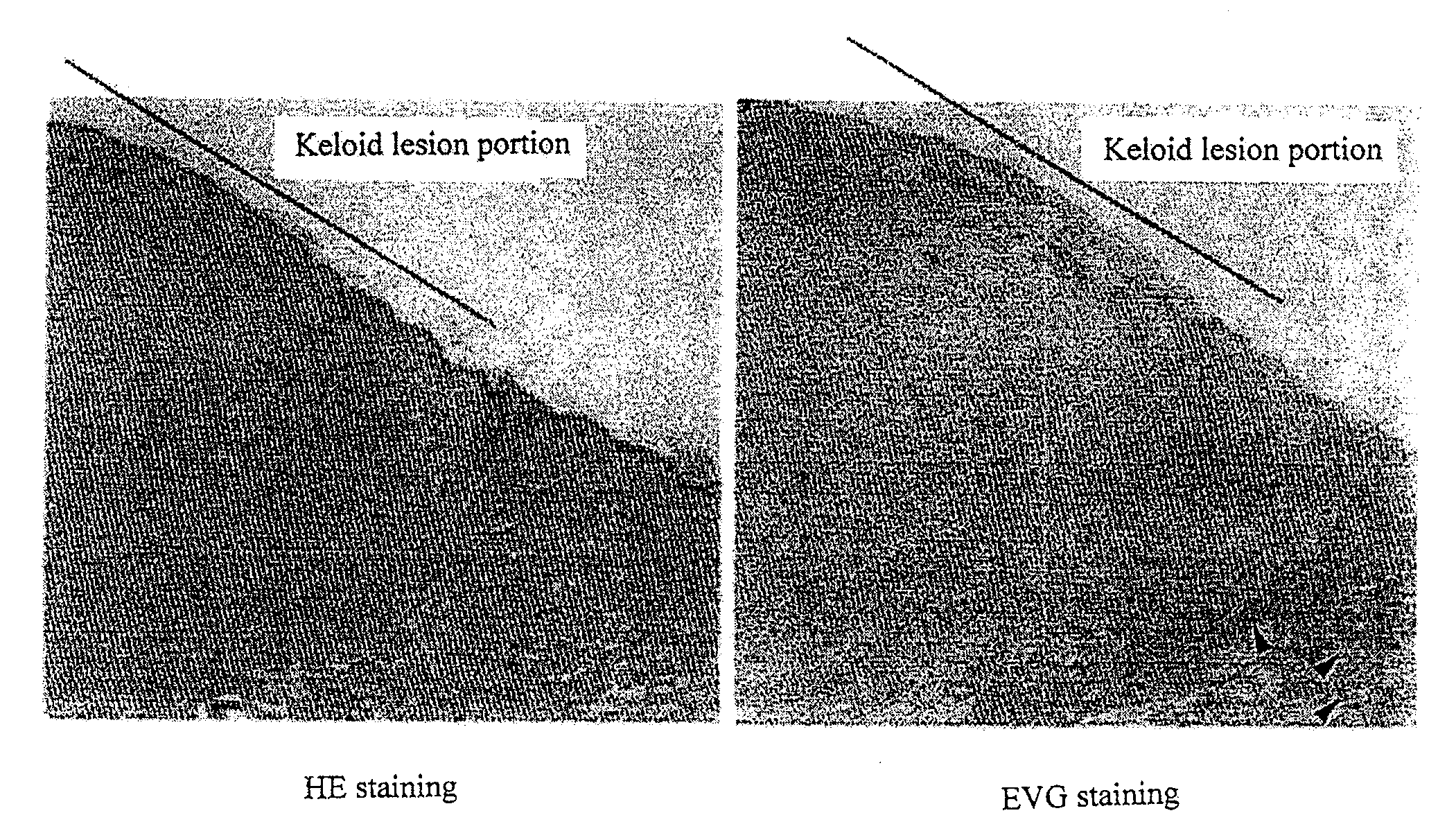
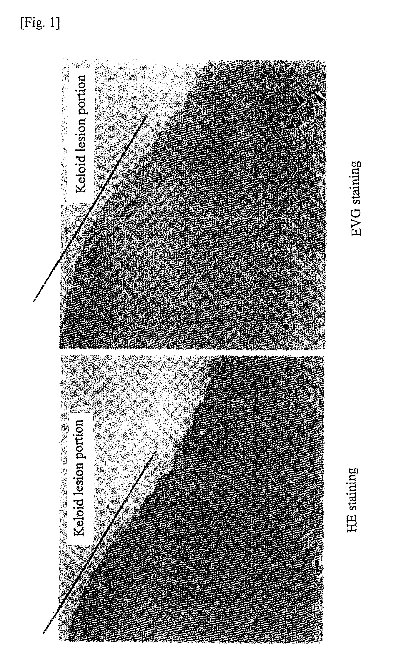
Observation of Keloid Lesion Portion and Normal Skin Part

[0092]As a tissue material, a human tissue sample containing a keloid lesion and normal skin part was extirpated from a keloid patient. The sample was fixed in 4% paraformaldehyde at 4° C. for 24 hours and subsequently embedded in paraffin to prepare a paraffin block from which a 3-μm paraffin section was prepared. After deparaffinization, hematoxylin and eosin (HE) staining and Elastica-van Gieson (EVG) staining were performed on the thus obtained paraffin section to prepare a specimen. The keloid tissue and normal skin tissue were observed under a microscope.

[0093]The results were shown in FIG. 1. The part which was indicated with a line is the keloid lesion portion and the adjacent part is the normal skin part. By EVG staining, the elastic fiber formation (indicated by arrows), which are stained in black, can be confirmed in the normal skin part other than keloid lesion portion. In contrast, hyalinization is observed in the...

reference example 2

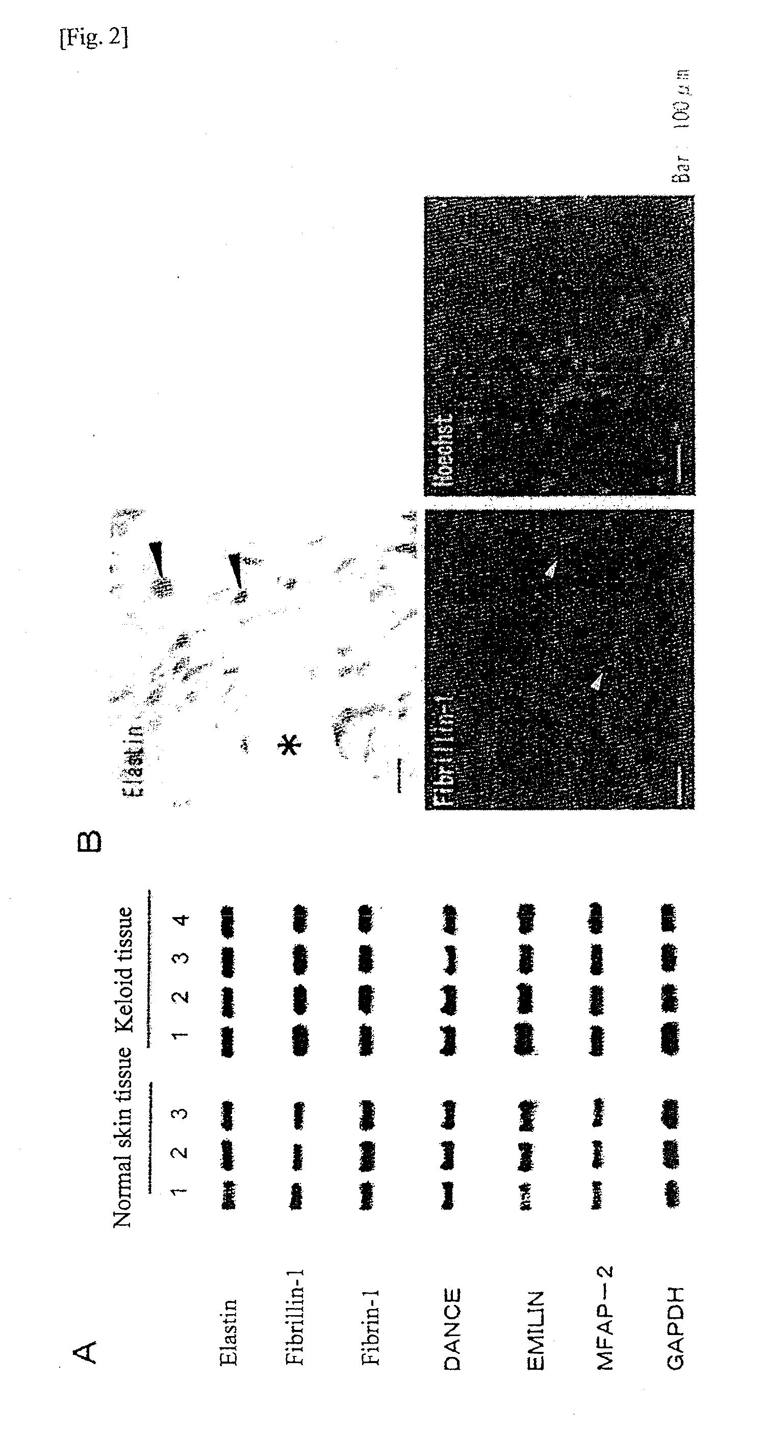
mRNA Expression of the Elastic Fiber Constituents in the Lesion Tissue of Keloid and Normal Skin Tissue

[0094]As tissue materials, human samples were extirpated from the keloid lesion tissues (4 individuals) and normal skin tissues (3 individuals). From these keloid tissues and normal skin tissues, total RNAs were extracted using RNeasy Plus kit (manufactured by QIAGEN). From 1 μg of the thus obtained total RNAs, cDNAs were synthesized using Advantage RT for PCR kit (manufactured by Becton, Dickinson and Company of Japan). For seven types of proteins that are the constituents of elastic fibers, the mRNA expressions thereof were examined by RT-PCR. The primers used in this PCR are shown in Table 1.

[0095]The specific sequences were amplified by performing PCR reactions using Blend Taq-plus (registered trademark) (manufactured by Toyobo Co. Ltd.), and the thus obtained PCR products were verified by electrophoresis. The PCR reactions were performed at the following conditions: denaturati...

reference example 3

Expressions of the Elastin and Fibrillin-1 Proteins in the Keloid Tissues

[0097]Reference Example 2 indicated that mRNAs of elastin and fibrillin-1 were expressed at a normal level in the keloid tissues. Subsequently, the expressions of elastin and fibrillin-1 at the protein level, as well as the localization thereof in the extracellular matrix, were examined by immunohistochemical staining. The procedures thereof were as follows.

[0098]Elastin staining: A human keloid tissue was extirpated and fixed in 4% paraformaldehyde at 4° C. for 24 hours. The thus fixed tissue was then embedded in paraffin and a 6-μm section was prepared therefrom. After deparaffinization, immunohistochemical staining was performed using LSAB / HRP kit (manufactured by Dako Japan, Inc.). As the primary antibody, an anti-elastin antibody (1:100; manufactured by Elastin Products Company, Inc., PR533) was used.

[0099]Fibrillin-1 staining: A human keloid tissue was extirpated, which was then immediately embedded in OC...


PUM

PropertyMeasurementUnit
sizeaaaaaaaaaa
pHaaaaaaaaaa
pHaaaaaaaaaa

Abstract

An effective therapeutic agent for keloids and/or hypertrophic scars is provided. Specifically, an elastic fiber regenerating agent consisting of chondroitinase ABC derived from Proteus vulgaris and a therapeutic agent for keloids and/or hypertrophic scars comprising the regenerating agent as an active ingredient are provided.

Description

TECHNICAL FIELD[0001]The present invention relates to an elastic fiber formation promoting agent and a radical therapeutic agent for keloids and / or hypertrophic scars.BACKGROUND ART[0002]The abbreviations used herein are as follows.[0003]GAG: glycosaminoglycan[0004]CS: chondroitin sulfate[0005]CS-A: chondroitin sulfate A[0006]CS-B: chondroitin sulfate B[0007]CS-C: chondroitin sulfate C[0008]CSPG: chondroitin sulfate proteoglycan[0009]GAGase: glycosaminoglycan lyase[0010]CSase: chondroitinase (chondroitin sulfate lyase)[0011]CSase-ABC: chondroitinase ABC[0012]CSase-B: chondroitinase B[0013]CSase-AC: chondroitinase AC[0014]Hypertrophic scars and keloids are characterized by, so to speak, an abnormality in wound healing, in which a fibrous tissue called “scar tissue” is formed, in the process of wound healing occurred on the skin, without regeneration of original normal tissue (non-Patent Documents 1 and 2). While a hypertrophic scar occur as a result of interference in wound healing, ...

Claims


Application Information

Patent Timeline
Patent Type & Authority Applications(United States)
IPC IPC(8): A61K38/51A61K38/43A61P17/02
CPCA61K38/00C12N9/88C07K14/24A61P17/02
Inventor SUZUKI, SHIGEHIKONAITO, MOTOKOIKEDA, MIKA
Owner SUZUKI SHIGEHIKO
Why Eureka
Try Eureka