Rapid Isolation of Monoclonal Antibodies from Animals

a monoclonal antibody and animal technology, applied in the field of rapid isolation of monoclonal antibodies from animals, can solve the problems of inability to detect important antibodies, inability to express important antibodies, and considerable technical complexity, and achieve the effect of being readily understandabl

Active Publication Date: 2011-12-22
BOARD OF RGT THE UNIV OF TEXAS SYST
View PDF0 Cites 50 Cited by
  • Summary
  • Abstract
  • Description
  • Claims
  • Application Information

AI Technical Summary

Benefits of technology

[0055]Other objects, features and advantages of the present invention will become apparent from the following detailed description. It should be understood, however, that the detailed description and the specific examples, while indicating preferred embodiments of the invention, are given by way of illustration only, since various changes and modifications within the spirit and scope of the invention will become apparent to those skilled in the art from this detailed description.

Problems solved by technology

Recently, B cell analysis has been expedited by microengraving techniques that utilize soft lithography for the high-throughput identification of antigen-specific B cells, however, this is at the cost of considerable technical complexity due to the need for antibody V gene amplification and cell expansion (Jin et al., 2009; Love et al., 2006).
Current use of display technologies coupled with library screening systems such as a phage display where antibodies are isolated by panning has a number of significant problems—in particular, some antibodies produced by a library may cause the death of the organism expressing them and therefore they simply cannot be detected.
There is a particular problem when one is searching for antibodies specific to an antigen from a pathogen which might be homologous to one produced by the host expression system (e.g., E. coli) then important antibodies cannot be expressed.
The use of E. coli to express libraries of human antibodies also suffers from the problem of codon usage—codons used by humans for specific amino acids can frequently not be the optimum ones for the same amino acid in E. coli or other host systems.
This means that an important antibody might not be expressed (or at least not in sufficient quantities) since the codons in its sequence are highly inefficient in E.
Coli, resulting in the E. coli being unable to read through and express it in full.
Codon optimization of antibody libraries is obviously not an option since the libraries would first have to be sequenced, which defeats the main advantages of using libraries.
Currently there are no methods available to identify the circulating antibodies in blood and to produce the antibodies that are specific to the disease agent and have a therapeutic effect.
A major limitation of the existing methods for isolation of monoclonal antibodies is that their application is limited to a very small number of species.
Such myeloma cell lines are difficult and time consuming to develop and therefore exist only for mice, primates, rabbits and sheep.
However, interspecies hybrids are generated with very low efficiency and are unstable, ceasing to produce monoclonal antibodies after a few passages.
Thus at present the production of monoclonal antibodies from the vast majority of animals that have an adaptive immunoglobulin system is a major challenge.
Moreover, even for species for which stable B cell fusions can be generated (rabbits, mice, sheep and primates) the isolation of monoclonal antibodies using the hybridoma technology is a lengthy process requiring 2-6 months after animal sacrifice.
Once again the utility of these methods is limited to the few species for which extensive information on their immunoglobulin repertoire is available, namely mice, primates and rabbits.
This in turn requires extensive information on the sequences of immunoglobulins expressed in a particular species and it is not available for the vast majority of animals that have an antibody-encoding, humoral immunity system.
Additionally it is not known whether the antibodies isolated by combinatorial library screening correspond to those that have been expanded by the immune system and produced in large amounts in animals.
Obviously all of these techniques are somewhat complex, inconvenient, and time consuming.

Method used


Image

  • Rapid Isolation of Monoclonal Antibodies from Animals
  • Rapid Isolation of Monoclonal Antibodies from Animals
  • Rapid Isolation of Monoclonal Antibodies from Animals

Examples


example 1

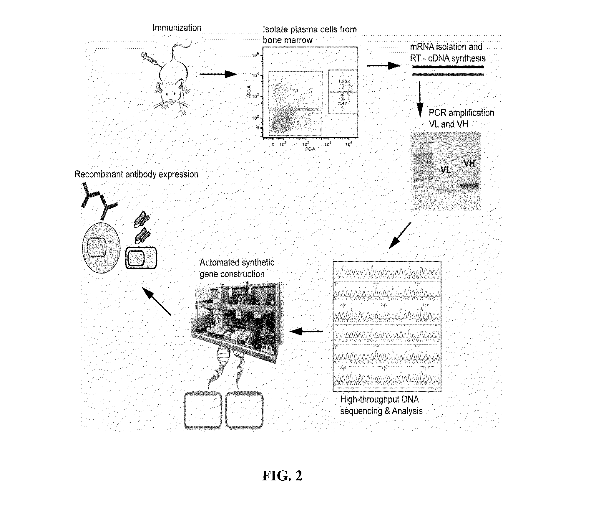
Immunization Protocol

[0177]Protein antigens (e.g., purified human complement protein C1s (CalBiochem), purified chicken egg ovalbumin (OVA, Sigma), or recombinant bacterially-expressed human B cell regulator of IgH transcription (BRIGHT) were resuspended in sterile-filtered phosphate buffered saline (PBS) at 1.0 mg / ml. On the day of primary immunization, 25 μl of antigen solution was thoroughly mixed with 25 μl of Complete Freund's Adjuvant (CFA, Pierce Biotechnology) and 50 μl of sterile PBS and stored on ice. Female Balb / c mice (Charles Rivers Laboratories) 6-8 weeks old were housed in conventional barrier space and were maintained on a normal chow diet. Prior to injections, mice were bled from the tail vein and approximately 25 μl of blood was collected and stored at −20° C. for later analysis. Day 1 was designated as the day primary immunizations were performed. 100 μl of the antigen-CFA mixture per mouse was injected with a 26-gauge needle subcutaneously into the backpad. Mice ...

example 2

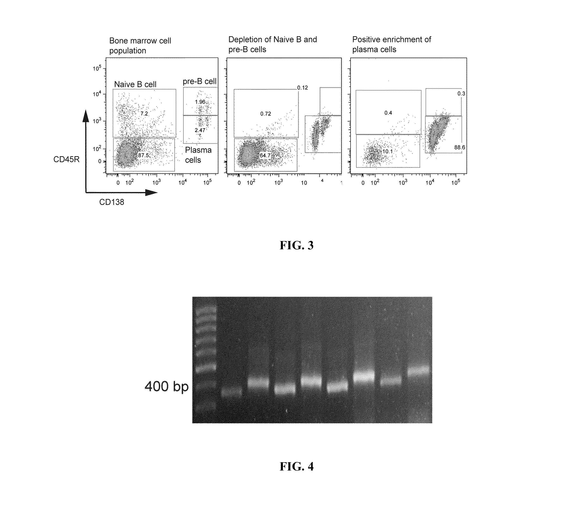
Isolation of the Plasma Cell (CD138+CD45R (B220)−CD49b−) Population from Murine Bone Marrow

[0179]The muscle and fat tissue was removed from tibias and femurs harvested from immunized mice. The ends of both tibia and femurs were clipped with surgical scissors and bone marrow was flushed out with a 26-gauge insulin syringe (Becton Dickinson, BD). Bone marrow tissue was collected in sterile-filtered Buffer#1 (PBS / 0.1% bovine serum albumin (BSA) / 2 mM ethylenediaminetetracetic acid (EDTA)). Bone marrow cells were collected by filtration through a 70-um cell strainer (BD) with mechanical disruption and washed with 20 ml of PBS and collected in a 50 ml tube (Falcon, BD). Bone marrow cells were then centrifuged at 1200 RPM for 10 min at 4° C. Supernatant was decanted and cell pellet was resuspended with 3.0 ml of RBC lysis buffer (eBioscience) and shaken gently at 25° C. for 5 minutes. Cell suspension was then diluted with 20 ml of PBS and centrifuged at 1200 RPM for 10 minutes at 4° C. Sup...

example 3

Isolation of Antibody Secreting Cells (ASCs) and Memory B Cells

[0184]Murine memory B cells. Secondary lymphoid organs (spleen and lymph nodes) are harvested from immunized mice following euthanization. Tissue was collected in sterile-filtered Buffer#1 (PBS / 0.1% bovine serum albumin (BSA) / 2 mM EDTA). Splenic and lymph node cells were collected by filtration through a 70-um cell strainer (BD) with mechanical disruption and washed with 20 ml of PBS and collected in a 50 ml tube (Falcon, BD). Splenic and lymph node cells were then centrifuged at 1200 RPM for 10 min at 4° C. Supernatant was decanted and cell pellet was resuspended with 3.0 ml of RBC lysis buffer (eBioscience) and shaken gently at 25° C. for 5 minutes. Cell suspension was then diluted with 20 ml of PBS and centrifuged at 1200 RPM for 10 minutes at 4° C. Supernatant was decanted and cell pellet was resuspended in 1.0 ml of Buffer#1.

[0185]Additionally whole blood is extracted by cardiac puncture. Whole Blood is added to his...


PUM

PropertyMeasurementUnit
concentrationaaaaaaaaaa

Abstract

Methods and compositions for identification of candidate antigen-specific variable regions as well as generation of antibodies or antigen-binding fragments that could have desired antigen specificity are provided. For example, in certain aspects methods for determining amino acid sequences of serum antibody CDR and abundancy level are described. In some aspects, methods for determining nucleic acid sequences of antibody variable region sequences and frequency are provided. Furthermore, the invention provides methods for identification and generation of antibody or antigen-binding fragments that comprise highly-represented CDR.

Description

CROSS-REFERENCE TO RELATED APPLICATIONS[0001]This application claims the benefit of U.S. Provisional Patent Application Nos. 61 / 345,538 and 61 / 377,816, filed May 17, 2010 and Aug. 27, 2010, respectively, the entirety of which are incorporated herein by reference.BACKGROUND OF THE INVENTION[0002]1. Field of the Invention[0003]The present invention relates generally to the field of antibody analysis and generation, such as antibody discovery from immunized animals. More particularly, it concerns novel methods and compositions for identification and / or production of desired antibodies or antigen-binding fragments. It also concerns identification of monoclonal antibodies from any mammal and more generally any animal that has an adaptive immune response that leads to the expression of soluble immunoglobulin and for which genomic information on its immunoglobulin locus is available.[0004]2. Description of Related Art[0005]Over the last 12 years, the development of cancer therapeutic antib...

Claims


Application Information

Patent Timeline
Patent Type & Authority Applications(United States)
IPC IPC(8): C40B20/00C12Q1/68C07K16/00G01N33/68C12Q1/34G01N33/53G16B40/30
CPCC07K16/065C07K2317/55C07K2317/56C07K2317/565C12Q2600/16C07K2317/92G01N33/6857C12Q1/6881C07K2317/622G16B40/00G16B40/30C12Q1/6869G01N33/48C40B30/04
Inventor REDDY, SAIGE, XINLAVINDER, JASONBOUTZ, DANIELELLINGTON, ANDREW D.MARCOTTE, EDWARD M.GEORGIOU, GEORGE
Owner BOARD OF RGT THE UNIV OF TEXAS SYST
Why Eureka
Try Eureka