Nanoparticle biosensor, method of preparing same and uses thereof

a biosensor and nanoparticle technology, applied in the field of nanoparticle biosensors, can solve the problems of high particle size distribution, destabilizing suspension, and limited detection limits of these techniques, and achieve the effects of reducing the sensitivity and detection limit of these techniques, and reducing the detection rang

Inactive Publication Date: 2013-02-14
CONSEJO SUPERIOR DE INVESTIGACIONES CIENTIFICAS (CSIC) +1
View PDF4 Cites 35 Cited by
  • Summary
  • Abstract
  • Description
  • Claims
  • Application Information

AI Technical Summary

Benefits of technology

[0022]A first aspect of the invention relates to a nanoparticle biosensor comprising: a magnetic nanoparticle, a silica layer, one or more outer metal layer or layers which can be of different types and deposited in an alternating manner and immobilized on its outer surface, and a layer of synthetic or natural organic or inorganic biosensor molecules that ca

Problems solved by technology

Conducting many of these assays in a homogeneous phase has a number of difficulties due to the fact that target DNA or RNA molecules are usually extremely diluted in natural samples, therefore the hybridization reaction is usually carried out on surfaces on which the PNA probe molecule has been previously immobilized.
This represents a limitation of the assay because the amount of probe is limited to a layer of PNA molecules on the surface in question, with which the sample to be analyzed must necessarily be placed in contact in order to give a positive signal.
The sensitivity and the detection limit of these techniques is therefore also limited.
However, this method, in addition to being expensive and slow, causes a high size distribution in the particles obtained.
Although the possibility that the particles are concentrated through the application of an externa

Method used


Image

  • Nanoparticle biosensor, method of preparing same and uses thereof
  • Nanoparticle biosensor, method of preparing same and uses thereof
  • Nanoparticle biosensor, method of preparing same and uses thereof

Examples


example 1

Synthesis of the Metalized Magnetic Nanoparticle Biosensors of the Invention

example 1.1

Gold Nanoparticle Biosensors

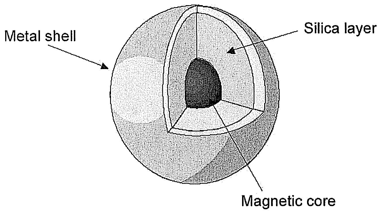
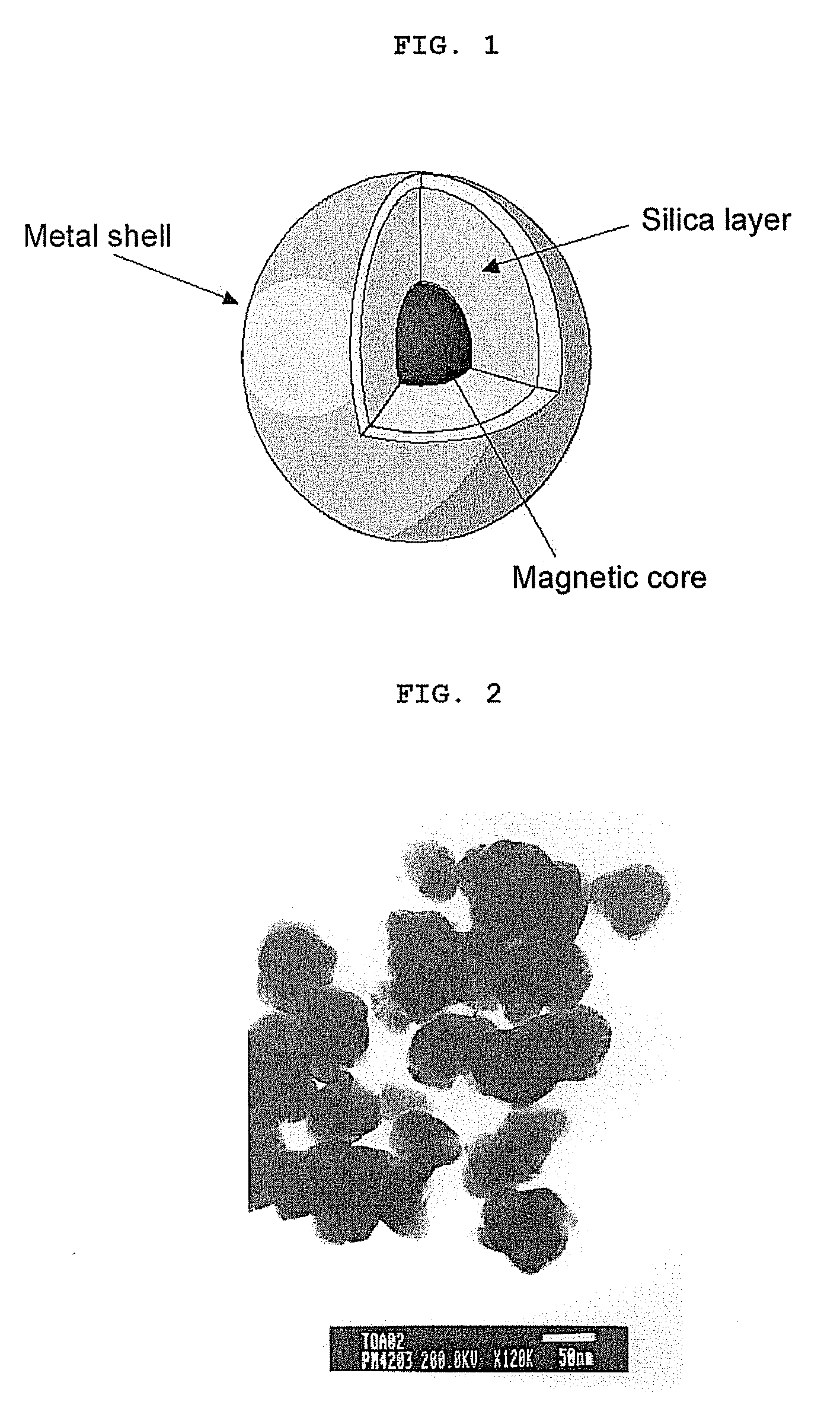
[0135]The initial object of this embodiment is to prepare a magnetic suspension formed by cobalt ferrite (CoFe2O4) nanoparticles with a uniform size, coated with a thin silica layer functionalized with amino groups, plus a final coating with a gold layer (as shown in FIG. 1) on which the thiolated PNA molecules will be chemisorbed as an example of a biosensor molecule. The silica coating takes place on the magnetic nanoparticles individually, also being able to obtain the coating several magnetic cores simultaneously. The nanoparticles thus prepared comply with the properties sought: they are stable in suspension for a long time period, but they sediment in the presence of a magnetic field. Furthermore, by removing said magnetic field, the nanoparticles must be dispersed upon applying a gentle stirring.

[0136]In one particular example, cobalt ferrite with an inverse spinel structure has been used, synthesized to a size of 17±3 nm, with suitable magnetic pr...

example 1.2

Synthesis of Gold / Silver Alloy Nanoparticle Biosensors

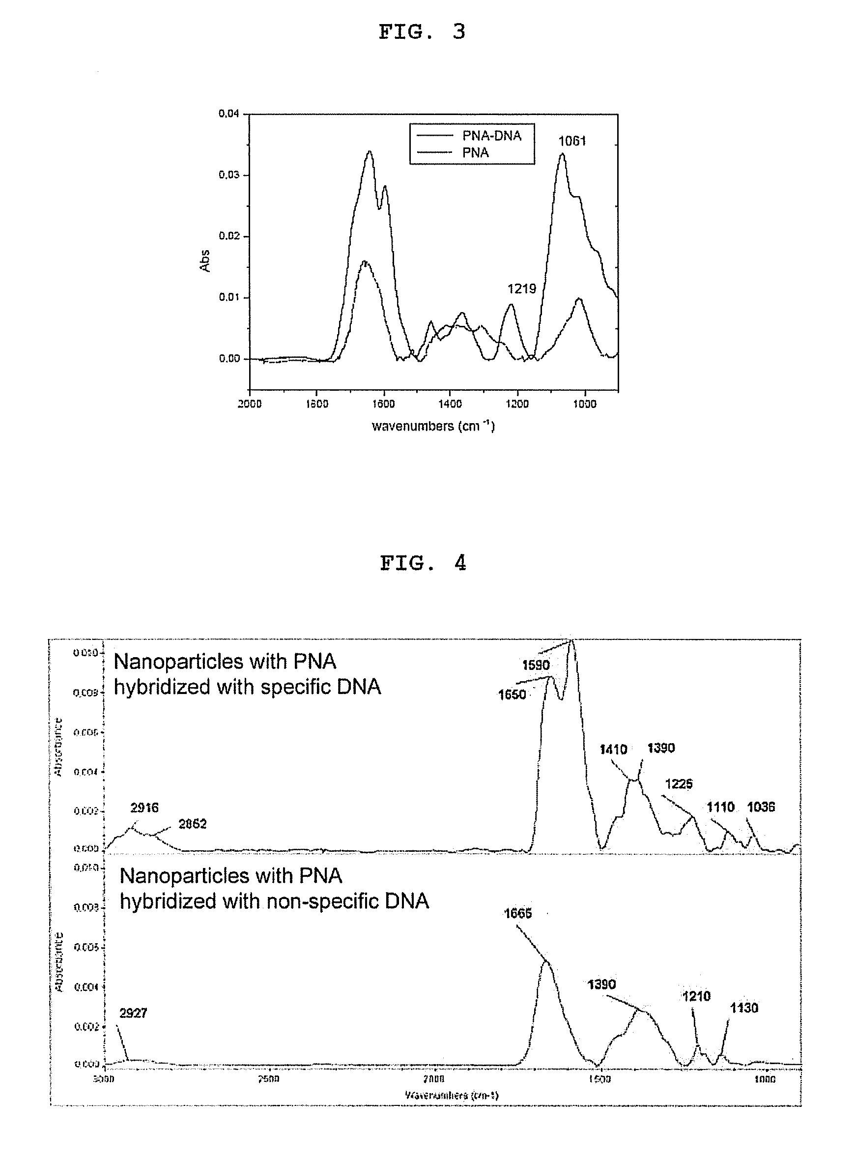
[0146]The process of obtaining nanoparticles coated by a Au / Ag alloy is based on reducing silver ion on the magnetic nanoparticles decorated with gold nanoparticles obtained as indicated in section 1.1. To that end, 500 μl of the ferrofluid decorated with gold particles, 400 μl of 0.5 M glycine, 600 μl of water (with Milli-Q purity), 20 W of 12 mM AgNO3 and 100 μl of 10 mM ascorbic acid were placed. This solution is left at room temperature and in darkness for 6 hours, the nanoparticles are washed by magnetic sedimentation three times, re-suspending in 500 μL of water each time, and the process is repeated (Huang et al., 2004). The immobilization of the PNA molecules is carried out as will be detailed in Example 1.4.


PUM

PropertyMeasurementUnit
Nanoscale particle sizeaaaaaaaaaa

Abstract

The invention relates to the field of biosensors and, more specifically, to nanoparticle biosensors comprising: a magnetic core, a silica layer, one or more outer metal layers which can be of different types and deposited in an alternating manner and immobilized on the outer surface, and a layer of synthetic or natural organic or inorganic biosensor molecules that can bind to biomolecules. The invention also relates to a method of obtaining the nanoparticle biosensors as well as to the different uses thereof.

Description

FIELD OF THE ART[0001]The present invention relates to the field of biosensors, specifically to the use of peptide nucleic acid (PNA) probes immobilized on nanoparticles for detecting the hybridization of natural nucleic acids (DNA or RNA) of specific sequences. The nanoparticles are particularly formed by a magnetic core and an outer metal layer, on which the relevant probes with base sequences complementary to the target object of identification and detection are immobilized. The magnetic, electric and optical properties of the nanoparticles are used to improve the method, being focused on the magnetic, optical or electric signal difference obtained from said nanoparticles before and after the modification involved in a positive hybridization assay. The types of tests which the present invention relates to can also be carried out spectroscopically on macroscopic metal surfaces.BACKGROUND OF THE INVENTION[0002]The progress of science in genetics, genomics and biotechnology has been...

Claims


Application Information

Patent Timeline
IPC IPC(8): C12Q1/68C12M1/34B05D7/00
CPCB01J2219/005B01J2219/00576B01J2219/00722G01N2446/20B82Y15/00B82Y30/00G01N33/54326B01J2219/00729G01N33/552G01N33/553
Inventor FERNANDEZ LOPEZ, VICTOR MANUELMARTIN GAGO, JOSE ANGELPITA MARTINEZ, MARCOSSERNA PEREDA, JOSE CARLOSBRIONES LLORENTE, CARLOSVAZ DOMINGUEZ, CRISTINAMATEO MARTI, EVA
Owner CONSEJO SUPERIOR DE INVESTIGACIONES CIENTIFICAS (CSIC)
Why Eureka
Try Eureka