Multi-lumen thoracic catheter and uses thereof

a thoracic catheter and multi-lumen technology, applied in the field of multi-lumen thoracic catheters, can solve the problems of negative pleural pressure, immediate lung collapse, lungs to remain inflated, etc., and achieve the effects of reducing pain, reducing pressure in the pleural space, and facilitating adhesion

Inactive Publication Date: 2013-06-13
STEVENS INSTITUTE OF TECHNOLOGY
View PDF22 Cites 21 Cited by
  • Summary
  • Abstract
  • Description
  • Claims
  • Application Information

AI Technical Summary

Benefits of technology

[0022]According to another aspect, the described invention provides a method for treating or preventing fluid accumulation or air accumulation in a body cavity of a subject using the multi-lumen catheter, the method comprising: (a) aseptically inserting through an incision at an insertion site of the subject the multi-lumen catheter comprising a main drainage lumen surrounded by a wall and at least one access lumen positioned in or on the wall; (b) securing the inserted multi-lumen catheter by closing the incision with a suture; (c) infusing a physiological solution through at least one access lumen of the multi-lumen catheter to dilute a drainage fluid; (d) connecting a distal end of a main drainage lumen of the multi-lumen catheter to a suction drainage system; and (e) applying a vacuum force to the suction drainage system to remove the diluted drainage fluid.
[0023]According to one embodiment of the method, the body cavity is a pleural cavity. According to another embodiment, the body cavity is a cranial cavity. According to another embodiment, the body cavity is a spinal cavity. According to another embodiment, the body cavity is a abdominal cavity. According to another embodiment, the body cavity is a pelvic cavity. According to another embodiment, the physiological solution in step (c) comprises a saline solution, Ringer's solution, 5% dextrose in water (D5W), or a mixture thereof. According to another embodiment, a thrombolytic agent is infused through an access lumen that exits into the main drainage lumen. According to another embodiment, the suction drainage system is a single-flow drainage system that only allows one direction of flow. According to another embodiment, the suction drainage system comprises a collection chamber, a water seal chamber, and a suction control chamber, wherein the collection chamber attaches the multi-lumen thoracic catheter to the subject; wherein the water seal chamber prevents air and fluid from returning to the pleural space; during inspiration; and wherein the suction control chamber controls the amount of suction allowed by the suction drainage system. According to another embodiment, the fluid accumulation or air accumulation in the pleural cavity of the subject results from a condition comprising pneumothorax, pleural effusion, chylothorax, empyema, hemothorax, hydrothorax, or a combination thereof. According to another embodiment, the fluid accumulation or air accumulation in the pleural cavity of the subject results from a condition selected from the group consisting of a pulmonary disease, a lung infection, a lung cancer, a breast cancer, and a surgery that affects a negative pressure in the pleural space. According to another embodiment, the insertion site is determined by reviewing clinical signs and chest imaging of the subject. According to another embodiment, the chest imaging comprises chest X-ray, chest fluoroscopy, computed tomography (CT), high-resolution computed tomography (CT), helical (spiral) computed tomography (CT), computed tomography (CT) angiography, magnetic resonance imaging (MRI), or ultrasonography. According to another embodiment, the insertion site is a lateral thorax, at a line drawn from an armpit to the nipple in male or to the side above the sternoxiphoid junction (lower junction of the sternum, or chest bone) in female. According to another embodiment, a size of the incision for the insertion of the multi-lumen catheter is similar to the diameter of the multi-lumen thoracic catheter being inserted. According to another embodiment, the method further comprises infusing a therapeutic agent through a second access lumen of the multi-lumen catheter. According to another embodiment, the therapeutic agent is a local anesthetic agent, and wherein the local anesthetic agent decreases pain associated with tissue irritation or tube insertion. According to another embodiment, wherein the local anesthetic is selected from the group consisting of benzocaine, lidocaine, and marcaine. According to another embodiment, the therapeutic agent is an anti-coagulant agent. According to another embodiment, infusing is performed as a bolus (single) infusion. According to another embodiment, infusing is performed as continuous infusion. According to another embodiment, the therapeutic agent is infused at a flow rate ranging from about 1 cc per hour to about 500 cc per hour. According to another embodiment, the therapeutic agent is an anti-infective agent comprising an antibiotic agent, an anti-tuberculin agent, an anti-fungal agent, or antiviral agent, wherein the anti-infective agent treats or prevents a localized infection. According to another embodiment, the therapeutic agent is anti-fungal agent. According to another embodiment, the therapeutic agent is anti-tuberculin agent. According to another embodiment, the therapeutic agent is a sclerotic agent, wherein the sclerotic agent induces adhesion between the parietal and visceral pleura. According to another embodiment, the sclerotic agent is infused as a bolus injection, and wherein the vacuum force is discontinued for an hour. According to another embodiment, the therapeutic agent is an anti-inflammatory agent, and wherein the anti-inflammatory agent decreases inflammation in the pleural space. According to another embodiment, the therapeutic agent is a thrombolytic agent, and wherein the thrombolytic agent dissolves clotted blood in the pleural space.

Problems solved by technology

Second, the surface tension of the pleural fluid creates a strong adhesive force between the parietal and visceral pleurae, binding the pleurae together.
This results in a negative pleural pressure (relative to the atmosphere), which causes the lungs to remain inflated.
However, if fluid accumulates at a rate greater than the lymphatic system can reabsorb, the pleural pressure becomes positive resulting in immediate lung collapse.
When a lung collapses, the lung is unable to expand during respiration leading to difficulty breathing and hypoxemia, a lack of oxygen in the blood (Marieb, E. and Hoehn, K., Human Anatomy and Physiology.
A major and common complication is occlusion.
Thoracic catheters often become partially or completely occluded with blood clots and other fibrous material.
These occlusions can lead to life-threatening complications, including tension pneumothorax and sepsis, and may require additional surgery if the resulting conditions cause loss of lung volume.
Several techniques can be tried to dislodge the occluding material, but each method has significant drawbacks and often does not succeed in removing the material.
One procedure is milking or stripping the thoracic catheter to attempt to manually dislodge the clot, but research has shown this can produce extremely high negative pressures in the thorax which are harmful to the patient.
This method creates a pneumothorax and is a severe sterility issue.
For these reasons, surgeons typically use large bore tubes to ensure patency, but even these tubes frequently become clogged (Shanaz, S. et al., “The Active Tube Clearance System,” International Society for Minimally Invasive Cardiothoracic Surgery 1st ser.
The use of large bore tubes raises another issue related to thoracic catheter insertion, as large tubes are associated with significant patient discomfort (Shanaz, S. et al., “The Active Tube Clearance System,” International Society for Minimally Invasive Cardiothoracic Surgery 1st ser.
Any size, but especially large bore, thoracic catheters irritate these areas causing patient discomfort, which can lead to other complications.
An uncooperative patient may move or turn, resulting in a dislodged tube or a broken seal, leading to reinsertion of the tube and sterility issues.
Another complication which can arise is a local or generalized infection from the procedure.
Clogging, pain and infection are three complications which frequently arise from the use of thoracic catheters.
However, the solutions currently offered and the ones being developed do not meet the needs of the patients and surgeons.
Because of this, surgeons are not able to access the pleural space or the interior of the main drainage lumen of the thoracic catheter without breaking the seal.
This causes a problem when complications arise.

Method used


Image

  • Multi-lumen thoracic catheter and uses thereof
  • Multi-lumen thoracic catheter and uses thereof
  • Multi-lumen thoracic catheter and uses thereof

Examples


example 1

Drain Testing Protocol

[0168]1.1 Rationale

[0169]A drain test is used to examine whether a multi-lumen thoracic catheter of the present invention is proficient in draining various materials from a simulated pleural space while avoiding clot formation within the catheter. The drain test also can test the efficacy of the catheter's drainage ability with a viscous substance, meant to simulate the materials that will need to be evacuated from the pleural space. The end result of the test is the appropriate saline flow rate into the main drainage tube to prevent tube occlusion.

[0170]1.2 Introduction

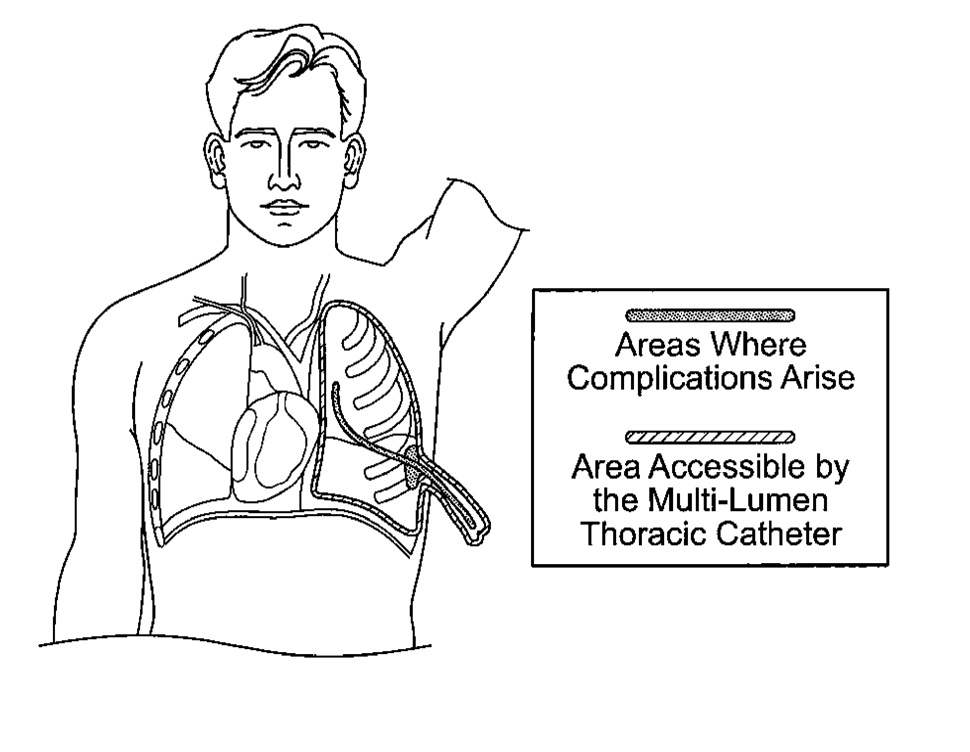
[0171]Any condition that causes fluid or air accumulation in the pleural space requires suction thoracic catheter insertion to ensure negative pleural pressure, prevent lung collapse, and to ensure efficient oxygen delivery. Tube occlusion often occurs when the blood's platelets bind to the surface of the catheter's inner diameter. These platelets form a plug, and along with a network of fibrin ...

example 2

Dye Movement in Pulmonary Model Testing Protocol

[0188]2.1 Rationale

[0189]The purpose of this test is to determine whether or not the access lumen is effective in the introduction of fluid into the pleural space. The test examines both infusion and injection methods of fluid injection through the access lumen. The test is designed to test whether or not the injected fluid can successfully diffuse not only to the wound site, but through the entire pleural space. This validates the efficacy of the ability of the access lumen to introduce fluid into the pleural space.

[0190]2.2 Introduction

[0191]Currently, physicians do not have direct access to the pleural space when a chest tube is placed, nor do they typically invade the area to administer fluid or drugs (such as anesthetic or anticoagulant). However, it is important to have an access to the pleural space, most specifically the pleural wound site. For fluids administered at this location, it is beneficial to have the fluid reach and i...


PUM


Abstract

A multi-lumen catheter for use in a cavity having a main lumen surrounded by a wall, and at least one access lumen positioned in or on the wall, the one access lumen conveys a solution to the cavity and said main lumen. A method for treating or preventing fluid or accumulation in a body cavity including (a) aseptically inserting through an incision at an insertion site the catheter comprising a main drainage lumen surrounded by a wall and the one access lumen positioned in or on the wall, (b) securing the inserted catheter by closing the incision with a suture, (c) infusing a physiological solution through the one access lumen to dilute a drainage fluid, (d) connecting a distal end of a main drainage lumen of the catheter to a suction drainage system, and (e) applying a vacuum force to the suction drainage system to remove the diluted drainage fluid.

Description

CROSS-REFERENCE TO RELATED APPLICATION[0001]This application is a Section 111(a) application relating to and claims the benefit of commonly owned, co-pending U.S. Provisional Application Ser. No. 61 / 557,276 entitled “MULTI-LUMEN THORACIC CATHETER AND USES THEREOF”, filed Nov. 8, 2011, the entirety of which is incorporated herein by reference.FIELD OF THE INVENTION[0002]The present invention relates to a multi-lumen thoracic catheter and its use for delivering therapeutic agents into or diagnosing a disease in a body cavity.BACKGROUND OF THE INVENTION[0003]1. Pathologic Conditions that Require Use of Thoracic Catheters[0004]In the human body the thoracic cavity, a hollow cavity, which is enclosed by the ribs, vertebral column, and the sternum and is separated from the abdominal cavity by the diaphragm, contains the lungs, the middle and lower airways, the heart, and various major blood vessels. Two thin membranes, known as pleurae, line the border of the cavity. Each pleura is contin...

Claims


Application Information

Patent Timeline
Patent Type & Authority Applications(United States)
IPC IPC(8): A61M1/00A61M5/14A61B5/00A61B10/04A61B5/01
CPCA61M1/008A61B10/04A61B5/01A61B5/0059A61B5/036A61B10/06A61M2025/0031A61M1/0084A61B5/6852A61M5/14A61M1/85
Inventor BUDAR, SARACARR, ZACHARY WAYNEGRIGGS, LAUREN JENNIFERRICCARDELLO, GERALDSPELMAN, STEPHANIEHAZELWOOD, VIKKIPEARLSTONE, DAVID B.RITTER, ARTHUR
Owner STEVENS INSTITUTE OF TECHNOLOGY
Why Eureka
Try Eureka