Devices and methods for long-term intracellular access

a long-term intracellular and cell technology, applied in the field of nano-scale devices, can solve the problems of difficult relationship with man-made biomaterials, inability to understand the interface formed between the interior edge of the membrane and the material surface, and less study of the bridging across the cell membrane itsel

Active Publication Date: 2014-12-04
THE BOARD OF TRUSTEES OF THE LELAND STANFORD JUNIOR UNIV
View PDF1 Cites 8 Cited by
  • Summary
  • Abstract
  • Description
  • Claims
  • Application Information

AI Technical Summary

Benefits of technology

[0027]The medial heterofunctional region may be formed of sequential layers of hydrophobic and hydrophilic bands (e.g., repeated medial heterofunctional regions separated by hydrophobic bands). This configuration may help allow cells to fuse to one or more bands of the medial heterofunctional region somewhat independently of the height / position of the medial heterofunctional region relative to the post. This may also allow use of the probes with cells of different dimension and configurations.
[0036]The layout of the probes may be designed to encourage more than one probe to be integrated into a single cell. For example, in some variations two or more probes may be spaced more closely than the average cell diameter such that both posts may contact, be near, and / or enter the same cell. For example, in some variations pairs or groups (e.g., groups of 2, 3, 4, 5, or more) of posts are spaced less than 50 μm apart, less than 25 μm apart, less than 20 μm apart, less than 15 μm apart, less than 10 μm apart, less than 5 μm apart, etc. This may provide additional benefit by allowing one post to perform current injection functions, while the other may record the voltage. This probe arrangement may provide an accurate way to reduce electrical recording artifacts and allow contact resistance to be removed from the signals. Alternatively, these probes could be used to record from different areas in the cell, reducing the amount of space-charge resistance caused by the resistivity of the surrounding fluid. More than two probes per cell can also be designed by producing many probes spaced less than the average cell diameter. For example, three or more probes could be used to triangulate the location of an electrical event within the cell. In some variations, probes are arranged in clusters or groups, and the groups are separated by more than a typical single cell diameter.
[0040]For these probe structures including an inner (e.g., recessed) region, a heterofunctional region at the distal edge for fusing to a cell membrane may generally be a hollow, elongate member (i.e. cylindrical) that projects up from a base region and is open at the distal end. The hollow, elongate structure may have a regular circular or oval cross-section (e.g., in a section parallel to the base), or it may have a polygonal (e.g., triangular, square, pentagonal, etc.) or irregular cross-section. The distal end of the hollow elongate member is typically open, may have a thin (“knife edge”) profile and be configured as a cell membrane adhering or fusing heterofunctional region. The hollow elongate member typically forms a lumen, and the proximal end of the lumen is typically closed, forming an inner recessed region. This inner lumen / recessed region may be the same depth as the base; thus the height of the inner wall of the hollow elongate member may be the same as the height of the outer wall of hollow elongate member, or it may be different (higher or lower). In general, the probe may form a structure around a highly sensitive electrode (or other sensor) within the lumen (recessed cavity) of the probe. The distal opening of the probe may include a heterofunctional region that can be configured to spontaneously attach to the surface of the nearest cell. This ring can be functionalized with a variety of molecules to promote cell attachment. This cell-attachment ring not only negates the effect of micromotions, it may provide a restricted volume between the cell and the electrodes for sensing and stimulation between the two. This design may restrict signal pick-up (electrical and chemical) to that specific cell only, roughly akin to using a tin can to listen at a door. This approach may also eliminate local environmental interference, such as the LFP, as well as chemicals released from surrounding cells. The environmental features can still be recorded using electrodes located outside of the ring, enabling direct comparison of the single cell behavior in the context of its surroundings. The probes described herein (including the probes forming a protective inner region) can be combined with ultra-sensitive transistors or other electronic devices. These devices have proven high signal-to-noise recording capability in-vivo and ultra-low inflammation, and with modification of the current transistor geometry will provide single cell sensitivity.
[0043]The direct contact between the cell membrane and the distal end / opening of the probes, and very small volumes created between the cell and any sensor (e.g., electrode) may also be highly advantageous for chemical sensing and manipulation. For example, 10 molecules released into a 2 um diameter, 1 um high inner probe volume would have a concentration of 5 nM, well within the range of a variety of chemical sensor technologies. Chemical sensing using microelectrodes directly proximal to single cells have been successfully developed before using electrodes mounted on micromanipulators, which are unfortunately impractical for parallel recordings. Thus, the methods, systems and devices described herein may be configured to incorporate sensors for dopamine, reactive oxygen species (ROS), or other molecules of interest for cell-specific detection. These chemical sensors may be placed beside the primary electrical transistor for facile integration. These methods, devices and systems may also be configured as a single-cell nucleotide (e.g. plasmid) delivery platform, for example by entrapping bioresorbable microparticles containing either DNA / viral particles within an electro-dissolvable matrix on the auxiliary electrode. This may enable genetic manipulation of the specific cell we are recording from, for instance to label it fluorescently for later imaging, or optogenetically modify it for multi-modal manipulation. Selective labeling may be particularly important for correlating optical experiments with electrical, as one can identify precisely the subset of cells that are being recorded from and manipulated. These features may enable gene manipulation specific to cells being examined.

Problems solved by technology

While surface modification techniques have been highly successful at controlling cell mobility, proliferation, and differentiation on two-dimensional surfaces, bridging across the cell membrane itself has been much less studied.
This relationship has been difficult to achieve with man-made biomaterials.
Although many man-made biomaterials are designed to regulate the interactions between artificial and natural surfaces, including across the cell membrane, when such materials are inserted through the cell membrane the interface formed between the interior edge of the membrane and the material surface is not well understood and poorly controlled.
However, these highly charged species can create holes leading to significant cytotoxicity, and the underlying lipid-cation interaction mechanism is still poorly understood.
New materials delivery systems such as DNA functionalized nanowires pierce the membrane and have had some success delivering cargo, but cells are unable to survive longer than several days following penetration.
While there are existing methods for gaining intracellular access, the techniques tend to be destructive (electroporation and patch-clamping), slow (microinjection and patch-clamping), or inefficient (liposomal delivery and endocytotic uptake).
For example, direct electrical access into the cell interior is required for low-noise recording of ion channel activity; yet conventional patch clamp techniques are destructive, leading to rapid cell death, while on-chip devices have poor seal resistances.
However, the rapid apoptosis of patched cells (approximately 2 h), small number of simultaneous measurements (e.g., 2-3), and inherently serial process have limited applicability of conventional patch-clamping for monitoring cell behavior over extended periods.
Although successful at accelerating the patch clamping process, these devices suffer from low gigaohm seal formation rates and the same rapid cell apoptosis as traditional patch-clamp methods.
Efforts to improve seal resistance have largely focused on homogeneously modifying the inorganic materials, including silicon oxide coated nitride membranes, silicon coated with plasma-enhanced chemical-vapor deposited oxide, silicon elastomers, polydimethylsiloxane, glass and quartz and varying the surface roughness, all with limited success.
Ideally, chip-based solid state electrodes could enter the cell without causing cell apoptosis, yet previous attempts could not achieve the high-resistance membrane seals necessary.
A key aspect to this arrangement is the formation of an unfavorable empty interstice, or void, where the bilayer splits, estimated to cost about 10 kbT per nm length of interstice.
In hemifused junctions between flexible lipid vesicles this energy can be reduced by increasing the local curvature and lipid splay, however in this case the bilayer must conform to the stiff probe surface and is thus largely unable to do so.
Related structures, such as membrane stalks, have been predicted to increase the rate of hole formation in the surrounding membrane, which could also destabilize this interface.
While at equilibrium holes in the membrane are unusual, they are commonly created by artificial means such as electroporation or mechanical tension, thus are not unlikely for penetrating probes.
However, to date, a fused seal such as shown in FIG. 1A has not been possible to reliably achieve.
Within the brain, interconnected networks of cells underlie all cognitive functions, yet monitoring the dynamic signaling networks at the single cell level have not been possible.
In particular, Local Field Potential (LFP) recordings only provide ensemble activity information, burying the individual cell contributions and making it impossible to pull out local cell function within the network.
However, there are significant challenges to this approach.
Even at cellular size scales and sensitivity, there is no guarantee that the electrode will pick up signal only from a single cell as the LFP is usually an order of magnitude larger.
Secondly, micromotions are problematic.
Micromotions are problematic even for larger-scale devices, but as the size scale of the sensor is reduced even more, nanoscale changes in the proximity between the sensor and the cell may drastically alter the recorded signal.
Finally, the neuron that the electrode is actually measuring electronically is typically not visible by other means, such as optical microscopy, limiting the integration with other techniques such as optogenetics.
Thus, while conventional microelectrodes could be scaled to single cell size and high density, these additional concerns make this simplistic approach impractical.
However, such devices have not yet achieved large-scale, single cell recordings, likely because of the way such electrodes interface to the surrounding cells.
Scaling the electrodes to neuronal dimensions (˜10 um for a single cell) and smaller than the inter-neuronal distance (˜25 um) also make these electrodes particularly prone to noise and nanoscale changes in the distance to the nearest cell.

Method used


Image

  • Devices and methods for long-term intracellular access
  • Devices and methods for long-term intracellular access
  • Devices and methods for long-term intracellular access

Examples


example 1

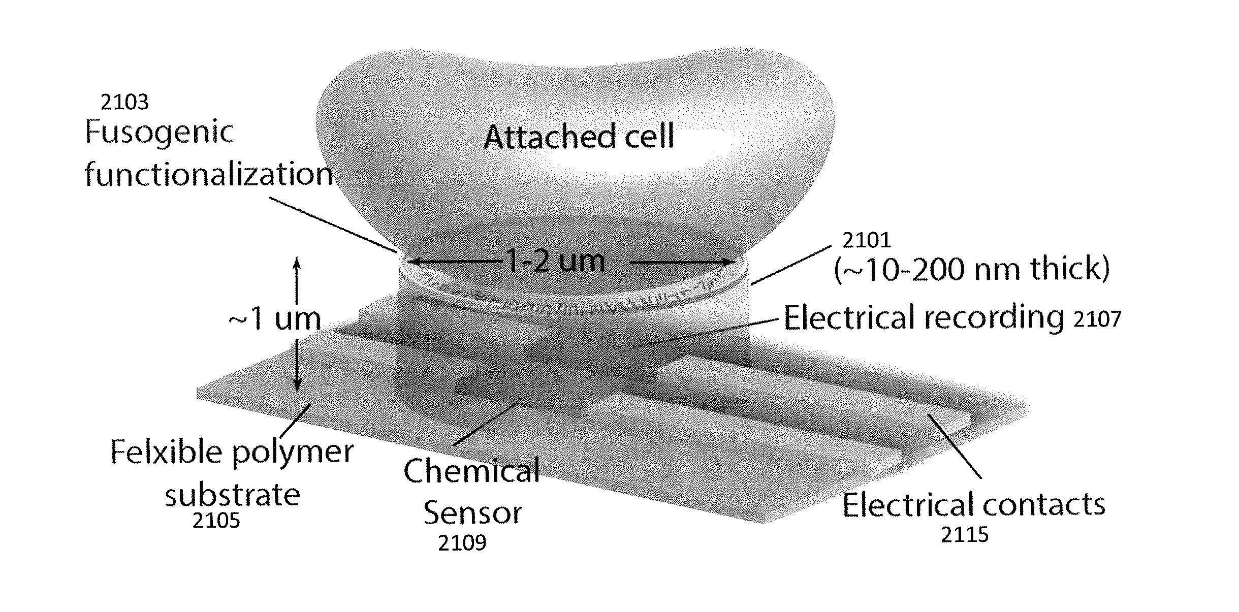
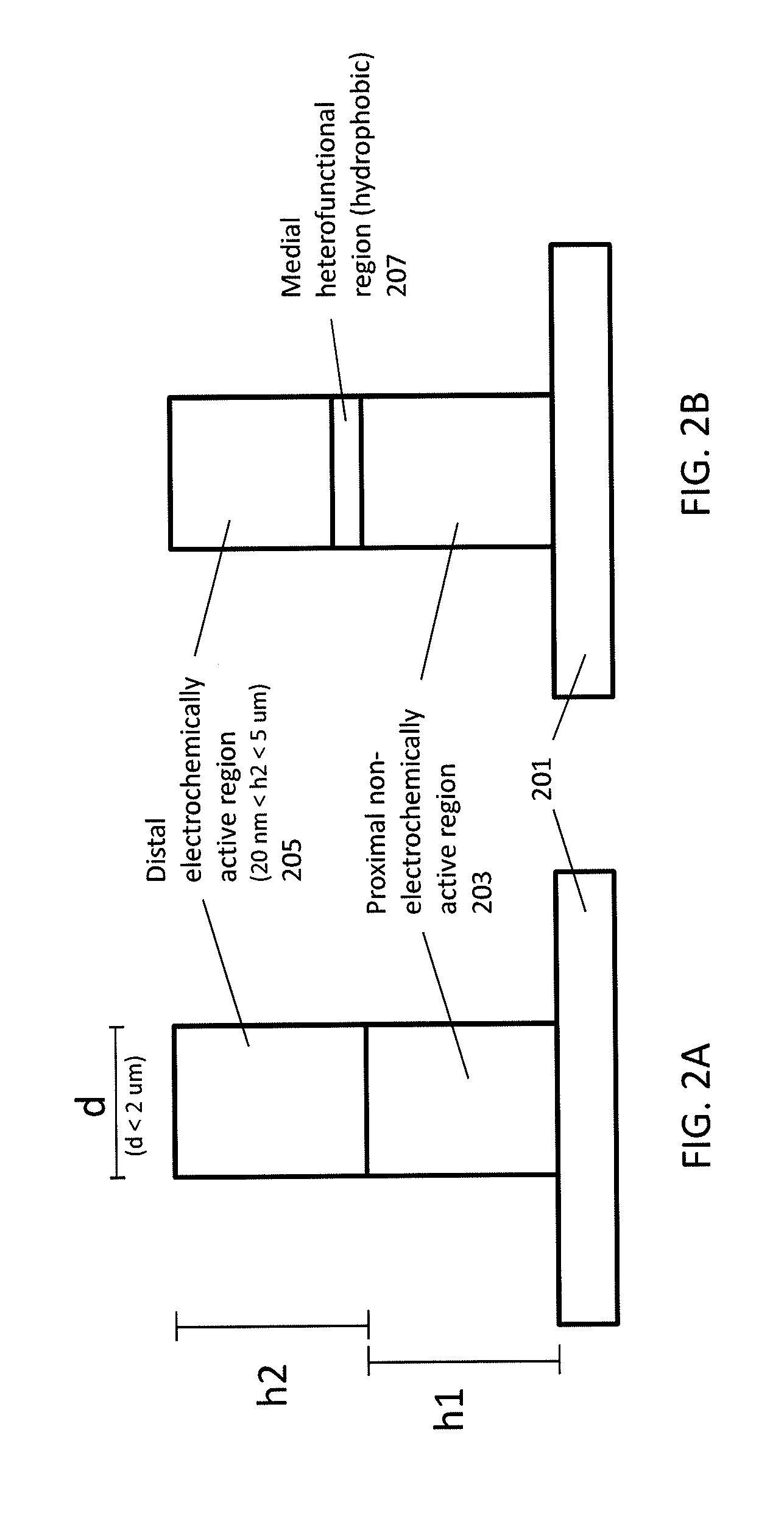
[0120]Here we present a chip-based device including a plurality of nanoscale probe electrodes formed as solid-state metallic nanoscale electrodes that spontaneously fuse with the cell membrane to form gigaohm seals. Instead of relying upon destructive formation of membrane holes and serendipitous surface adhesion, these metallic electrodes spontaneously insert into the hydrophobic membrane core by mimicking the hydrophobic banding of transmembrane proteins, forming a well-defined bioinorganic lateral junction. For example, FIG. 3A-3B schematically illustrates attachment of a cell to the probe electrodes described herein. These “stealth” probes consist of hydrophilic posts with a 5-10 nm hydrophobic band (medial heterofunctional region) formed by molecular self-assembly onto the exposed edge of a Au layer. Due to hydrophobic interactions between the band and the lipid membrane core, a tight interface impermeable to polar molecules and charged ions is formed between the probe and the ...

example 2

[0127]As mentioned above, some variation of nanoscale probes may include a medial heterofunctional region that is more hydrophobic than adjacent regions on the probe and allow for insertion of the probe into the lipid membrane. Here we demonstrate that by replicating the nanometer-scale hydrophilic-hydrophobic-hydrophilic architecture of transmembrane proteins in an artificial nanoscale probe, the probe can be made to spontaneously insert and anchor within the lipid bilayer core, forming a high-strength interface. These nanometer-scale hydrophobic bands are fabricated on metallic probes by functionalizing the exposed sidewall of an ultrathin evaporated Au metal layer rather than by lithography. Penetration and adhesion forces for butanethiol and dodecanethiol functionalized probes were directly measured using atomic force microscopy (AFM) on thick stacks of lipid bilayers to eliminate substrate effects. The penetration dynamics were starkly different for hydrophobic versus hydrophil...

example 3

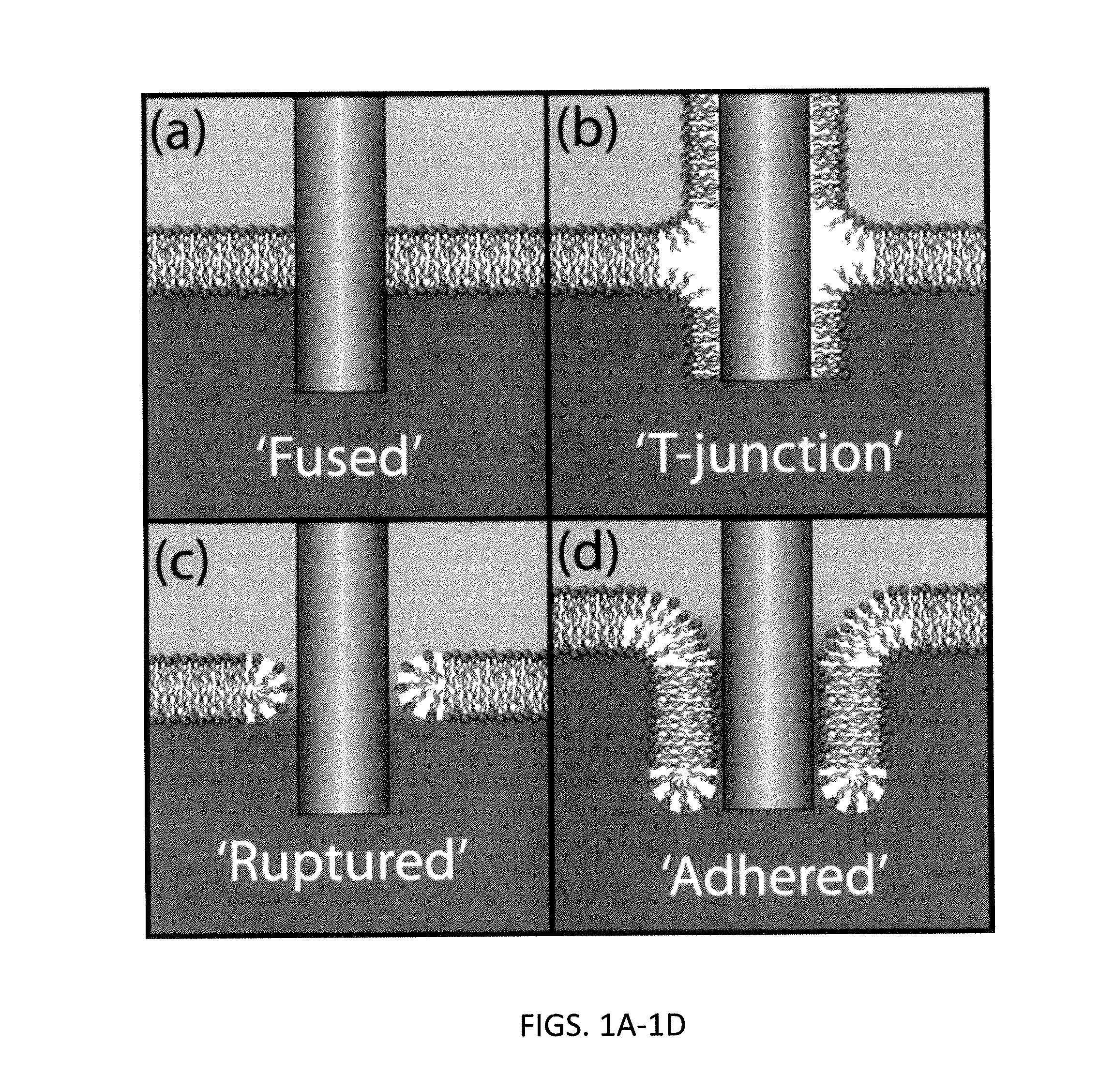
[0151]In variations of probes (e.g., nanoscale probes) including a medial heterofunctional region, the thickness of the heterofunctional region (which may be a band or annular region) may be selected to enhance the interaction with the membrane into which the probe is to be inserted. In this example we describe the role of nanoscale patterning on the strength of biomembrane-inorganic interfaces. AFM measurements show that inorganic probes functionalized with hydrophobic bands with thicknesses complimentary to the hydrophobic lipid bilayer core exhibit strong attachment in the bilayer. As hydrophobic band thickness increases to 2-3 times the bilayer core the interfacial strength decreases, comparable to homogeneously hydrophobic probes. Analytical calculations and molecular dynamics simulations predict a transition between a ‘fused’ interface and a ‘T-junction’ that matches the experimental results, showing lipid disorder and defect formation for thicker bands. These results show tha...


PUM

PropertyMeasurementUnit
heightaaaaaaaaaa
heightaaaaaaaaaa
diameteraaaaaaaaaa

Abstract

Nanoscale probes for forming stable, non-destructive seals with cell membranes. The probes, systems including these probes, and methods of fabricating and using the probes described herein may be used to sense from, stimulate, modify, or otherwise effect individual cells or groups of cells. In particular, described herein are nanoscale cellular probes that may be used to fuse with, and in some variations span, the lipid membrane of a cell to provide stable and long lasting contact to the cell. Thus, the probes described herein may be used as part of a system, method or device that would benefit from stable, non-destructive access to and across a cell membrane. In some variations the nanoscale probe devices or systems described herein may be used as part of a drug screening procedure.

Description

CROSS REFERENCE TO RELATED APPLICATIONS[0001]This patent application is a continuation-in-part of U.S. patent application Ser. No. 13 / 412,543, filed on Mar. 5, 2012, and titled “DEVICES AND METHODS FOR LONG-TERM INTRACELLULAR ACCESS,” which will issue on Aug. 19, 2014 as U.S. Pat. No. 8,808,516, which claims priority to U.S. Provisional Patent Application No. 61 / 449,615, filed on Mar. 4, 2011, and titled “DEVICES AND METHODS FOR LONG-TERM INTRACELLULAR ACCESS.” Each of these patents and patent applications are herein incorporated by reference in their entirety.STATEMENT OF GOVERNMENT SUPPORT[0002]This invention was made with government support under 0425897 awarded by the National Science Foundation. The Government has certain rights in the invention.INCORPORATION BY REFERENCE[0003]All publications and patent applications mentioned in this specification are herein incorporated by reference in their entirety to the same extent as if each individual publication or patent application w...

Claims


Application Information

Patent Timeline
Patent Type & Authority Applications(United States)
IPC IPC(8): G01N27/327
CPCG01N27/327C12M35/00G01N33/48728
Inventor MELOSH, NICHOLAS ALEXANDERVERMA, PIYUSHALMQUIST, BENJAMIN DAVID
Owner THE BOARD OF TRUSTEES OF THE LELAND STANFORD JUNIOR UNIV
Why Eureka
Try Eureka