Rapid detection and quantitation of pathogen-specific biomarkers using nanoporous dual- or multi-layer silica films

a biomarker and nanoporous technology, applied in the field of molecular biology and medicine, can solve the problems of increasing the difficulty of identifying tb in the highly vulnerable human-immunodeficiency virus population, and the difficulty of tb detection in pediatric patients, so as to facilitate the fractionation and digestion of samples on the chip, and facilitate the detection and quantification of such biomarkers. the effect of high detection accuracy

Inactive Publication Date: 2015-09-17
THE METHODIST HOSPITAL
View PDF0 Cites 22 Cited by
  • Summary
  • Abstract
  • Description
  • Claims
  • Application Information

AI Technical Summary

Benefits of technology

[0017]Importantly, the present invention provides label-free, highly reproducible diagnostic tests that are cost-effective, and capable of identifying active TB disease including the more dangerous multi-drug-resistant tuberculosis. The disclosed methods provide rapid diagnosis of TB infections (typically within one hour from sample collection to diagnosis), and importantly, can be used to distinguish between active TB disease and latent TB infection.
[0018]The present invention utilizes nanoporous silica chips that are constructed with a dual-layered film, engineered with properties that facilitate on-chip fractionation and digestion of samples, exclusion of the abundant proteins that norm

Problems solved by technology

With the increase of international travel and immigration, control of infectious disease such as tuberculosis (TB) is more challenging than any time in the history of public health.
Identification of TB in pediatric patients and the highly vulnerable human-immunodeficiency virus (HIV) population is even more challenging with the paucibacillary nature of the disease, non-specific symptoms, and difficulties in obtaining clinically relevant specimens.
Unfortunately, this method generally requires 6 to 8 weeks to complete (Dunlap et al., 2000; Scarpellini et al., 2004).
In addition to the significant delay in receiving results, conventional TB tests are unable to detect some types of active TB disease, such as tuberculous meningitis, which does not actively shed bacteria.
The method has a number of drawb

Method used


Image

  • Rapid detection and quantitation of pathogen-specific biomarkers using nanoporous dual- or multi-layer silica films
  • Rapid detection and quantitation of pathogen-specific biomarkers using nanoporous dual- or multi-layer silica films
  • Rapid detection and quantitation of pathogen-specific biomarkers using nanoporous dual- or multi-layer silica films

Examples


example 1

ESAT-6 Detection

[0163]Fabrication and Characterization of Nanoporous Silica Thin Films.

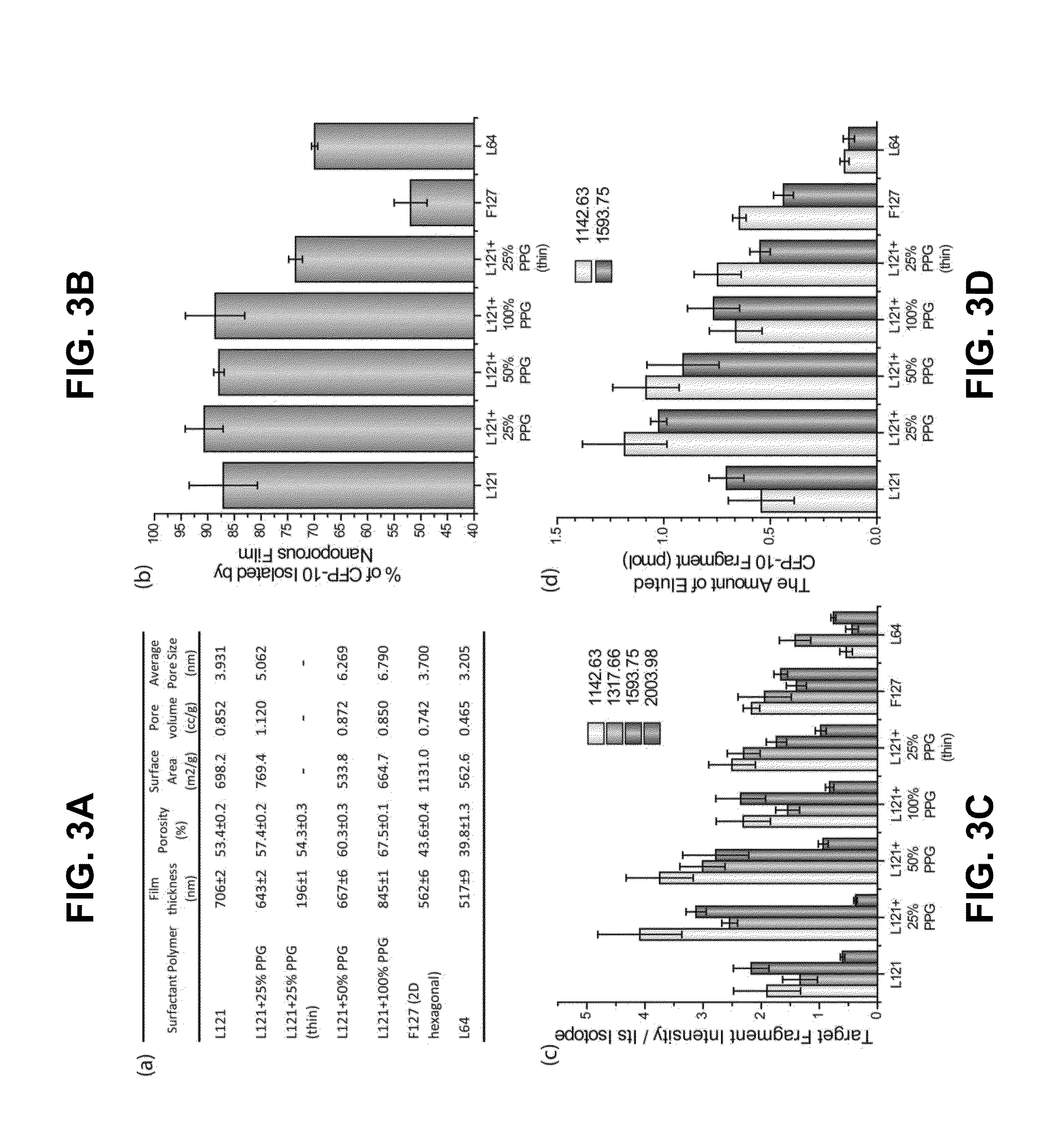
[0164]The general methods for fabrication of nanoporous silica thin films are described in the art (see e.g., Bouamrani et al., 2010; Hu et al., 2010; Hu et al., 2011; and U.S. Patent Appl. Publ. No. 2011 / 0065207). Briefly, the coating silicate sol was prepared by adding 14 mL of tetraethyl orthosilicate (TEOS) into a mixture of 17 mL of ethanol, 6.5 mL of distilled water, and 0.5 mL of 6 M HCl and stirred for 2 hrs at 80° C. to form a clear silicate sol. After cooling to room temperature, 10 mL of silicate sol was added to a mixture of 1.2 g of Pluronic L121, 10 mL of ethanol, and differing amounts of polypropylene glycol (PPG). The coating solution was stirred at room temperature for 2 hr, and deposited on a Si(100) wafer by spin-coating at a spin rate of 2500 rpm for 20 sec. To increase the degree of polymerization of the silica framework in the films, and to further improve their thermal stabi...

example 2

On-Chip Fractionation and Digestion of ESAT-6

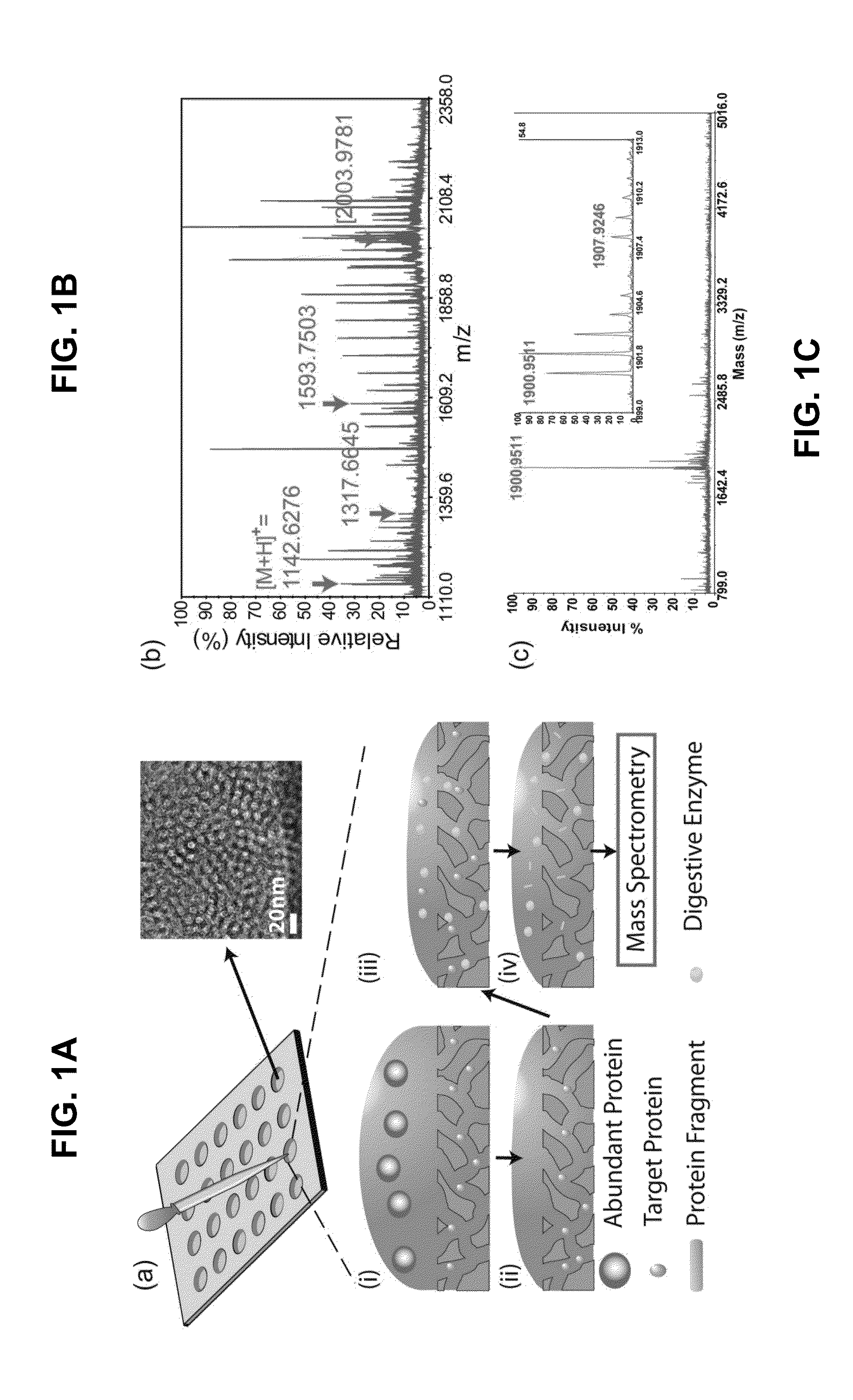
[0166]Normal human serum was obtained from Valley Biomedical (Winchester, Va., USA). Recombinant ESAT-6 was purchased from Diagnostics, Inc. (Woburn, Mass., USA). As shown in FIG. 1A, FIG. 1B, and FIG. 1C, 6 μL of protein solutions was pipetted onto the silica nanoporous film and incubated for 30 min in a humidified chamber at room temperature. The protein solution was then discarded, and 10 μL of deionized water was applied onto the silica porous film to wash away larger proteins. The washing process was then repeated three times. For enzymatic digestion, 10 μL of 0.01 mg / mL trypsin dissolved in 100 mM sodium bicarbonate was applied onto the silica nanoporous film and incubated overnight at 37° C. 10 μL of elution buffer (0.1% trifluoroacetic acid [TFA]+50% acetonitrile [ACN]) was then pipetted to extract the protein fragments. The elution buffer was then removed and stored in microcentrifuge tubes for MALDI-TOF MS analysis. To test the ...

example 3

ESAT-6 / CFP-10 Detection Analyses

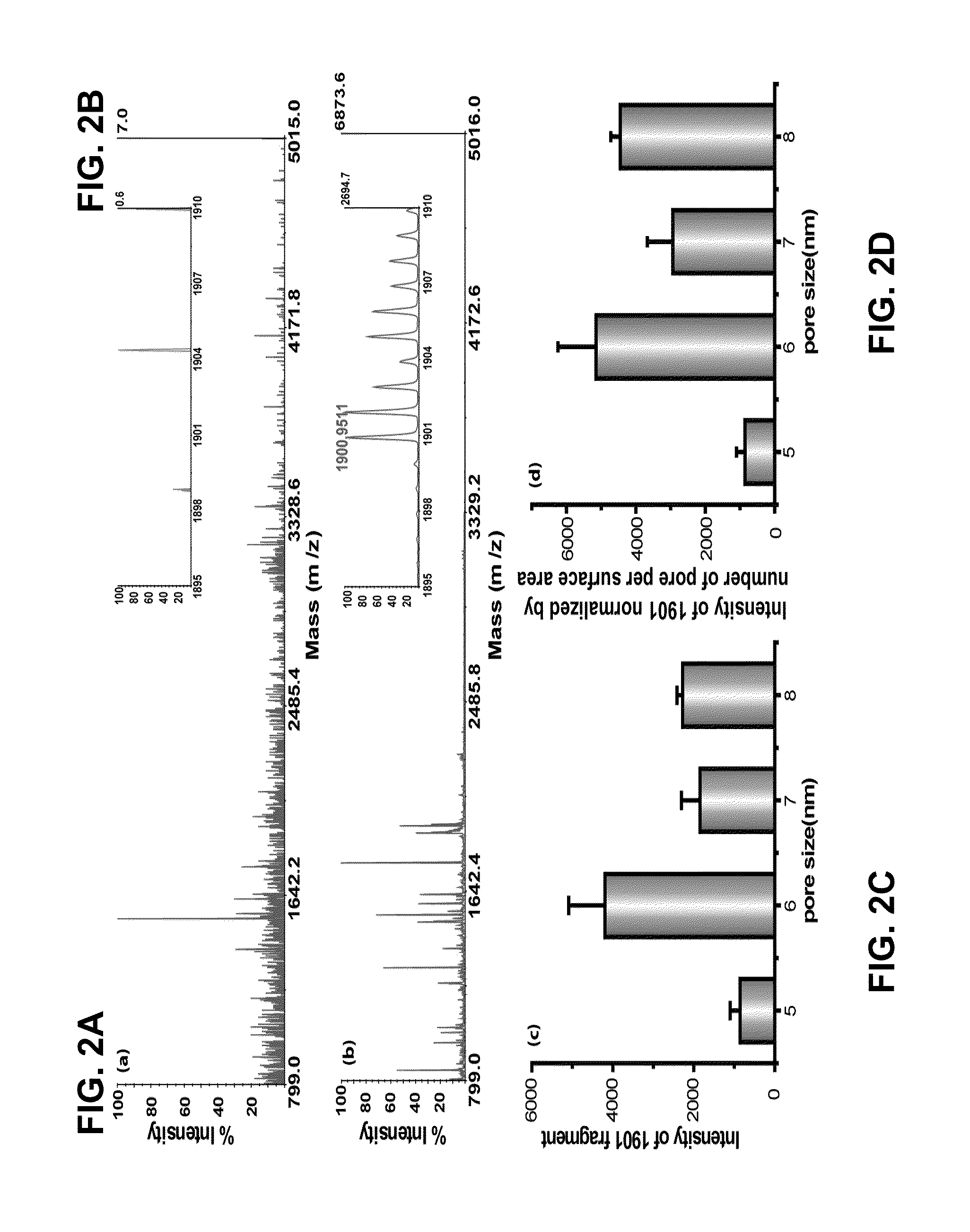
[0174]An exemplary detection procedure in accordance with one aspect of the present invention is illustrated in FIG. 1A. The biological samples were applied to a silicone gasket culture well (3-mm diameter and 1-mm height) attached on top of the nanoporous silica film. The fractionation process was completed after serial washes as described in Examples 1 and 2. Because the silica films were fixed on the flat substrate, the fractionation process was easily applied without any inconvenient washing procedure, such as sedimentation steps usually required in particle-based systems. The relatively small size of ESAT-6 (MW=10 kDa) allowed it to be captured by the silica nanopores. To identify ESAT-6 protein from biological fluids with MALDI-TOF MS, a mass fingerprinting must first be established. Because smaller protein or peptides species provide higher signals and resolution in MS and full-length ESAT-6 (10 kDa) is relatively large for MALDI-TOF MS under l...


PUM


Abstract

Improved methods for detecting active tuberculosis are disclosed. A method comprises enriching at least one M. tuberculosis-specific biomolecule from a sample by contacting the sample with a nanoporous film; and detecting the presence of the M. tuberculosis-specific biomolecule or fragment(s) thereof. The method may further comprise digesting the enriched M. tuberculosis-specific biomolecule with an enzyme to produce a digestion product comprising at least one fragment of the M. tuberculosis-specific biomolecule. Improved sensitivity and speed achieved.

Description

CROSS-REFERENCE TO RELATED APPLICATIONS[0001]The present application claims priority to PCT Intl. Pat. Appl. No. PCT / US2013 / 072416; filed Nov. 27, 2013 (pending; Atty. Dkt. No. 37182.163), which claims the benefit of U.S. Prov. Pat. Appl. No. 61 / 732,266, filed Nov. 30, 2012 (expired; Atty. Dkt. No. 37182.162), the contents of each of which is specifically incorporated herein in its entirety by express reference thereto.STATEMENT REGARDING FEDERALLY SPONSORED RESEARCH OR DEVELOPMENT[0002]This invention was made with government support under Grant No. W81XWH-11-2-0168 awarded by the United States Department of Defense, and Grant No. U54-CA-151668 awarded by the National Institutes of Health. The government has certain rights in the invention.NAMES OF THE PARTIES TO A JOINT RESEARCH AGREEMENT[0003]Not Applicable.BACKGROUND OF THE INVENTION[0004]1. Field of the Invention[0005]The present invention relates to the fields of molecular biology and medicine. In particular, the invention prov...

Claims


Application Information

Patent Timeline
IPC IPC(8): G01N33/569G01N33/552
CPCG01N33/5695G01N2333/35G01N2469/10G01N33/552G01N33/569Y02A90/10
Inventor HU, YEMA, XINWU, HUNG-JENFAN, JIASUN, TONG
Owner THE METHODIST HOSPITAL
Why Eureka
Try Eureka