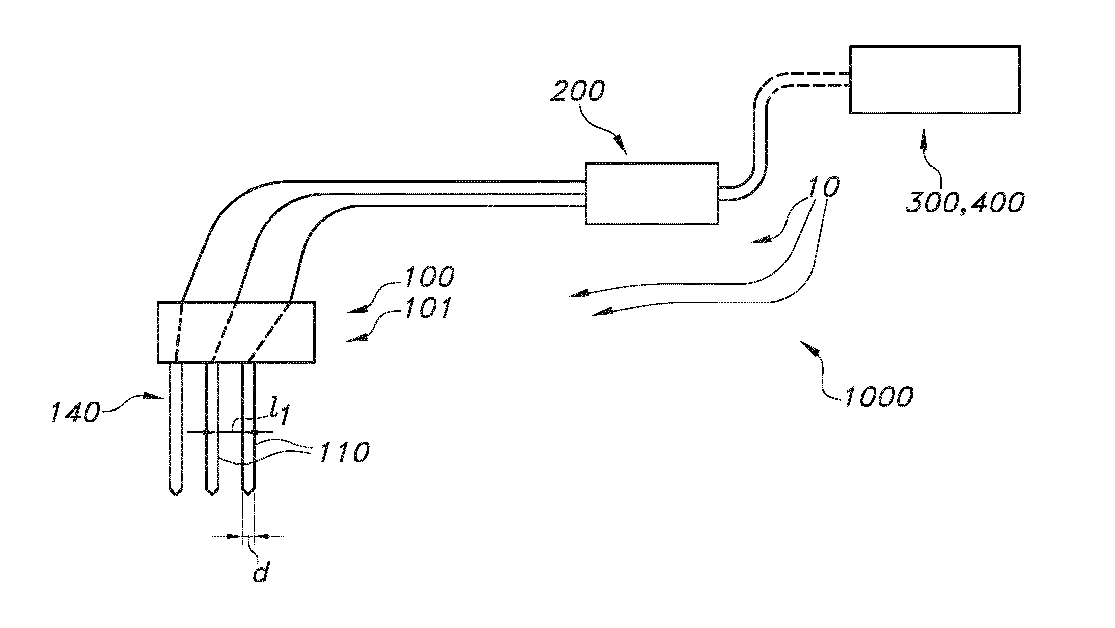
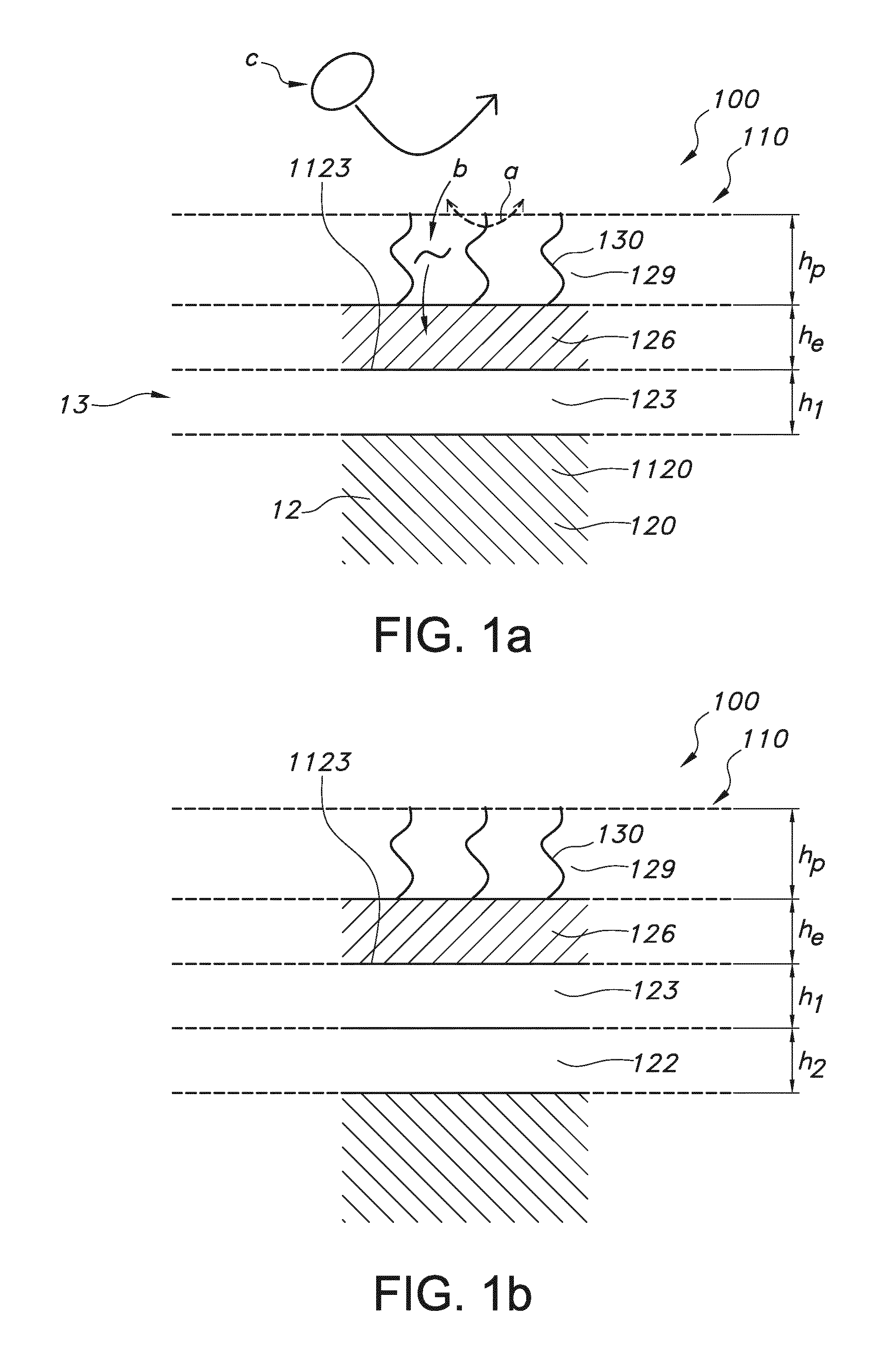
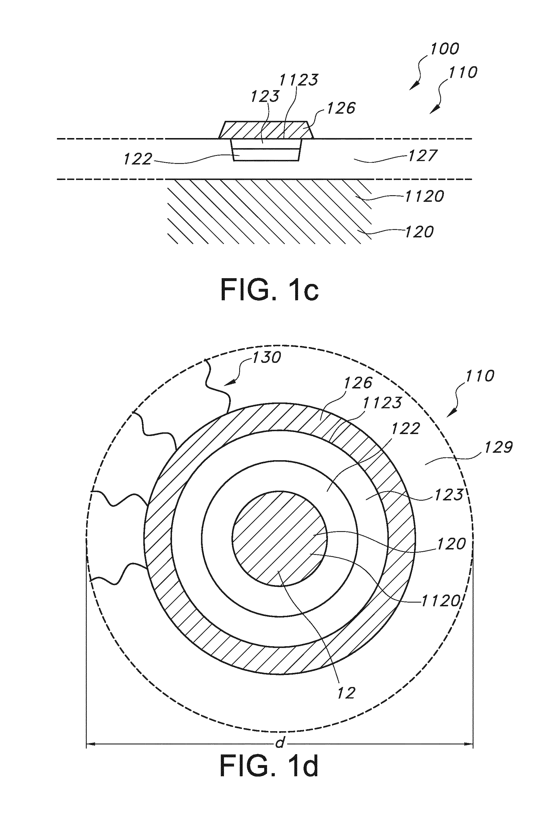
Rod shaped implantable biosensor
a biosensor and rod-shaped technology, applied in the field of brain implantable biosensors, can solve the problems of inability to detect noise, lack of temporal resolution or specificity of non-electrochemically active analytes, and fragility of sensor materials such as carbon and platinum, so as to facilitate connection, improve electrical conductivity, and improve electrical conductivity
- Summary
- Abstract
- Description
- Claims
- Application Information
AI Technical Summary
Benefits of technology
Problems solved by technology
Method used
Image

Examples
example 1
Sensitivity of Pt Coated Tungsten Sensor to Peroxide
[0043]In one experiment the H202 sensitivity of platinum coated tungsten wires was tested, in order to study whether it would be possible to use them as biosensors. Table 1 shows the responsiveness of 50-micrometer thick (diameter) platinum-coated sensors to H202, in comparison to conventional 200 micrometer thick platinum wire sensors. The production procedure for the novel platinum-coated tungsten sensors is described in appendix A. These 50-micrometer thick (diameter) platinum-coated sensors have good responsiveness to H202 as Table 1 shows. In addition, platinum-coated tungsten is very rigid, so remains intact during insertion in the tissue, such as brain at diameters below 50 micrometers. Platinum wire electrodes, by comparison, typically do not survive these insertions, especially not after insertion in freely moving animals.
TABLE 1Sensitivity of Pt coated tungsten sensors to H2O2Pt200-GluS1Pt200-GluS2PtTungsten50PtTungsten50...
example 2
Assembly of a Sensor
[0044]
Order form Assembly of standard coiled biosensorStep 1: Part assembling sensorDay 1 Assembling1.1Cutting: TSP075150; L = 12 mmCut Pt Plated tungsten wire (P069) (OD = 0.05 mm; L = 16 mm) in L = 15 mFill the silica tube with wire.1.2Prepare two component epoxy glue in 1 ml syringe. Fill the silica tubing containing Pt wirewith epoxy glue. Make sure that there are no air bubbles inside the silica tubing.Day 2Put the glued shaft for a minimum of 24 hours in the stove at 70° C.1.3Take out the glued silica tubes from the oven. Check if the PT wire is fixed well inside bypulling the wire.Cut one end of the Pt wire to 1 mm and 2 mm preciselyClean the sensors by immersing in:1. 5 minutes in acetone2. 10 minutes in UP water3. Dry the sensors at 40° C. for 15 minutes1.4Prepare Ag wire (OD = 0.05 mm) in a roll and place it on a stick to hold the round. Underthe microscope place a small drop of pattex glue on silica tube opposite end of the sensor tip.Place Ag wire ver...
example 3
[0045]Gold plated fetal scalp electrodes for CTG were used for the experiments. Electrodes were cleaned by immersing for 10 minutes in acetone followed by 10 minutes in ultrapure water. The sensors were dried at room temperature for 1 hour. The sensors were coated by nafion for 5 minutes by repeated immersing in 5% nafion (sigma), followed by 20 sec of drying on air. The nafion was baked for 2 times 3 min at 170 degrees and left for at least 2.5 hrs. before further use.
[0046]Lactate oxidase was dissolved in ultrapure water to a concentration of 1 unit per microliter (stock solution). Glutardialdehyde was used as linker together with bovine serum albumin (BSA) as an additive to stabilize enzyme coating. 6.25 mg BSA was dissolved in 0.5 ml ultrapure water. 1.6 microliter of 50% Glutardialdehyde was added and the solution was gently mixed. The solution was left for 5 min. 137 microliter of the BSA-linker solution was...
PUM
| Property | Measurement | Unit |
|---|---|---|
| shear strength | aaaaa | aaaaa |
| shear strength | aaaaa | aaaaa |
| length | aaaaa | aaaaa |
Abstract
Description
Claims
Application Information
 - R&D
- Intellectual Property
- Life Sciences
- Materials
- Tech Scout
- Unparalleled Data Quality
- Higher Quality Content
- 60% Fewer Hallucinations
Browse by: Latest US Patents, China's latest patents, Technical Efficacy Thesaurus, Application Domain, Technology Topic, Popular Technical Reports.
© 2025 PatSnap. All rights reserved.Legal|Privacy policy|Modern Slavery Act Transparency Statement|Sitemap|About US| Contact US: help@patsnap.com