Methods of cell separation

a cell and cell technology, applied in the field of cell separation, can solve the problems of reducing the recovery of desired cells and/or contamination with undesired cell types, and reducing the yield of desired cell subsets

Active Publication Date: 2016-02-25
CELLS4LIFE GRP
View PDF2 Cites 0 Cited by
  • Summary
  • Abstract
  • Description
  • Claims
  • Application Information

AI Technical Summary

Problems solved by technology

Hypotonic lysis, while useful in low volume in vitro studies, can be impractical for the large volumes of blood tissues processed for cellular therapies.
Consequently, particular cell types can become distributed throughout a density-gradient medium rather than precisely segregating at a discrete area in the density medium, resulting in reduced recovery of desired cells and/or contamination with undesired cell types.
In procedures that enrich for rare blood cell types such as hematopoietic progenitor cells, density-gradient sedimentation can lead to loss or reduced yields of desired cell subsets.
For example, using conventional density-gradient methods to isolate progenitor cells from umbilical cord blood results in a significant loss of the desired stem cells e.g., CD34+ hematopoietic stem cells (HSCs) or VSELs (Very Small Embryonic Like Stem Cells) (Wagner, J. E., Am J Ped Hematol Oncol 15:169 (1993)).
Using conventional density-gradient methods to isolate lymphocytes can result in s

Method used


Image

  • Methods of cell separation
  • Methods of cell separation
  • Methods of cell separation

Examples


example 1

General Cell Separation Method

[0184]Umbilical cord blood was collected into standard Baxter 250 ml (nominal) blood bags containing 35 ml of a citrate phosphate dextrose adenine (CPDA) anticoagulant solution.

[0185]A solution containing dextran and a further constituent selected from dimethyl sulphoxide (DMSO), dimethyl glycine (DMG), L-valine, L-proline, β-alanine, leucine, isoleucine and glycine, in phosphate-buffered saline (PBS), was prepared. This solution was added to a blood sample at a ratio of 1:1, this was then mixed thoroughly. Separation of the erythrocyte and nucleated cell fractions took place at room temperature within a time period of 15 or 30 minutes.

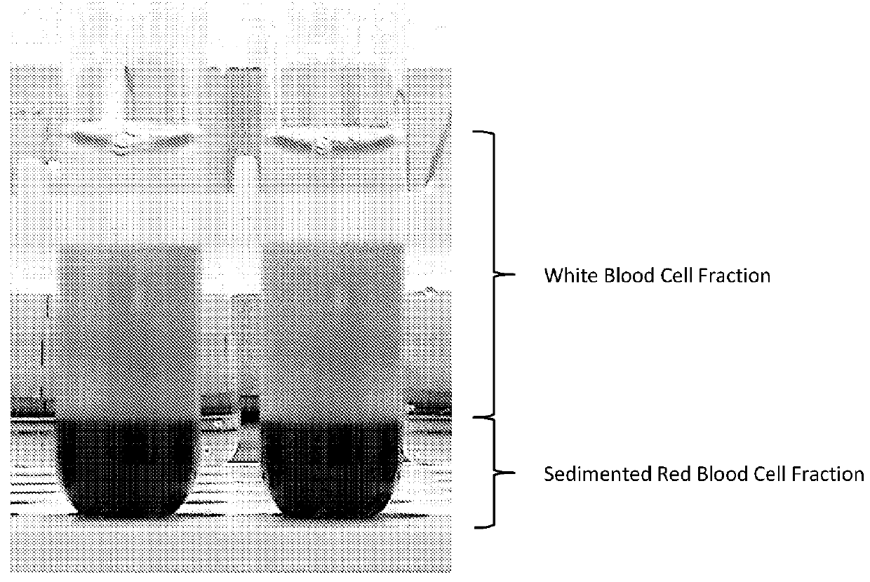
[0186]Separation of the cell fractions could be observed visually. FIG. 1 shows clear separation of the erythrocytes (bottom of test tube) from the nucleated cells (top of test tube). This image was taken 30 minutes after exposure to dextran (500 molecular weight (Mw)) at a final concentration of 2.5% w / v and DMSO at a fi...

example 2

Erythrocyte Volume Fraction (Haematocrit) Study

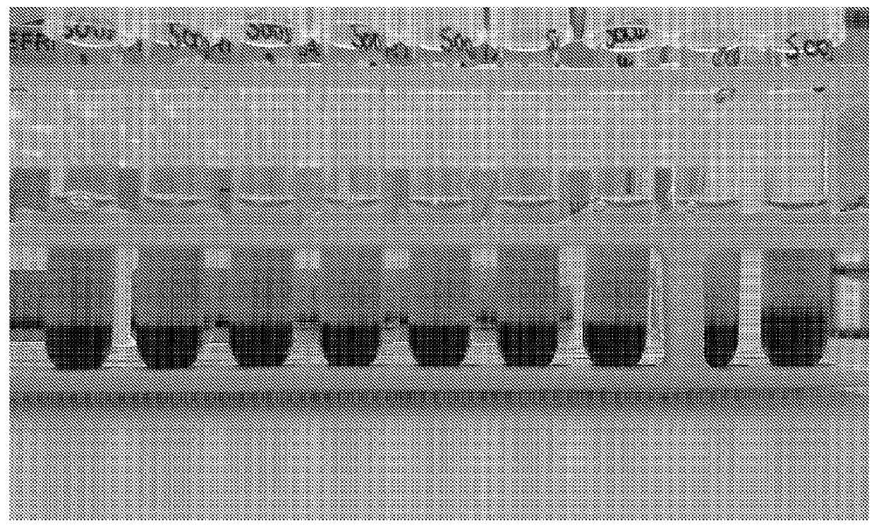
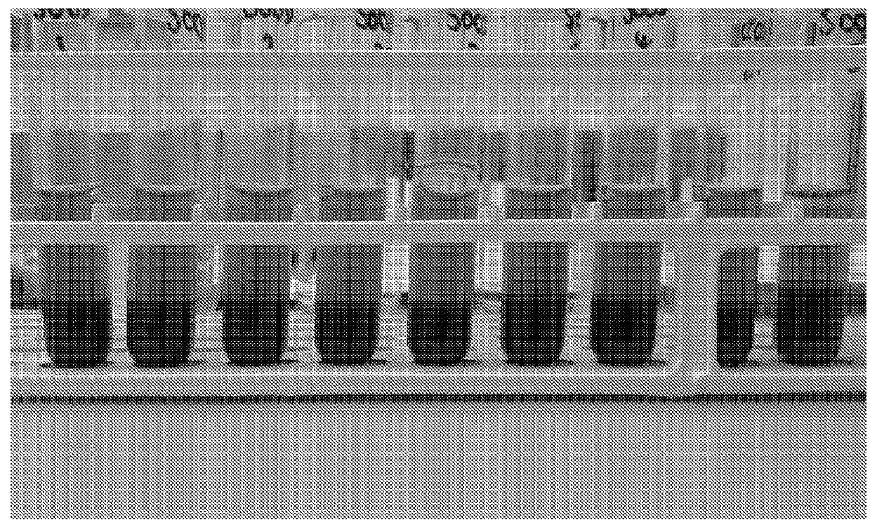
[0191]In this Example, the levels of erythrocyte volume fraction remaining in the nucleated cell fraction after cell separation were determined. Three tubes were prepared for the separation: (i) blood sample mixed at a ratio of 1:1 with PBS only (control), (ii) blood sample mixed at a ratio of 1:1 with a solution containing 500 Mw dextran at a concentration of 5% w / v in PBS (final concentration of dextran of 2.5% w / v), (iii) blood sample mixed at a ratio of 1:1 with a solution containing 500 Mw dextran at a concentration of 5% w / v and DMSO at a concentration of 5% v / v in PBS (final concentration of dextran of 2.5% w / v and of DMSO of 2.5% v / v). Samples were left to separate for 30 minutes at room temperature.

[0192]It was observed that the cell fractions in sample (iii) separated at a faster rate than in sample (ii) and, after 30 minutes, sample (iii) had developed a more compact erythrocyte fraction compared to sample (ii) (data not show...

example 3

Assessment of Cell Viability and the Presence of Hematopoietic Stem Cells (HSCs) using Flow Cytometry

[0195]A blood sample was mixed at a ratio of 1:1 with a solution containing 500 Mw dextran at a concentration of 5% w / v and DMSO at a concentration of 5% v / v in PBS (final concentration of dextran of 2.5% w / v and of DMSO of 2.5% v / v). Samples were left to separate for 30 minutes at room temperature.

[0196]A sample of the resulting nucleated cell fraction was then analysed using flow cytometry. In particular, analysis was carried out using a “Stem Cell Enumeration Kit” obtained from Becton, Dickinson and Company. Analysis was carried out as per the Application Guide provided with the Kit and the templates used are based on a method featured in the Clinical and Laboratory Standards Institute H42-A2 approved guideline (Enumeration of Immunologically Defined Cell Populations by Flow Cytometry; Approved Guideline-Second Edition. Wayne, PA: Clinical and Laboratory Standards Institute; 2007....


PUM


Abstract

The present invention relates to the use of a combination of: (i)a macromolecular erythrocyte sedimentation enhancer, and (ii) dimethyl sulphoxide (DMSO), dimethylglycine (DMG) and/or valine; to recover non-erythrocyte blood cells from a blood cell-containing sample and/or to prime non-erythrocyte blood cells to protect their integrity in subsequent cryopreservation step(s).

Description

[0001]This invention relates to methods and compositions for separating and / or priming cells, and more particularly to methods and compositions for separating erythrocytes from peripheral blood, bone marrow, umbilical cord blood and related blood tissues.[0002]Isolation of white cells from blood for in vitro studies or in cellular therapy usually incorporates an initial separation of blood mainly based on the bulk depletion of erythrocytes, which comprise >99% of the cellular mass.[0003]Techniques for erythrocyte removal are based on hypotonic lysis of erythrocytes, density gradient separation, enhanced centrifugal sedimentation using hydroxyethyl starch or mixtures (containing amongst other things antibodies) that promote accelerated sedimentation of the erythrocytes.[0004]Hypotonic lysis, while useful in low volume in vitro studies, can be impractical for the large volumes of blood tissues processed for cellular therapies. In cell therapy procedures, erythrocyte hypotonic lysis...

Claims


Application Information

Patent Timeline
IPC IPC(8): C12N5/00A01N1/02C12N5/078
CPCC12N5/0087C12N5/0641C12N2500/32C12N2500/62A01N1/0221C12N5/0634C12N2500/34
Inventor DREW, JEFFREY
Owner CELLS4LIFE GRP
Why Eureka
Try Eureka