
Medical devices with reduced thrombogenicity

a medical device and thrombogenic technology, applied in the field of biomaterials, can solve the problems of reducing the clinical application range of effective biomaterials available for clinical vascular applications, affecting the clinical application range of biomaterials, and dapt carrying an inherent risk of bleeding, and achieves robust application, low thrombogenicity, and resistance to delamination.

Active Publication Date: 2016-03-17
HEART RES INST LTD
View PDF2 Cites 3 Cited by
  • Summary
  • Abstract
  • Description
  • Claims
  • Application Information

AI Technical Summary

Benefits of technology

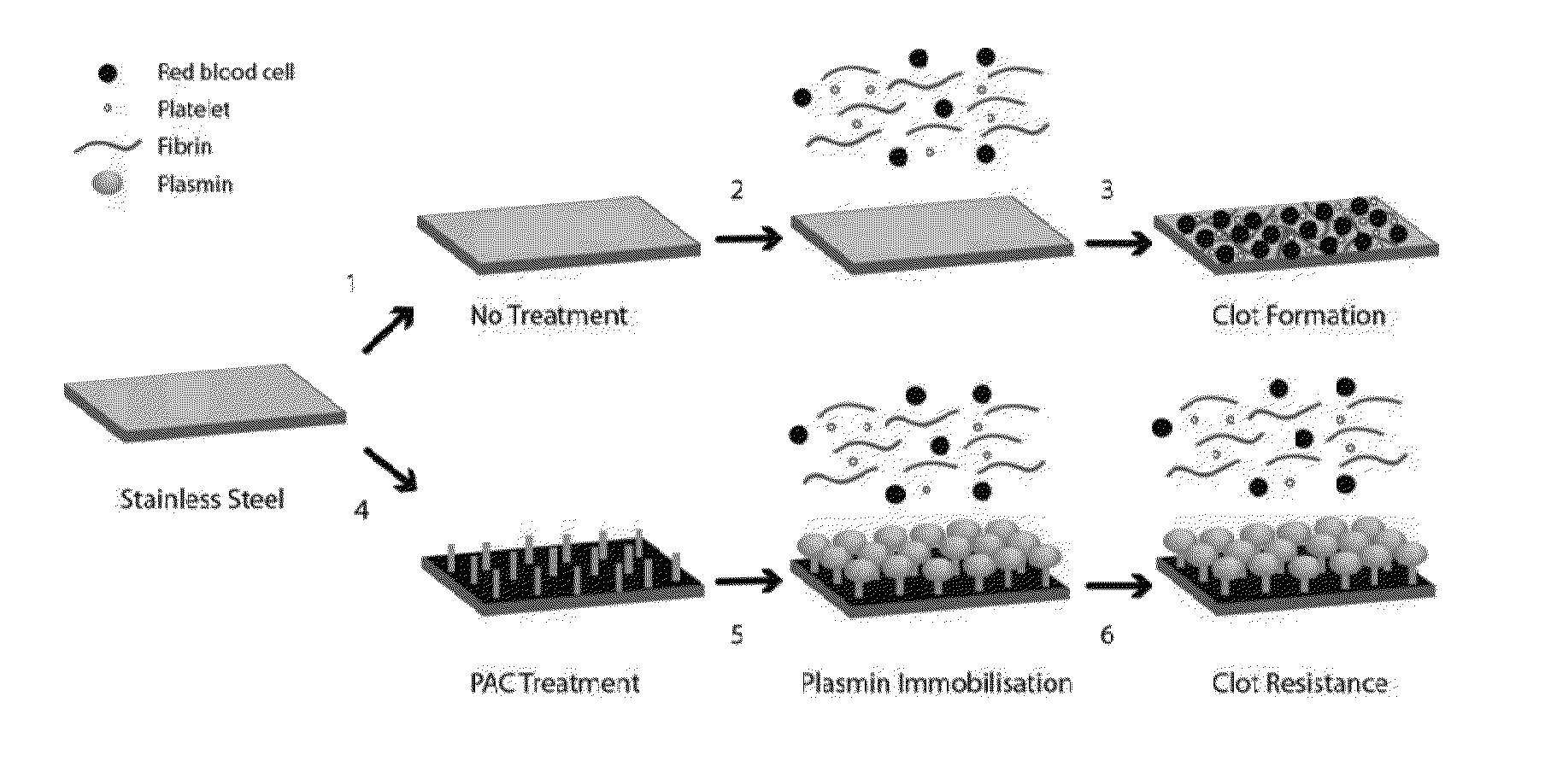
The plasma-activated coating (PAC) process can attach enzymes and other proteins to medical devices, making them more effective in inhibiting thrombosis and anticoagulation. The process is robust and can be stored for long periods without losing effectiveness. The resulting devices can be safely used in vascular applications.

Problems solved by technology

Despite the increasing incidence of cardiovascular disease, there are few effective biomaterials currently available for clinical vascular applications.
Currently available synthetic biomaterials such as polyethylene terephthalate have a range of vascular applications including endovascular grafting, heart valve replacement, vascular / myocardial patches and vascular closure devices, but perform poorly as vascular surgical conduits.
Confounding this issue, DAPT carries an inherent risk of bleeding, significantly increasing if therapy is continued for 12 months or longer (Valgimigli et al, Circulation, 125:2015 (2012)).
This work highlights important challenges in the adaptation of fibrinolytic agents to enhance biomaterial hemocompatibility, including the need for complex linker chemistry schemes for robust attachment and the long-term retention of biologically active substrates.

Method used


Image

  • Medical devices with reduced thrombogenicity
  • Medical devices with reduced thrombogenicity
  • Medical devices with reduced thrombogenicity

Examples


example 1

Covalent Immobilization of Plasmin to PAC

[0084]Materials and Methods

[0085]Reagents: All reagents were purchased from Sigma-Aldrich, St Louis and used without further purification unless otherwise noted. Human umbilical vein endothelial cells (HUVECs) were harvested enzymatically from umbilical cords. Endothelial cells from passages 2-4 were used.

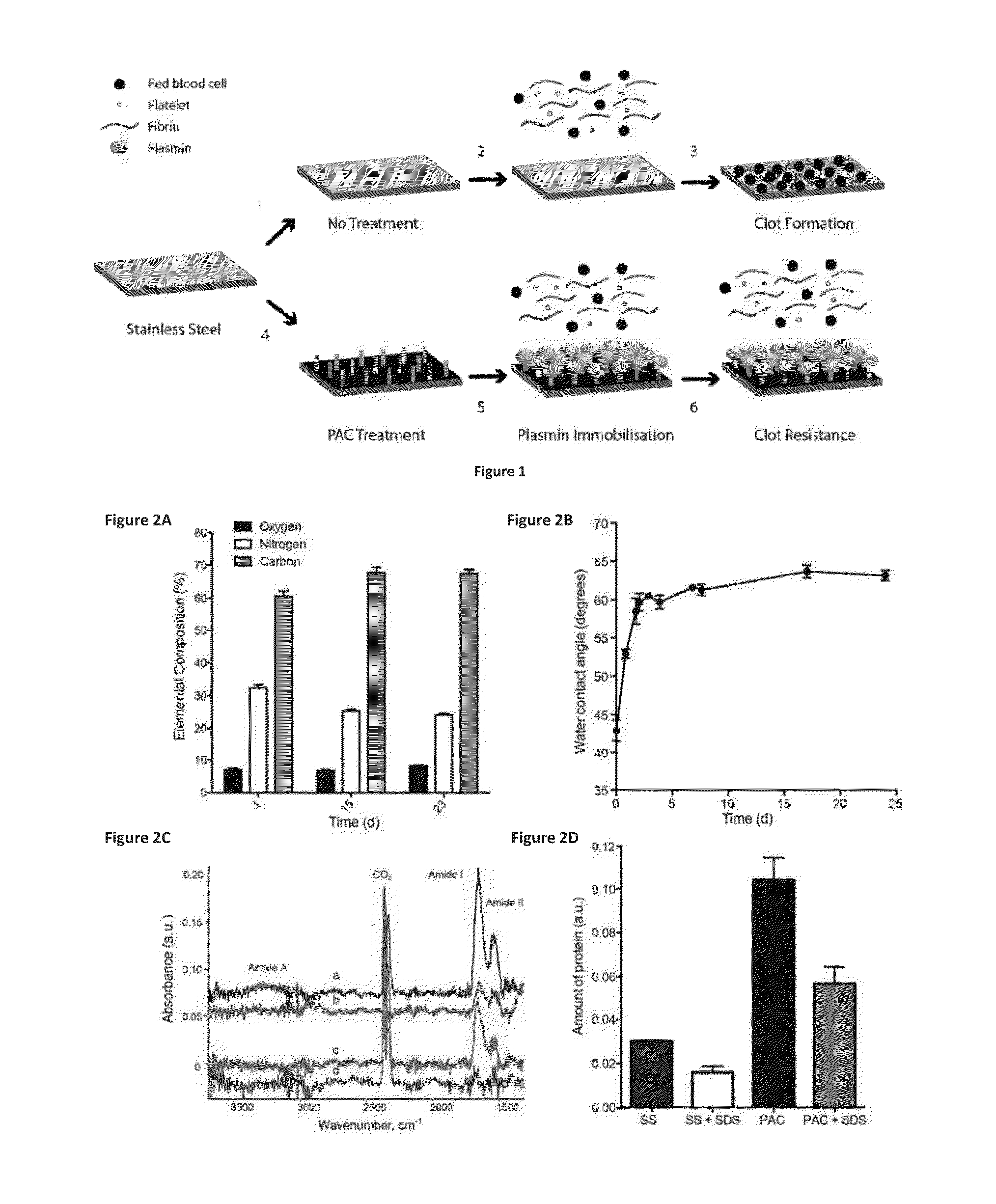
[0086]Sample Preparation: The substrates were 316L , stainless steel foil (SS) 25 μm thick (Brown Metals), or 3.0×10mm 316LVM stainless steel stents (Laserage, Calif., USA). Plasma-activated coating on 316L stainless steel (PAC) surfaces were generated from acetylene in, argon mixed with nitrogen. Stainless steel stents were imaged with a Zeiss EVO 50 scanning electron microscope. Samples were incubated with increasing concentrations of plasmin (0.1, 1.0 and 10 μg) in PBS at 37° C. overnight and washed in PBS prior to use.

[0087]Surface Characterization: The contact angle between PAC and de-ionized water was measured using a Kruss contact an...

example 2

Covalently Bound Plasmin Retains Bioactivity.

[0091]Materials and Methods

[0092]Covalent attachment: Samples were washed with water to remove salt and dried prior to accumulation of spectra using a Digilab FTS7000 FTIR spectrometer fitted with an attenuated total reflection (ATR) accessory with a trapezium germanium crystal at incidence angle of 45°. To obtain sufficient signal / noise ratio and resolution of spectral bands, 500 scans with a resolution of 1 cm−1 were taken. Difference spectra were used to detect changes associated with the presence of plasmin, and analysis carried out. Unbound protein was removed by aspiration and the surfaces were washed with PBS. Non-covalently bound protein was removed by SDS-washing. Samples were treated with 5% (w / v) SDS for 1 hat 80° C. Following the SDS treatment, samples were washed with PBS and distilled water.

[0093]Bioactivity assay: The enzymatic activity of plasmin was monitored using a commercially available kit. One unit of activity is def...

example 3

Effect on Cell Attachment and Proliferation

[0096]Materials and Methods

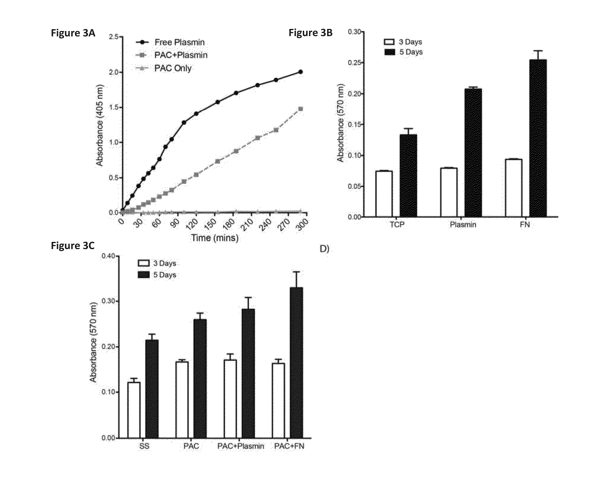
[0097]Endothelial Cell Interactions: For proliferation assays, HUVECs (20,000 cells / mL) were plated in 24-well plates for 3 and 5 days. Attachment and proliferation of cells to and on plasmin-coated wells was analyzed in comparison to tissue culture plastic alone and to wells coated with fibronectin (10 μg / well). Cells were quantified at 3 and 5 days post-seeding using the MTT (3[4,5-dimethylthiazol-2-yl]-2,5 diphenyl tetrazolium bromide) assay according to manufacturer's instructions. Dimethyl sulfoxide (DMSO) was used to dissolve insoluble formazan crystals, and the absorbance at 540 nm was measured using a spectrophotometer (Biorad).

[0098]Results

[0099]After 3 days of incubation, cell numbers on TCP, plasmin and fibronectin (FN) were not significantly different (FIG. 3B). At day 5, cell proliferation on plasmin was 56.40±3.2% higher than TCP alone (p<0.001), but remained statically less than then FN positive con...


PUM

PropertyMeasurementUnit
water contact angleaaaaaaaaaa
thickaaaaaaaaaa
thickaaaaaaaaaa

Abstract

A plasma-activated coating (PAC) process covalently binds enzymes in their bioactive state, has low thrombogenicity and can be robustly applied to medical devices, resisting delamination when deployed in vivo. Applying this process to attachment of proteins such as enzymes that inhibit thrombosis and anticoagulants such as heparin or heparin fragments, one can produce medical devices and other materials for use in vascular applications having a number of benefits including covalent attachment, not requiring intermediate linkers or chemistry; substrate independent works on polymers, metals, ceramics, 3D shapes like stents, valves, etc.; bioactivity is retained; surface may retain greater bioactivity over time in vivo; Simultaneously supports endothelialisation; can be stored for long periods, following freeze drying, and retains effectiveness when rehydrated and; surface is able to bind many fibrinolytic enzymes such as streptokinase, urokinase, tPA, plasmin).

Description

CROSS REFERENCE TO RELATED APPLICATION[0001]This application claims benefit of and priority to U.S. Provisional Application No. 62 / 049,879, filed Sep. 12, 2014, all of which is incorporated by reference in its entirety.FIELD OF THE INVENTION[0002]The present disclosure relates generally to biomaterials with improved blood compatibility, made by immobilizing proteins such as blood enzymes for reduced surface thrombogenicity using plasma activation of the substrate for attachment.BACKGROUND OF THE INVENTION[0003]Despite the increasing incidence of cardiovascular disease, there are few effective biomaterials currently available for clinical vascular applications. Currently available synthetic biomaterials such as polyethylene terephthalate have a range of vascular applications including endovascular grafting, heart valve replacement, vascular / myocardial patches and vascular closure devices, but perform poorly as vascular surgical conduits.[0004]An increasing number of implantable devic...

Claims


Application Information

Patent Timeline
Patent Type & Authority Applications(United States)
IPC IPC(8): A61L33/00A61L33/06B05D7/00
CPCA61L33/0011B05D7/58A61L33/0047A61L33/068A61B2017/00893A61L2300/608A61F2210/00A61F2/82A61F2/24A61L2300/42A61B17/12109A61B17/12122A61B17/12177A61B2017/00526A61B2017/00575A61L27/34A61L31/10A61N1/056B05D1/62
Inventor WISE, STEVEN G.NG, MARTIN K.C.
Owner HEART RES INST LTD
Learn More