

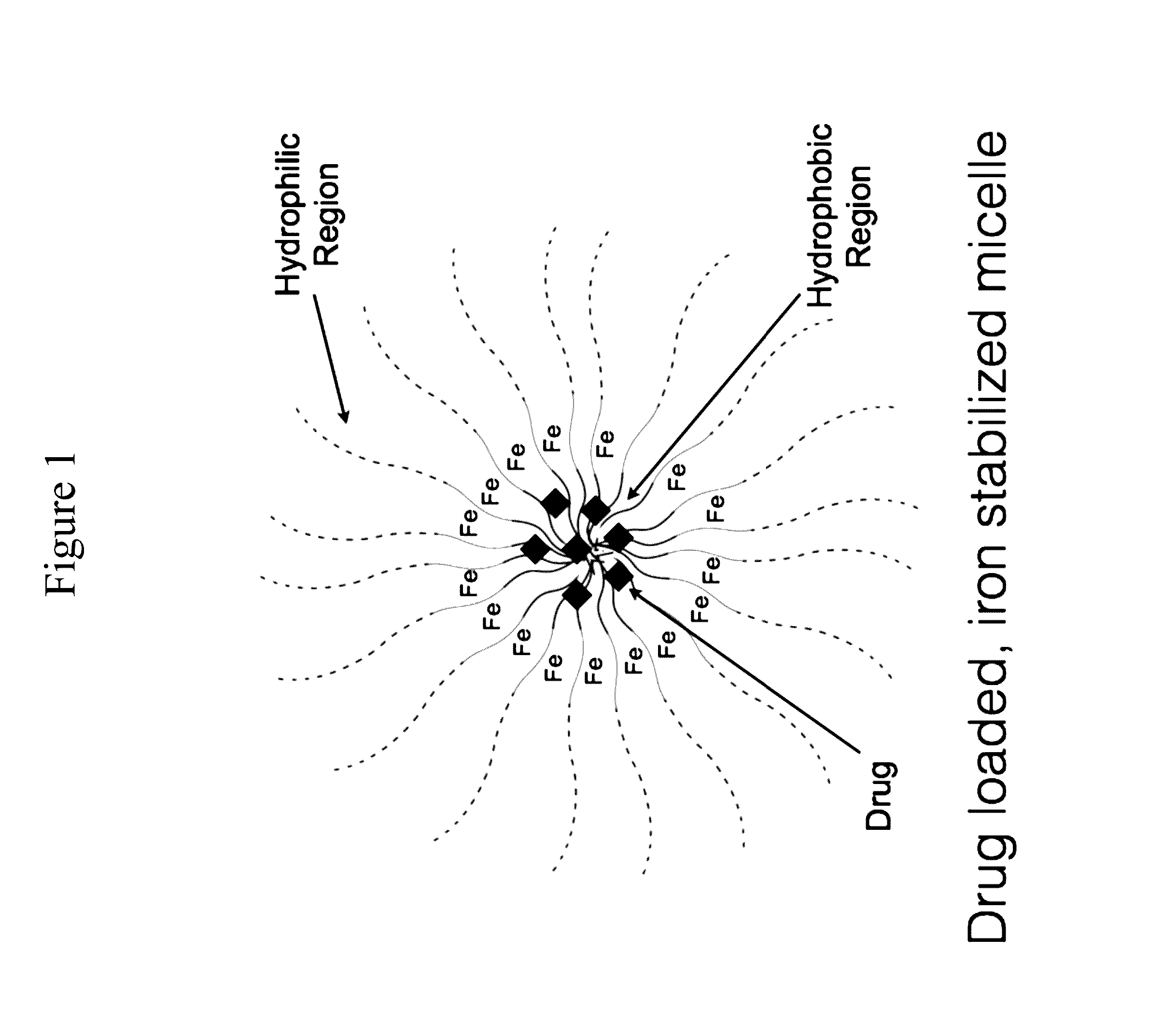
Iron stabilized micelles as magnetic contrast agents
a technology of iron stabilized micelles and magnetic contrast agents, which is applied in the field of polymer chemistry, can solve the problems that many other organs and tissues cannot be easily imaged without contrast enhancemen
- Summary
- Abstract
- Description
- Claims
- Application Information
AI Technical Summary
Benefits of technology
Problems solved by technology
Method used
Image

Examples
example 1
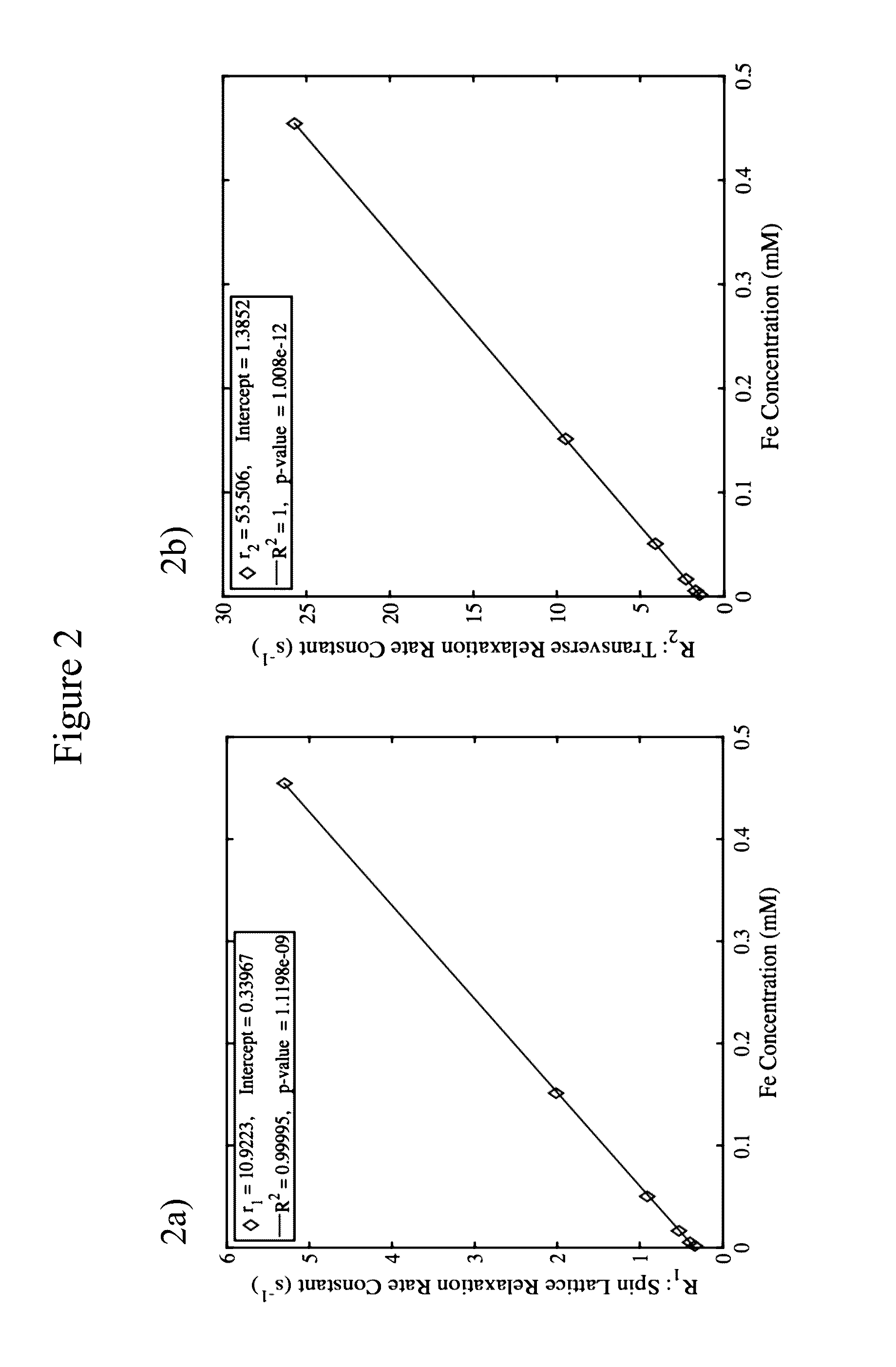
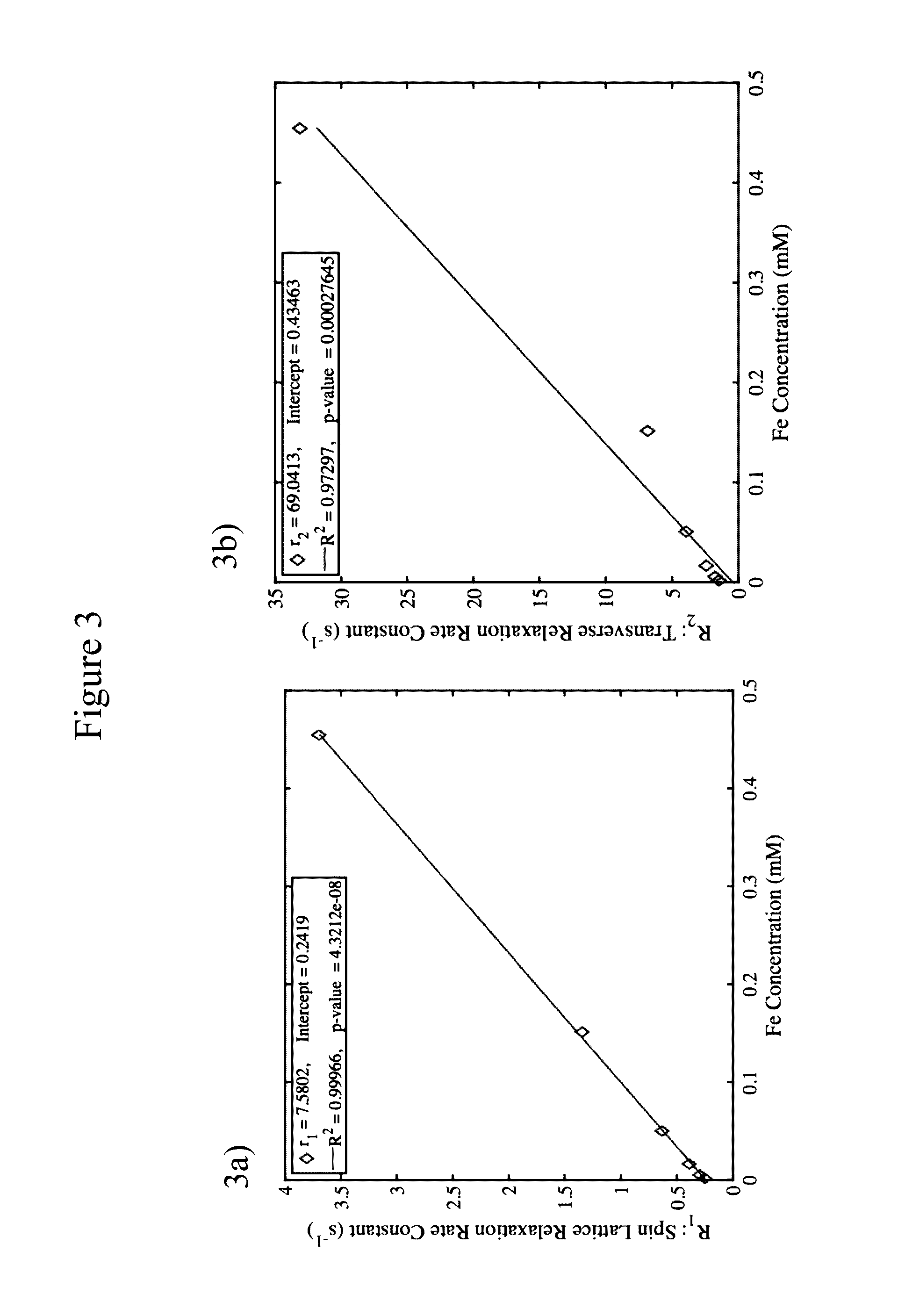
[0229]In vitro phantom measurements were performed to determine spin-lattice (r1) and spin-spin (r2) relaxivity values. Multiple concentrations of each nanoparticle were prepared at values ranging from 0.002 mM to 0.6 mM and repeated in triplicate in a 96-well plate cut into two. This was placed directly in a 72 mm ID birdcage coil in a horizontal bore magnet at 7 Tesla (Agilent ASR 310) and spin echo images (sems) were acquired at either multiple TR values (progressive saturation) or followed by a train of 180° pulses for collection of multiple spin echoes (mems). These images were collected with a field of view of 8×4 cm2 and a matrix size of 256×128. Mean values were obtained from regions of interest within each voxel and used to fit the relaxivity with a nonlinear least squares fit using the Levenberg-Marquardt algorithm. Estimates of the each relaxivity parameter (n=1 for spin lattice or n=2 for spin-spin) were determined by linear regression of the expression rn=(Rn−Rn,0) / [Fe]...
example 2
[0230]All in vivo imaging experiments were done in a 7T horizontal magnet (ASR 310, Agilent Technologies, Inc.) with 205 / 120 / HDS gradients and 310 mm bore, using a 35-mm Litzcage coil (Doty Scientific). Mice were anesthetized with 2% isoflurane and restrained in a specific holder. Whole body coronal slices were acquired using a multislice spin-echo (SEMS) sequence with TR / TE 315 / 7.43 ms, 17 slices, 1 mm slice thickness and 2 averages, FOV=80×40 mm 256×128 pixels. Images were acquired before drug injection, and again at multiple intervals post administration to monitor nanoparticle distribution and clearance. Tumors were manually segmented using a Matlab script to calculate mean and standard deviation of each entire tumor as well as tumor histograms. Regions of Interest (ROIs) in kidneys, liver, muscle were also drawn manually with the same Matlab script to monitor drug clearance.
[0231]MRI imaging of aHCT 116 cell line human colon cancer xenograft mouse was performed using a 7T Varia...
example 3
[0232]Transmission electron microscopy was performed on HCT-116 cell line human colon cancer xenograft mouse tissue. SN-38 loaded, iron stabilized micelles were administered by tail vein injection to a mouse possessing an HCT-116 human colon cancer xenograft tumor. After 1 hour, the animal was sacrificed, and the tumor tissue collected. The tumor tissued was fixed, cut into 70-80 nm thick sections with a microtome, then stained with osmium tetroxide, lead citrate, and uranyl acetate for microscopy. Cross sections were placed on a copper grid then imaged with a transmission electron microscope. Representative images are shown in FIG. 16, FIG. 17, and FIG. 18. Arrows indicate the presence of vacuoles that contain SN-38 loaded, iron stabilized micelles. One skilled in the art will appreciate that these images indicate that the micelles are taken into tumor cells and tumor macrophages while they are intact (e.g. micelles accumulate in the tumor, then are taken up as a micellar nanoparti...
PUM
 Abstract
Description
Claims
Application Information