Aqueous contrast agents for dynamic MRI and mra

Inactive Publication Date: 2017-03-30
BOSTON MEDICAL CENTER INC
View PDF6 Cites 7 Cited by
  • Summary
  • Abstract
  • Description
  • Claims
  • Application Information

AI Technical Summary

Benefits of technology

[0015]Gadolinium-based contrast agents can have risks including nephrogenic systemic fibrosis, allergic reactions and limitation of use during pregnancy. Normal saline (NS) is a nontoxic sodium chloride water solution that can significantly increase the MR relaxation times of blood via transient hemodilution. The examples report the results of in-vivo tests in the head of the potential of NS as a safer, exogenous perfusion contrast agent. In a HIPAA compliant prospective study approved by the local IRB: twenty patients were scanned with T1-weighted inversion recovery (IR) sequences. The IR pulse sequence was run during and after the NS injection for up to 5min: 100 ml of NS was power injected via antecubital veins at 3-4 ml/s. Images were processed to map maximum enhancement area-under-the-curve, time-to-peak, and mean-transit-time.

Problems solved by technology

Nonetheless, exogenous MRI contrast agents offer additional diagnostic information that is not easily obtainable with standard nonenhanced MRI techniques.
While GBCAs have many useful properties, GBCAs also have clinical limitations.
Data has also demonstrated that GBCAs may accumulate in several organ systems of the body and the possible detrimental effects of this are of concern.
Use of GBCAs also adds significantly to the total cost of MRI procedures.
This increases the economic burden of MRI and may discourage its use in some instances.
Furthermore, these investigators did not perform dynamic MRI scanning with the saline inj

Method used


Image

  • Aqueous contrast agents for dynamic MRI and mra
  • Aqueous contrast agents for dynamic MRI and mra
  • Aqueous contrast agents for dynamic MRI and mra

Examples


example 1

Phantom Experiments

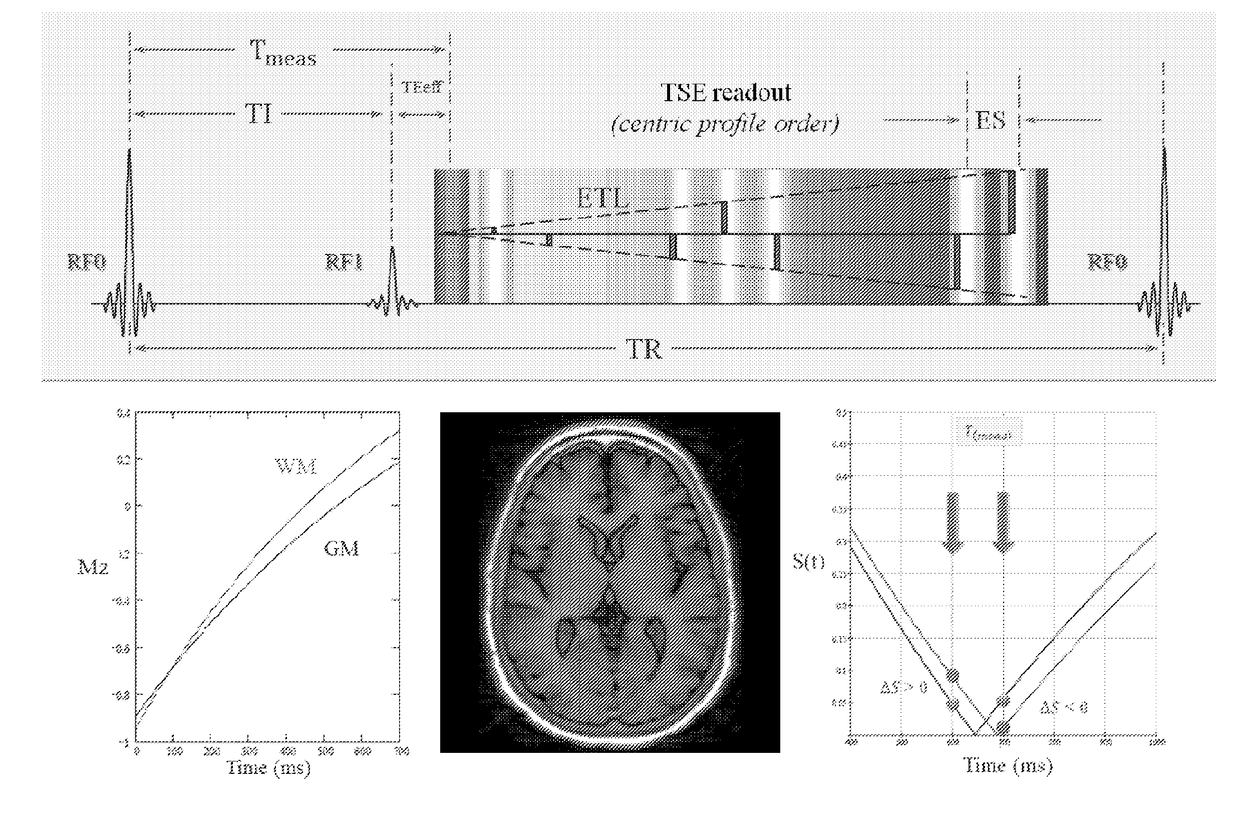
[0136]The measured qMRI parameters of NS are listed in FIG. 10 along with those of several head tissues and blood (5, 9, 11, 16-20): PD, T1, and T2 of NS are nearly identical to those of pure water and substantially different from the values of blood and brain tissues (21): gray matter (GM) and white matter (WM). The scan of the agarose phantom with the dynlR-TSE pulse sequence showed a very high temporal stability both with the large ROIs in several areas of the phantom as well as at the vial interfaces. Overall, temporal pixel intensity variations of less than 1% over the whole phantom during the five-minute scan were achieved with the dynlR-TSE pulse sequence.

example 2

In Vivo Findings

[0137]The twenty enrolled subjects successfully completed the NS injection and the five-minute dynamic-MRI scan without experiencing any adverse effects or expressing any discomfort associated with the research portion of the MRI exam. This was a multivariable exploratory study geared at detecting a small perfusion related imaging effect in vivo and prospectively: as a result three different pulse sequences were tested and only twelve of the twenty subjects were scanned with the dynlR-TSE pulse sequence at 1.5T, which proved to be the most effective technique for detecting THD perfusion effects. The other eight subjects, which are not discussed further in this paper, were scanned with either an IR prepared T1-weighted pulse sequence based on echo planar imaging (n=3), or with standard T2-weighted TSE sequence (n=4) or at 3T (n=1).

example 3

THD Signal Dynamics

[0138]As shown in FIGS. 5 and 6, which correspond to two different patients, the brain tissue THD signals changed slowly time, extended over long times that far exceeded the injection time, and several (FIG. 7) even persisted past the five-minute observation time allowed in our protocol. With the injection parameters and imaging conditions used herein, the THD signal amplitudes were in the ranges of 10-15% and 2-5% for GM and WM respectively (FIGS. 5 and 6). Much stronger THD signals with amplitudes in the 20-30% range were observed in the nasal mucosa, which is consistent with the known higher vascularity of mucosal tissue relative to brain tissue. Other tissues that showed strong THD signals include the retina, choroid plexus, cranial bone marrow, and muscles. Pulsating arterial blood showed oscillatory signals that are likely modulated by inflow phenomena.


PUM


Abstract

In a first aspect this invention provides methods comprising administering an aqueous contrast agent to the vascular system of a subject, and performing a magnetic resonance scan to detect the MR signal enhancement effects of the aqueous contrast agent. In embodiments the magnetic resonance scan comprises applying at least one pulse sequence selected from a PD-weighted pulse sequence, a T1-weighted pulse sequence, a T2-weighted pulse sequence, and a D-weighted pulse sequence. In embodiments the magnetic resonance scan comprises applying a T1-weighted pulse sequence. A system for performing perfusion MRI comprising an aqueous contrast solution and an injection apparatus configured to provide a maximum injection rate of the aqueous contrast solution to a subject vascular system of at least about 5 ml/s. is also provided.

Description

[0001]This application claims priority to U.S. Provisional Application No. 62 / 000,048, filed May 19, 2014, which is hereby incorporated herein.INTRODUCTION[0002]MRI contrast agents are a group of contrast media used to improve the visibility of soft tissues, organs and vasculature with magnetic resonance imaging (MRI). MRI offers a wide array of intrinsic tissue contrast mechanisms for aiding in the diagnosis of disease and these can be explored by simple manipulation of the imaging parameter values; a.k.a. pulse sequence parameters. Nonetheless, exogenous MRI contrast agents offer additional diagnostic information that is not easily obtainable with standard nonenhanced MRI techniques. In particular, gadolinium-based contrast agents (GBCA) are routinely used in the practice for diagnosing tissue pathology via contrast enhanced (CE) MRI, for assessing the vascular supply and the capillary bed integrity of biological tissue via magnetic resonance perfusion (MRP), and for providing hig...

Claims


Application Information

Patent Timeline
IPC IPC(8): G01R33/56A61B5/055A61B5/00G01R33/563
CPCG01R33/5601G01R33/5602G01R33/5635A61B5/0042G01R33/56366A61B5/055A61B5/0044G01R33/56341
Inventor JARA, HERNANMIAN, ASIMANDERSON, STEPHANSAKAI, OSAMUNORBASH, ALEXSOTO, JORGE
Owner BOSTON MEDICAL CENTER INC
Why Eureka
Try Eureka