

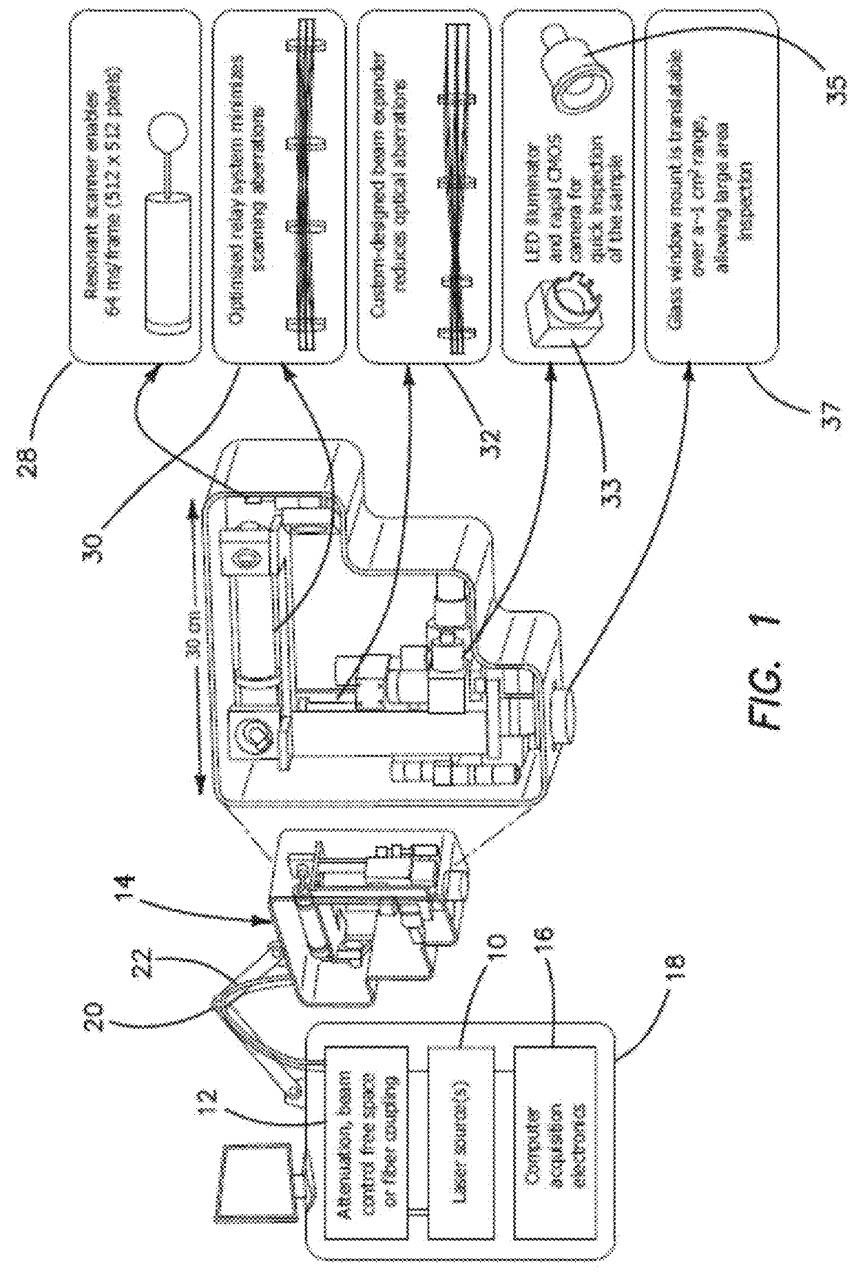
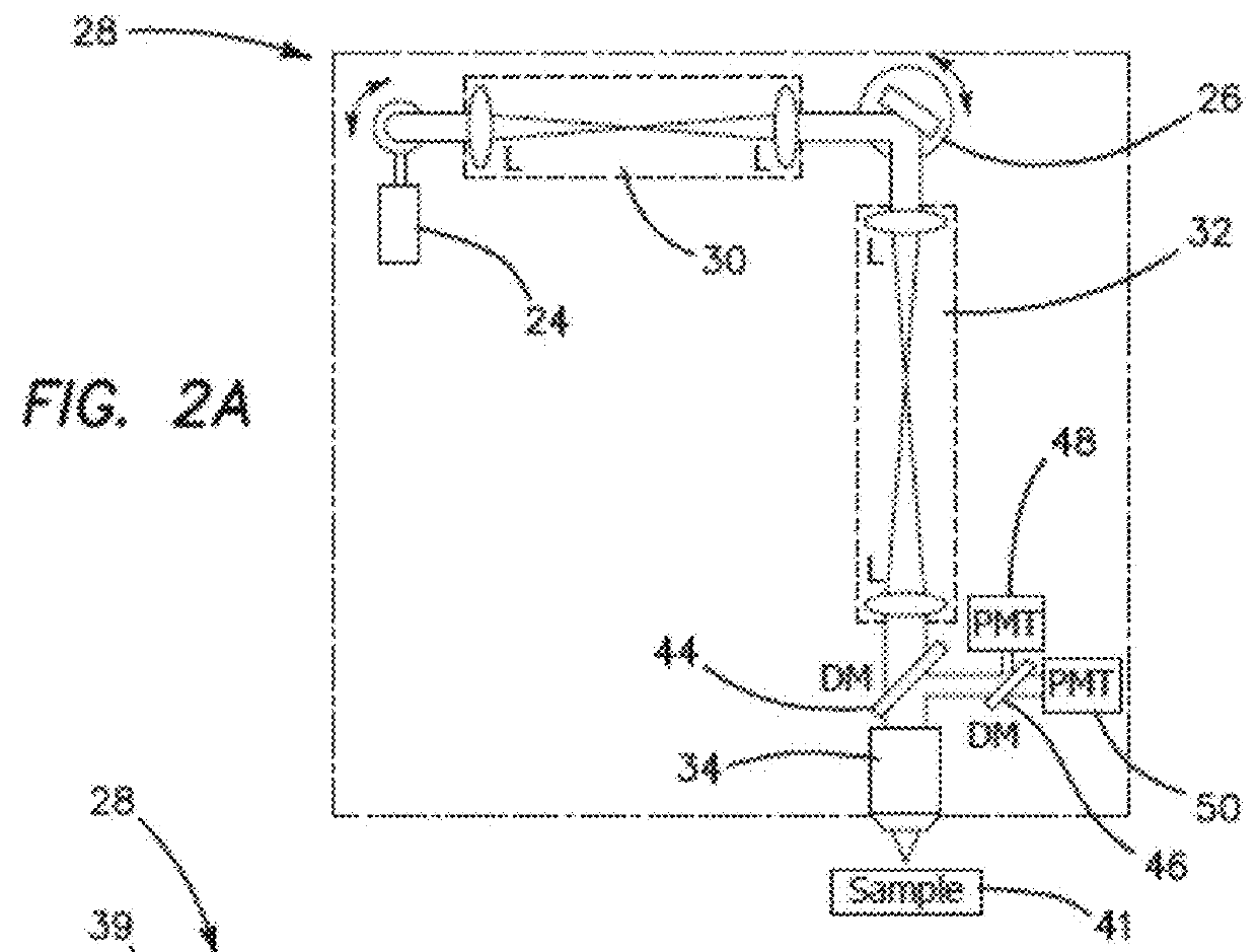
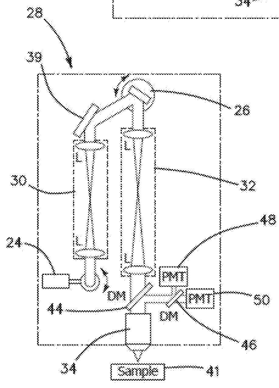
Imaging platform based on nonlinear optical microscopy for rapid scanning large areas of tissue
- Summary
- Abstract
- Description
- Claims
- Application Information
AI Technical Summary
Benefits of technology
Problems solved by technology
Method used
Image

Examples
Embodiment Construction
[0052]The feedback provided by the clinical studies involving a conventional prior art microscope reveals that such a design lacks the potential to provide non-invasive diagnosis. In order for a clinical microscope to be efficiently used for disease diagnosis and treatment guiding, namely for clinical-decision making at the bedside, four requirements are necessary:[0053]1. Sub-micron resolution for identifying cellular and sub-cellular features in a similar manner with histopathology, the gold-standard diagnosis method.[0054]2. Large FOV assessment. Lesions often show irregularity in color and appearance in general, especially when they start to progress towards malignancy. Imaging of limited FOV of the lesion can easily result in false negative diagnosis.[0055]3. Fast scanning speed. Long acquisition times introduce motional artifacts in the images and also negatively affect the patient recruitment process.[0056]4. Reduced barriers-to-access and cost effectiveness, such as reduced ...
PUM
 Abstract
Description
Claims
Application Information