Method and device for detecting circulating tumor cell

a tumor cell and detection method technology, applied in the field of biological and medical detection, can solve the problems of bringing a great technical challenge for detection, unable to fully and accurately reflect the current status of tumor focus, and the risk of metastases, so as to achieve the effect of effective and rapid detection of the degree of malignancy of free ctcs

Active Publication Date: 2018-09-13
View PDF1 Cites 6 Cited by
  • Summary
  • Abstract
  • Description
  • Claims
  • Application Information

AI Technical Summary

Benefits of technology

[0006]The object of the present invention is to provide a method and detection equipment, which can effectively and rapidly detect the degree of malignancy of free CTCs in the blood from a tumor patient, without affecting the subsequent sequencing or in vitro culturing for the identified tumor cells.

Problems solved by technology

However, CTC is extremely rare in the blood.
One milliliter of blood contains 5 billion red blood cells and nearly 10 million white blood cells, but it only has several to several tens of CTCs, thereby bringing a great technical challenge for the detection.
However, only counting the entire CTCs cannot fully and accurately reflect the current status of tumor focus and the risk of metastases.
At present, there is still a lack of technology and equipment for testing CTC function to determine the degree of malignancy and the metastasis possibility of CTC.
In addition, the current detection method for CTC basically adopts the strategy of enriching CTC in blood and then performing detection, such as the CellSearch system, but this type of CTC detection method has several defects.
Firstly, the enrichment method of the CellSearch system is not sensitive enough, the detection rate is low, and a considerable part of the CTC is lost during the enrichment process.
Actually the epithelial-derived cells but not tumor cells are identified, and the latest research shows that epithelial-derived cells can also be detected in the blood from patients with benign disease and even healthy people, so there is a possibility of false positives for this method of identification.
At the same time, studies have found that epithelial-mesenchymal transition (EMT) of tumor cells that occurs during metastasis will result in non-expression of CTCs or low expression of epithelial markers and may be missed.
Thirdly, because fixation and nuclear staining are used in the immunofluorescent staining-based CTC identification method described above, it is difficult for the CTCs after immunofluorescence staining to be further used for sequencing analysis and in vitro culture, whereas sequencing is currently the most important molecular detection method for tumors.
Single cell sequencing for CTCs can further clarify the properties of tumor cells and identify molecular targets for targeted drug therapy by sequencing the driver genes, while fixation and nuclear staining will interfere with genomic amplification for a single cell, thereby affecting the follow-up sequencing.
Fourthly, the study has found that CTCs have a great functional heterogeneity.
Technologies, including CellSearch, cannot functionally identify the degree of malignancy and metastatic potential of CTCs.
At present, almost all the CTC detection methods have similar defects with the CellSearch system.
The enrichment procedures are cumbersome and complicated, and the CTCs are lost.
However, the immunofluorescence staining-based CTC identification method can not accurately identify tumor cells, and the subsequent gene sequencing analysis and in vitro culture are limited.

Method used


Image

  • Method and device for detecting circulating tumor cell
  • Method and device for detecting circulating tumor cell
  • Method and device for detecting circulating tumor cell

Examples


example 1

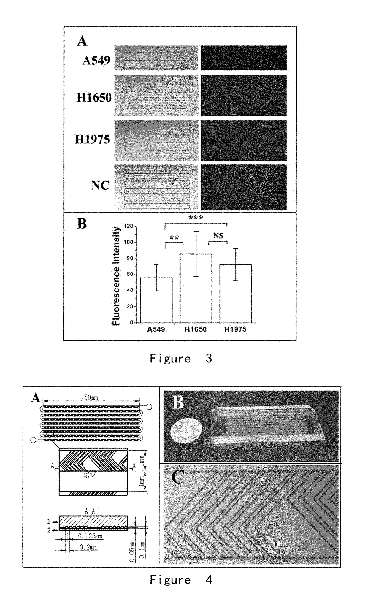
[0202]Detection of Glucose Analogue Uptake for a Very Small Number of Cells

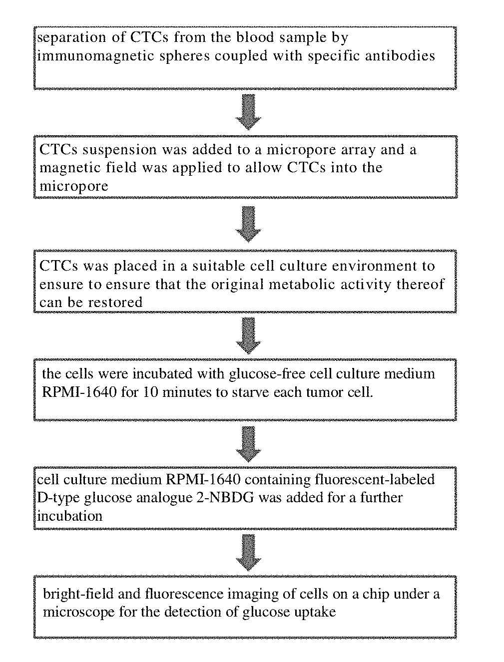
[0203]A microwell array PDMS chip was provided, and its structure is shown in FIG. 1. 30 lung cancer cells A549, H1650, and H1975 were obtained respectively, wherein the surface of each cell was combined with immunized magnetic beads, and the cells were suspended in approximately 200 microliters of PBS, respectively and placed in the microwell array PDMS chip (FIG. 1) to ensure each of them enter and fix in the microwells by manipulating the magnetic field.

[0204]As shown in FIG. 2, the cells were incubated with glucose-free cell culture medium RPMI-1640 for 10 minutes to starve each tumor cell. Then, cell culture medium RPMI-1640 containing 2-NBDG (the concentration was 0.3 mM) was added and the cells were incubated for another 20 minutes. After the incubation, the cells were washed completely and repeatedly with PBS at 4° C. on ice, and the cells were fixed in the microwells by applying a magnetic field. Fin...

example 2

[0207]Detection of Glucose Analog Uptake of CTC in Peripheral Blood

[0208]In this example, the method comprises the following steps:

[0209](1) For 2 ml of peripheral blood samples from lung cancer patients (volunteers), the upper platelet-rich plasma was first removed by low speed centrifugation (200 g) for 5 minutes, and the remaining cells were resuspended in 2 ml with Hank's balanced salt solution (HBSS), and a group of biotin-labeled antibodies (targeted antigens were EpCAM, EGFR, HER2, MUC1, respectively, with a final concentration of 1 μg / mL for each antibody) was added and co-incubated for 1 hour. The excess antibodies were centrifuged (300 g, 5 minutes) and removed, and then streptavidin-labeled magnetic balls (0.8 μm) were added for co-incubation for 30 min, excess magnetic balls were removed by centrifugation (300 g, 5 min), and the cells were resuspended in 5 ml of HBSS to form the pretreated sample.

[0210](2) A fishbone chip was provided, with a structure shown in FIG. 4.

[0...

example 3

[0220]Example 2 was repeated, wherein the peripheral blood sample used was the same batch of peripheral blood samples from the same lung cancer patient (volunteer). The difference is that in step (5), the upper conditioned medium in the culture dish of lung cancer cell H1650 and the upper conditioned medium in the lung fibroblast culture dish were combined in a ratio of 1:1 to form a new conditioned medium for CTC culturing in microchip.

[0221]As a result, similar results as in Example 2 were obtained, and significant fluorescent signals were detected in 6 CTCs.

[0222]EXAMPLE 4-6

[0223]Example 2 was repeated with the following differences:

[0224]In Example 4, in the resuscitation treatment, the addition of the blocking agent of bovine serum albumin was also included to block cells and microwell chips, thereby eliminating the non-specific adsorption of 2-NBDG (Example 4);

[0225]In Example 5, in the resuscitation process, the addition of a fluorescent-modified antibody (e.g., anti-CD45-FIT...


PUM

PropertyMeasurementUnit
diameteraaaaaaaaaa
diameteraaaaaaaaaa
lengthaaaaaaaaaa

Abstract

Provided are a method for detecting a free rare tumor cell in a human biological fluid sample and assay kit. Specifically, the method of the invention determines the existence of an active tumor cell in a biological fluid sample of a cancer patient by detecting and confirming the existence of glucose absorption capability of a karyocyte and expression of a leukocyte marker (CD45).

Description

TECHNICAL FIELD[0001]The present invention relates to the field of biological and medical detection. More specifically, the present invention relates to a detection method and device for the metabolic activity of circulating tumor cells in human peripheral blood. The method and device of the present invention can perform metabolic function typing on CTC captured in the human peripheral blood and can be used to predict the degree of malignancy of these CTC.BACKGROUND[0002]The circulating tumor cells (CTC) in the human peripheral blood refer to the tumor cells that spread from the tumor focus into the peripheral blood circulation and can develop into metastatic tumor focus under a certain condition. Since more than 90% of cancer deaths are caused by metastases, whereas CTC is a direct source of tumor metastasis, therefore, the separation of the CTC from the blood and molecular detection therefor has attracted an increasing attention. However, CTC is extremely rare in the blood. One mi...

Claims


Application Information

Patent Timeline
Patent Type & Authority Applications(United States)
IPC IPC(8): C12M1/34G01N33/569C12Q1/02
CPCC12M1/34G01N33/569C12Q1/02G01N33/56966G01N33/56972G01N33/57492G01N2333/70589
Inventor SHI, QIHUILU, SHUNDENG, YULIANGTANG, YIN
Why Eureka
Try Eureka