Automated quantitative restoration of bright field microscopy images
a bright field microscopy and automatic restoration technology, applied in image data processing, instruments, computer peripheral equipment, etc., can solve the problems of poor contrast of unstained cells' bfm images, difficult quantitative measurements of cellular shape and size, and lack of necessary contrast in bfm images of unstained cells
- Summary
- Abstract
- Description
- Claims
- Application Information
AI Technical Summary
Benefits of technology
Problems solved by technology
Method used
Image
Examples
example 1
[0051]Using a conventional BF microscope operating under Koehler illumination, we collected z-stacks of background-free images of E. coli cells immobilized on a coverslip. An operational pPSF was generated based on Eq. (1) and three-dimensional (3D) deconvolution was performed on stacks of cell images using an available deconvolution routine.
pPSF(x,y,z)=∫01J0[kNAρ[x2+y2]1 / 2] sin [knz[[1−(NAρ / n)2]1 / 2−1]]dρ, (1)
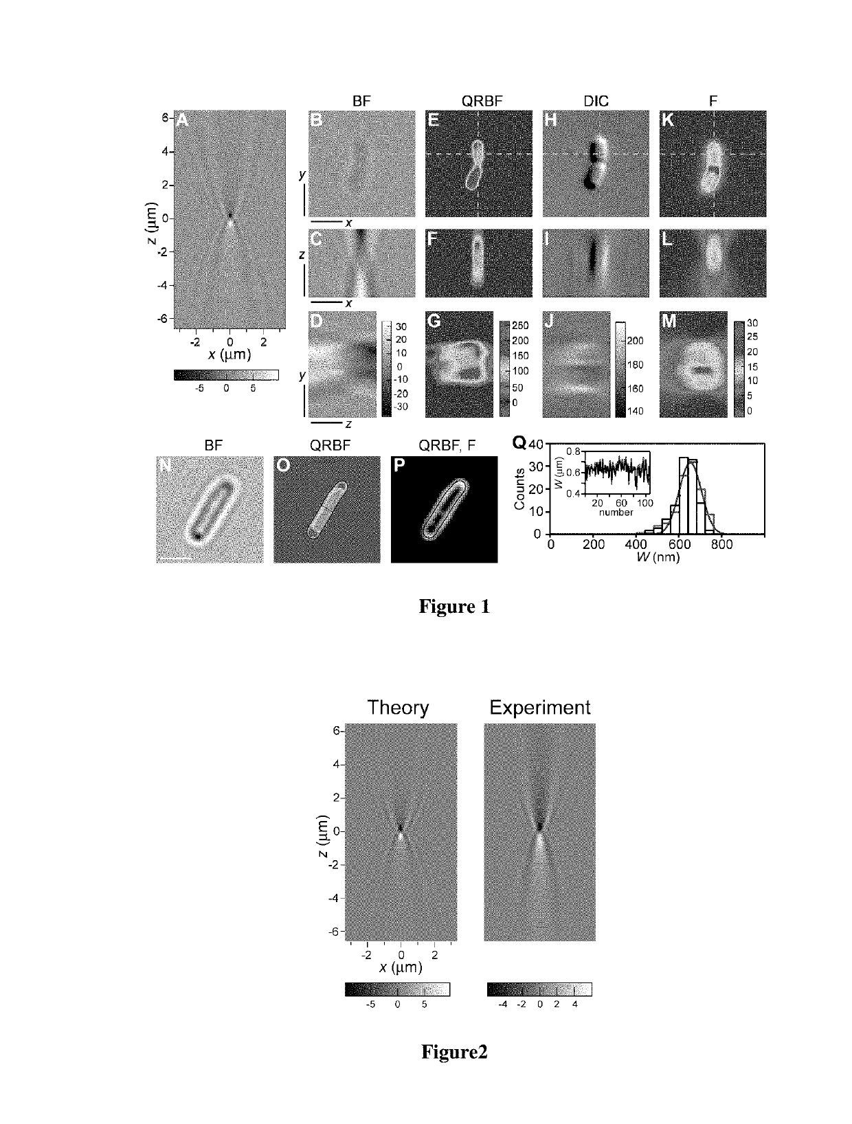
[0052]where J0 is the zeroth-order Bessel function of the first kind, k=2π / λ, λ is the wavelength of illumination, NA is the numerical aperture of the microscope objective, n is the index of refraction of the medium between the objective and the coverslip, (x,y) are transversal image coordinates and z is the amount of defocusing. FIG. 1A shows the resulting intensity distribution of the BF pPSF.
[0053]FIG. 1 shows how QRBF effectively restores images in 3D, turning an original BF image that features an ill-defined object (FIG. 1B,C,D) into the image of a cell undergoing divisio...
example 2
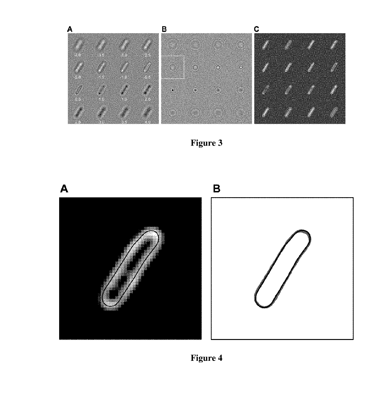
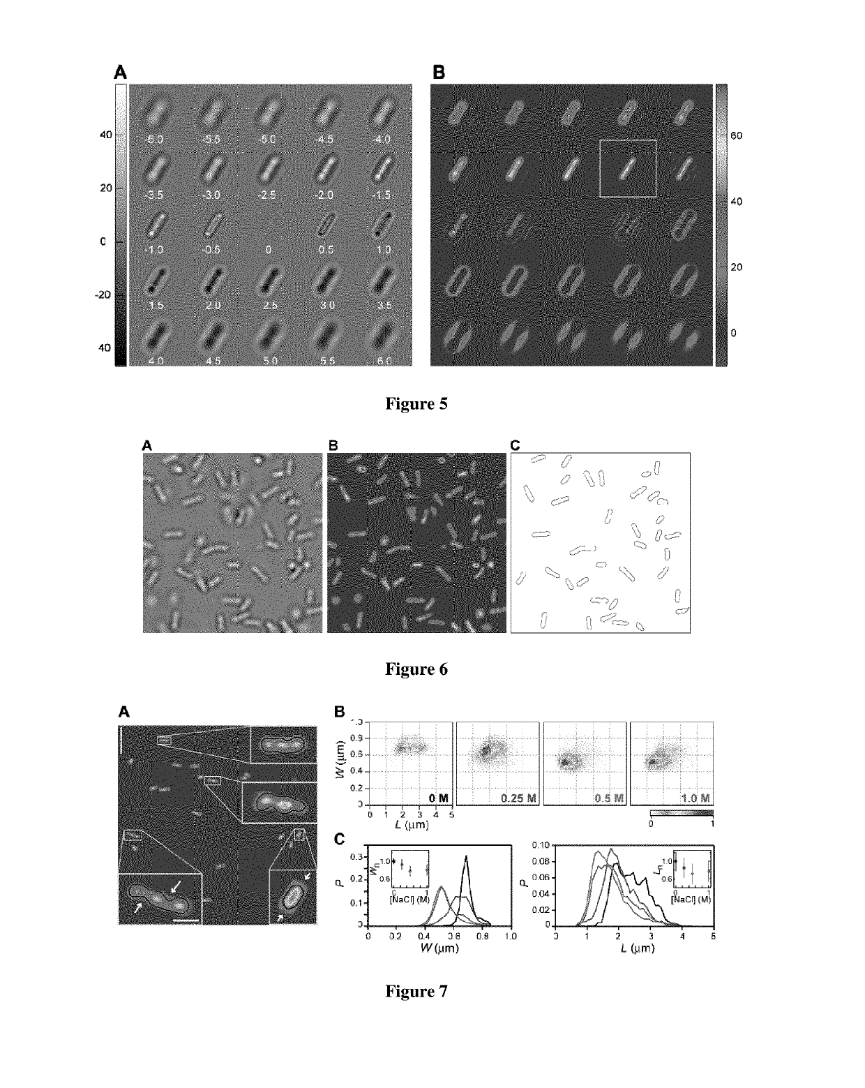
[0055]A large number of applications involving the study of cellular morphology would benefit from reliable, fast 2D-imaging of cells. To address this point, we implemented two-dimensional QRBF (2D-QRBF), where a single BF image of a defocused cell (FIG. 3A) is acquired and then deconvolved using the corresponding pPSF image at the same amount of defocus (FIG. 3B). The resulting 2D-QRBF image effectively restores cellular shape (FIG. 3C). Close inspection of cellular contour in 2D-QRBF images at various z-values above and below focus revealed that excellent agreement of cellular morphology is still maintained with respect to fluorescence microscopy (FIG. 4). Furthermore, we found that 2D-QRBF performs best when the amount of defocus is the same for both BF and pPSF images (FIG. 5), indicating that restoration is optimum at a given plane. We took advantage of this feature by applying 2D-QRBF on BF images of cells found both attached to a coverslip and free in solution (FIG. 6A). The ...
example 3
[0057]We applied 2D-QRBF in a new type of image cytometer to evaluate bacteria size changes in response to hyperosmotic shock. In the presence of hypertonic media bacteria modify their size and shape, developing plasmolysis—a process where water lost form the cytoplasm causes the inner plasma membrane to shrink away from the cell wall (P. Scheie, 1969. “Plasmolysis of Escherichia coli B / r with Sucrose”, J. Bacteriol. 98, 335-340). Here, non-growing E. coli cells were deposited on coverslips, subjected to increasing external concentrations of the solute NaCl, and imaged using 2D-QRBF. The deconvolved images of osmotically challenged bacteria show cells with deformations and voids in the cell body (FIG. 7A) that are in remarkable similarity with fluorescence (M. C. Konopka, I. A. Shkel, S. Cayley, M. T. Record, J. C. Weisshaar. 2006. “Crowding and confinement effects on protein diffusion in vivo”, J. Bacteriol. 188, 6115-6123) and electron microscopy (H. Schwarz, A. L. Kocht, 1995, “P...
PUM
 Abstract
Description
Claims
Application Information
 - R&D
- Intellectual Property
- Life Sciences
- Materials
- Tech Scout
- Unparalleled Data Quality
- Higher Quality Content
- 60% Fewer Hallucinations
Browse by: Latest US Patents, China's latest patents, Technical Efficacy Thesaurus, Application Domain, Technology Topic, Popular Technical Reports.
© 2025 PatSnap. All rights reserved.Legal|Privacy policy|Modern Slavery Act Transparency Statement|Sitemap|About US| Contact US: help@patsnap.com


