

Developable Hyaluronic Acid Microspherical Embolic Agent, Preparation Method and Use Thereof
- Summary
- Abstract
- Description
- Claims
- Application Information
AI Technical Summary
Benefits of technology
Problems solved by technology
Method used
Image

Examples
example 1
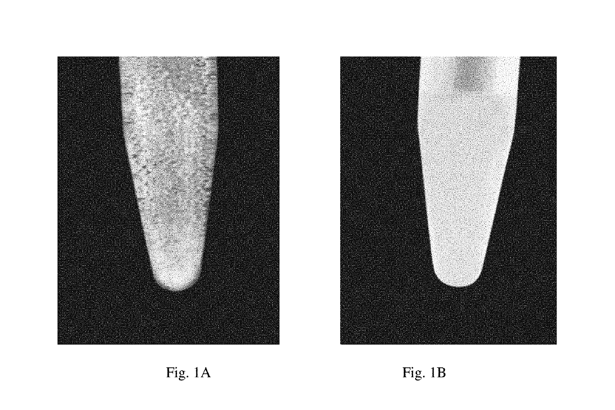
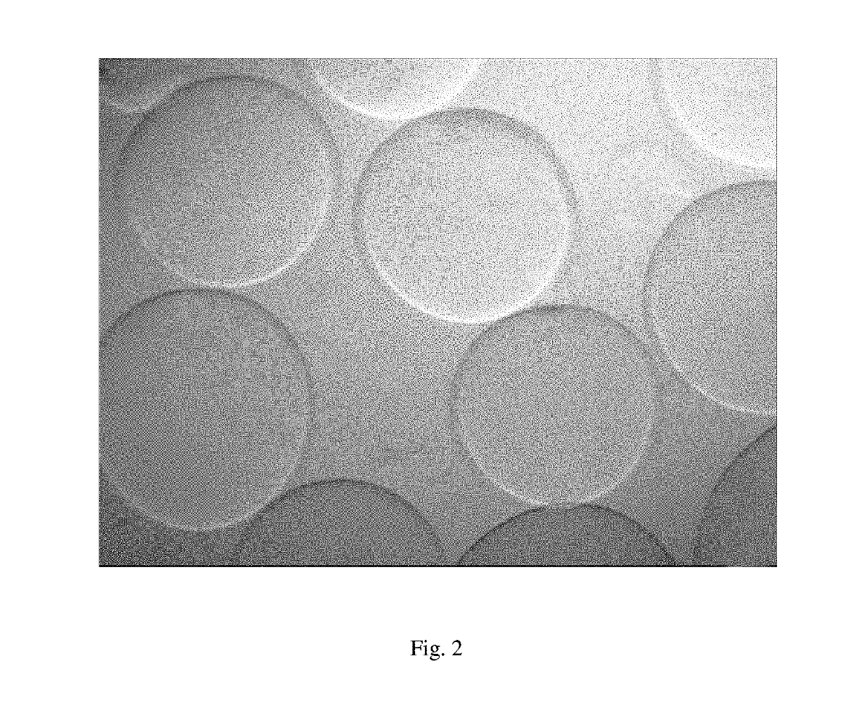
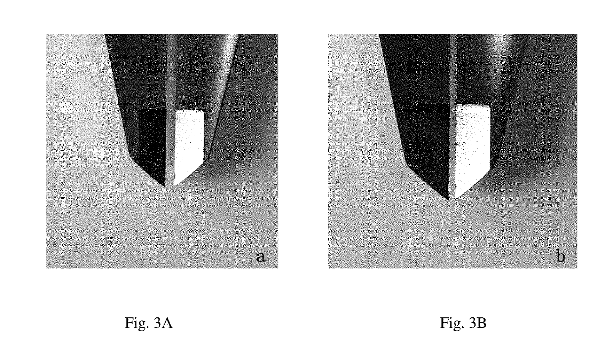
on of Hyaluronic Acid Microspherical Embolic Agent Containing Iohexol
[0027]A hyaluronic acid solution having a concentration of 0.1 g / mL was prepared by using 8.04 g of solid power of sodium hyaluronate and 0.5% of sodium hydroxide solution. The hyaluronic acid solution contained 0.07 g / mL of iohexol. The hyaluronic acid solution was added to an oil phase containing 200.71 g of liquid paraffin and 3.41 g of Span 80. The resultant mixed phase was emulsified and dispersed for 10 minutes by using a shearing machine at 1000 rpm, to obtain a water-in-oil hyaluronic acid microspheres emulsion. 1% of 1,4-butanediol diglycidyl ether was added to the emulsion and the resultant mixture was stirred for 4 hours at room temperature for cross-linking reaction. After the stirring was finished, the mixture was kept still overnight to leave the microspheres to settle. The oil phase at the upper layer was poured away, and then acetic acid was added to adjust the pH value of the remaining mixture to b...
example 2
on of Hyaluronic Acid Microspherical Embolic Agent Containing Iohexol
[0028]A hyaluronic acid solution having a concentration of 0.1 g / mL was prepared by using 8.02 g of solid power of sodium hyaluronate and 0.5% of sodium hydroxide solution. The hyaluronic acid solution contained 0.07 g / mL of iohexol. The hyaluronic acid solution was added to an oil phase containing 201.01 g of liquid paraffin and 3.45 g of Span 80. The resultant mixed phase was emulsified and dispersed for 10 minutes by using a shearing machine at 1000 rpm, to obtain a water-in-oil hyaluronic acid microspheres emulsion. 1% of 1,4-butanediol diglycidyl ether was then added to the emulsion and the resultant mixture was stirred for 4 hours at room temperature for cross-linking reaction. After the stirring was finished, the mixture was kept still overnight to leave the microspheres to settle. The oil phase at the upper layer was poured away, and the remaining mixture was washed in turn with ethyl acetate and absolute e...
PUM
| Property | Measurement | Unit |
|---|---|---|
| Time | aaaaa | aaaaa |
| Mass | aaaaa | aaaaa |
| Density | aaaaa | aaaaa |
Abstract
Description
Claims
Application Information