

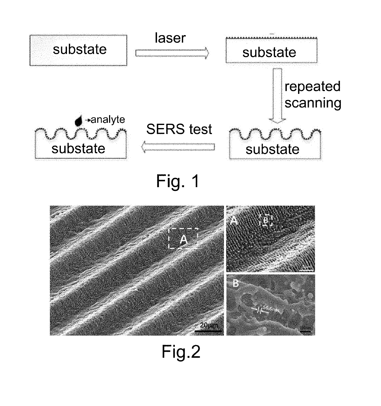
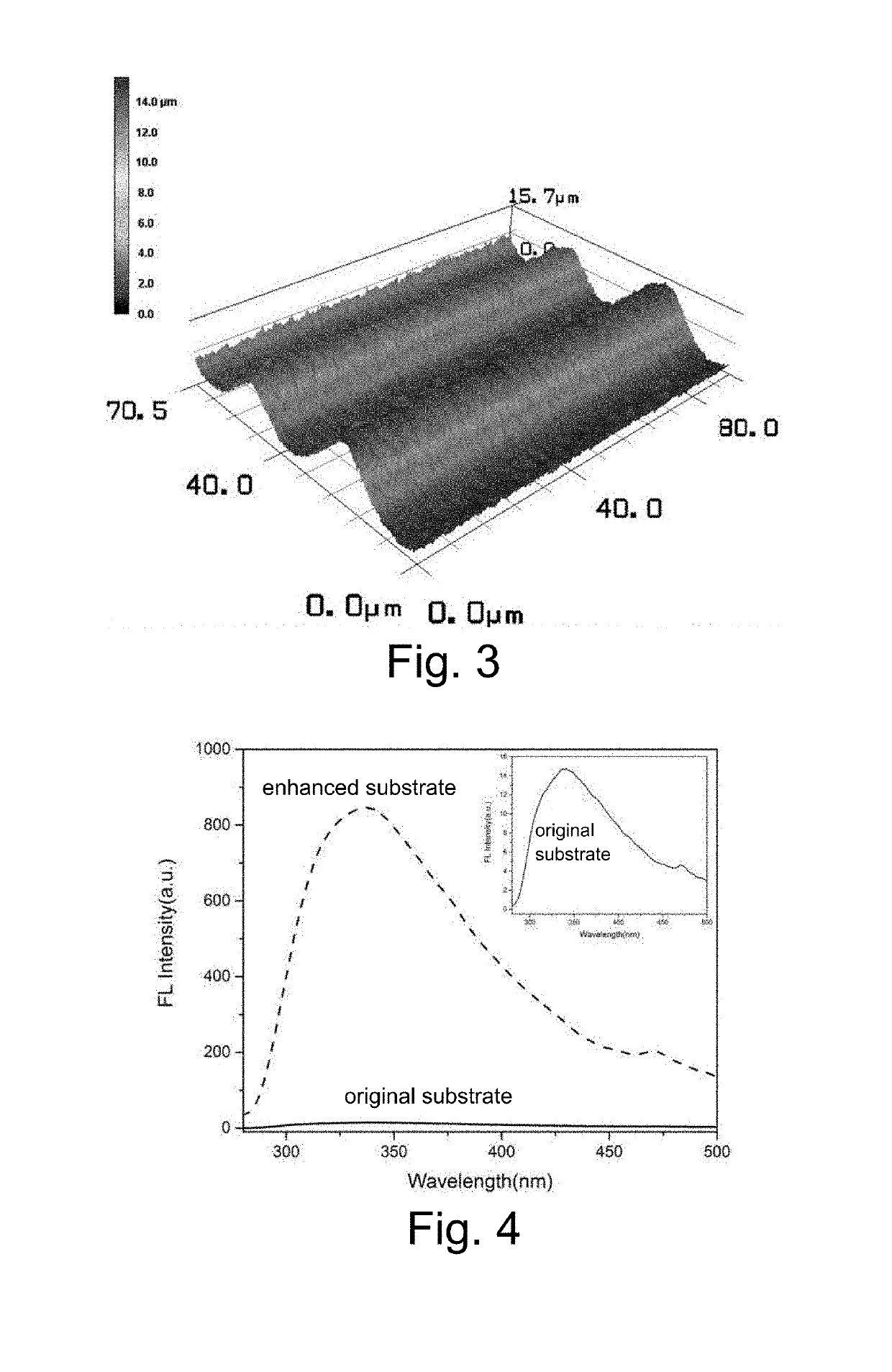
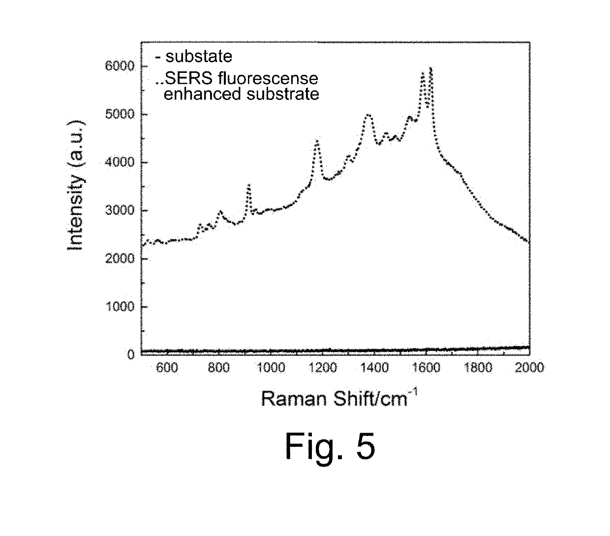
Fabrication of Fluorescence-Raman Dual Enhanced Modal Biometal Substrate
a biometal substrate and enhanced modal technology, applied in metal working devices, manufacturing tools, instruments, etc., can solve the problems of affecting the detection of fluorescence, so as to achieve the effect of easy production
- Summary
- Abstract
- Description
- Claims
- Application Information
AI Technical Summary
Benefits of technology
Problems solved by technology
Method used
Image

Examples
embodiment 1
[0039](1) Preparing a TC4 specimen with an area of 10*10 mm and thickness of 2 mm; cleaning the substrate in the 100% alcohol; grinding the surface of the specimen in a sequence of 360 mesh, 600 mesh, 800 mesh, 1000 mesh, 2000 mesh and 4000 mesh sandpaper in turn; cleaning the grinded specimen within ultrasonic bath for 20 s;
[0040](2) Placing the cleaned TC4 specimen on the stage of an ultrashort pulse laser system (the wavelength is 1030 nm; the beam diameter is 35 μm; the pulse width is 800 fs); wherein the laser parameters are set as the following: power 2 W; frequency: 300 KHz; scan rate: 1500 mm / s; scanning time: 15 times; scanning area: 800*800 μm; the scanning route is one-direction parallel line; starting the laser processing system;
[0041](3) When the process is completed, the three-dimensional periodic micro-nano structure for SERS and fluorescence substrate is achieved.
[0042]FIG. 2 is the SEM image of three-dimensional micro-nano structures formed by adopting a preparing m...
embodiment 2
[0043](1) Preparing a copper specimen with an area of 10*10 mm and thickness of 2 mm; cleaning the substrate in the 100% alcohol; grinding the surface of the specimen in a sequence of 360 mesh, 600 mesh, 800 mesh, 1000 mesh, 2000 mesh and 4000 mesh sandpaper in turn; cleaning the grinded specimen within ultrasonic bath for 20 s;
[0044](2) Placing the cleaned copper specimen on the stage of an ultrashort pulse laser system (the wavelength is 800 nm; the beam diameter is 35 μm; the pulse width is 600 fs); wherein the laser parameters are set as the following: power 1 W; frequency: 200 KHz; scan rate: 1500 mm / s; scanning time: 20 times; scanning area: 800*800 μm; the scanning route is one-direction parallel line; starting the laser processing system;
[0045](3) When the process is completed, the three-dimensional periodic micro-nano structure for SERS and fluorescence substrate is achieved.
embodiment 3
[0046](1) Preparing an aluminum specimen with an area of 10*10 mm and thickness of 2 mm; cleaning the substrate in the 100% alcohol; griding the surface of the specimen in a sequence of 360 mesh, 600 mesh, 800 mesh, 1000 mesh, 2000 mesh and 4000 mesh sandpaper in turn; cleaning the grinded specimen within ultrasonic bath for 20 s;
[0047](2) Placing the cleaned aluminum specimen on the stage of an ultrashort pulse laser system (the wavelength is 532 nm; the beam diameter is 35 μm; the pulse width is 600 fs); wherein the laser parameters are set as the following: power 0.5 W; frequency: 600 KHz; scan rate: 2500 mm / s; scanning time: 20 times; scanning area: 800*800 μm; the scanning route is one-direction parallel line; starting the laser processing system;
[0048](3) When the process is completed, the three-dimensional periodic micro-nano structure for SERS and fluorescence substrate is achieved.
PUM
| Property | Measurement | Unit |
|---|---|---|
| Length | aaaaa | aaaaa |
| Length | aaaaa | aaaaa |
| Length | aaaaa | aaaaa |
Abstract
Description
Claims
Application Information