Microdroplet Based Bioassay Platform

a bioassay and microdroplet technology, applied in the field of microdroplet based bioassay platforms, can solve the problems of significant morbidity, significant gap in the effective management of microbial infections, and presents a significant challenge to uti management, and achieve high throughput testing of the susceptibility of single cells and efficient monitoring of bacterial infection and cell function

Active Publication Date: 2020-04-16
NORTHEASTERN UNIV
View PDF0 Cites 4 Cited by
  • Summary
  • Abstract
  • Description
  • Claims
  • Application Information

AI Technical Summary

Benefits of technology

[0011]The invention provides a fast, highly sensitive, direct-from-patient, microfluidic droplet-based bioassay platform that offers single cell based analysis for efficient monitoring of bacterial infection and cell function, as well as for high throughput testing of the susceptibility of single cells to antibiotics and other pharmaceutical agents. The methods and devices provided can be used to monitor bacteria and perform antibiotic susceptibility testing for urinary tract infection directly from patient samples with no or minimal pre-processing steps.
[0012]The bioassay platform is based on analysis of an array of single droplets using a microfluidics device coupled with a fluorescence microscopy imaging system. Cells are encapsulated in aqueous microdroplets suspended in an oil medium and directed, sorted, and arranged by the microfluidic device for analysis, which may include identification and quantification of the cells as well as characterization of cell phenotype, such as function, gene expression, presence or absence of biomarkers, or susceptibility to antibiotics or other drugs. Various reagents for the analysis can be added to the droplets at formation or by later merger of droplets. Such reagents include antibody-conjugated microspheres, fluorescent detection antibodies, cell viability indicators, antibiotics or drugs, and other cells for cell-cell interaction studies. The analysis of single droplets provides high sensitivity and short reaction times. A further advantage is the use of an on chip docking array, where up to hundreds or thousands of microdroplets can be maintained stably for long-term culture, such as to assess time-variant growth dynamics of encapsulated bacteria at single cell resolution or to study T-cell mediated killing of tumor cells, for example. Tracking individual cells such as bacteria over time can provide critical information on cellular heterogeneity based on characteristics such as cell division, density, morphology and antibiotic resistance.
[0014]Furthermore, the present microfluidics-based technology can be deployed with inexpensive portable instrumentation and disposables. The portability and low cost of consumables should permit distribution to sites of patient care in hospitals, primary care, and resource-limited settings, thereby accelerating personalized therapy and improving patient outcomes. The same qualities also make the technology ideal to identify infected patients efficiently for clinical trial enrollment.

Problems solved by technology

The rapid emergence of antibiotic resistance, and decline in discovery of new antibiotics, has created a significant gap in the effective management of microbial infections.
Infection may involve both lower (bladder, cystitis) and upper urinary tract (kidney, pyelonephritis) and lead to significant morbidity, including permanent kidney damage (chronic pyelonephritis) or life threatening bacterial sepsis.
However, the rapid emergence of antibiotic resistance in recent years presents a significant challenge for UTI management.
(4-8) In particular, carbapenem-resistant and extended-spectrum β-lactamase producing Enterobacteriaceae are an immediate public health threats, as they express resistance to commonly used empiric antimicrobial regimens.
(13) However, using traditional technology, susceptibility results are generally not available for 48-72 hours.
This means that for the first three days of suspected infection, therapy is largely unguided.
These newer platforms offer several benefits over traditional culture methods; however, they also have significant limitations.
However, lengthy culture pre-amplification is still required, since analysis can only be performed on isolated bacterial colonies.
(17) Also, MS-based AST only marginally decreases the time needed to identify organisms and does not address the need for rapid AST.
Lastly, MS equipment is expensive ($200,000-300,000), bulky, complex to operate, and requires regular maintenance.
(16) Therefore, MS systems must be placed in a centralized laboratory, away from the site of patient care, further delaying access to diagnostics.
(18-21) However, they fall short in their ability to yield a comprehensive resistance profile.
Therefore, even highly multiplexed NAATs cannot accurately predict phenotypic susceptibility.
This results in biological noise, which can be further enhanced by minor differences in environmental stimuli, variations in cell state and polyfunctional responses.
However, it cannot provide time-varying spatiotemporal resolution of signaling dynamics in the same cell.
Many of these techniques suffer from limitations of throughput and complicated operation.
However, active mechanisms such as dielectric forces can negatively impact cell viability; additionally, the throughput achieved with these methods is generally low.
However, non-adherent cells could potentially be lost from their holding sites over time with this technology.
However, a common limiting feature of most of these microfluidic approaches is the lack of isolation of a cell from its neighbors, leaving room for paracrine effects.

Method used


Image

  • Microdroplet Based Bioassay Platform
  • Microdroplet Based Bioassay Platform
  • Microdroplet Based Bioassay Platform

Examples


example 1

re-Based Assay for Simultaneous Analytical Detection of Common Uropathogens

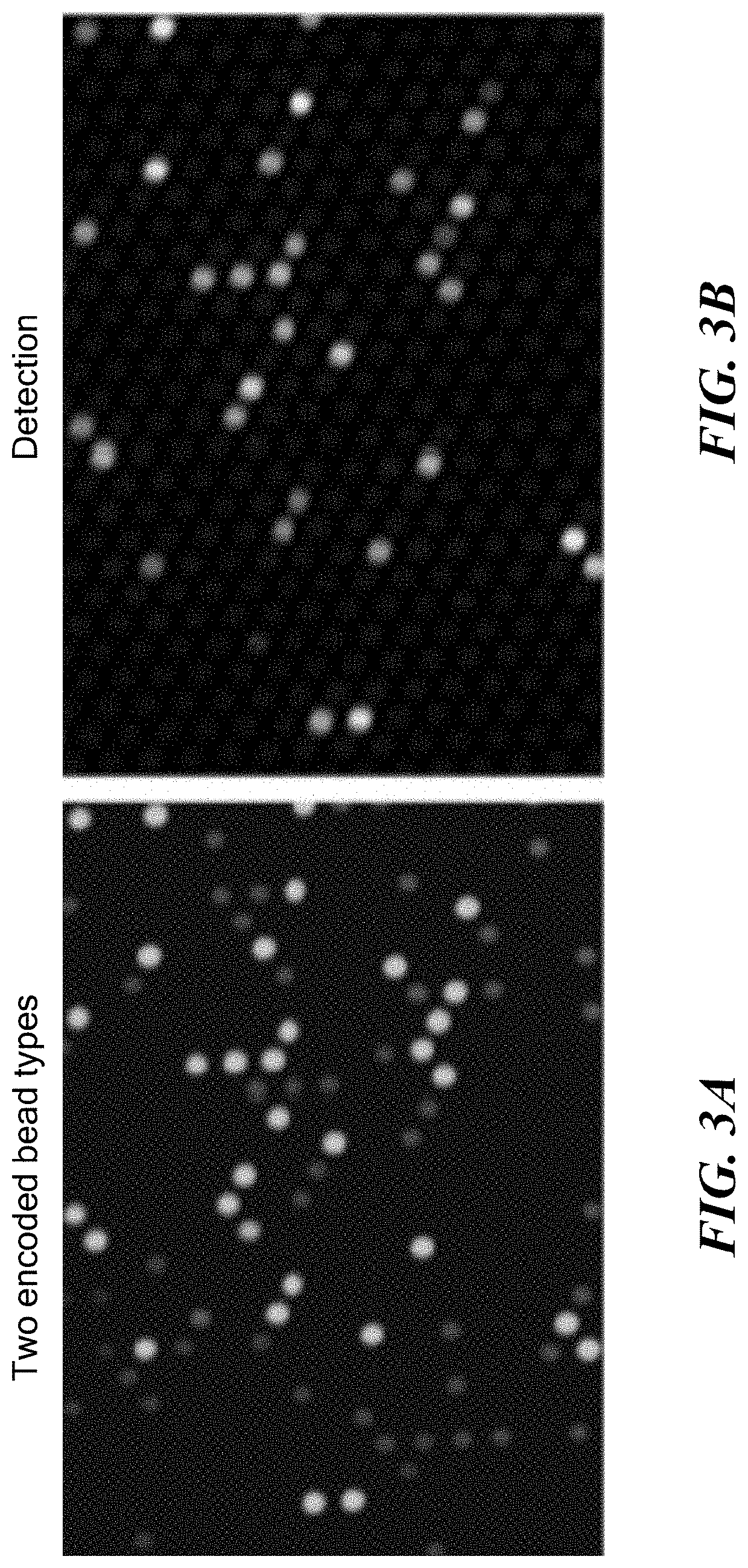
[0050]Common causative agents for UTI, including Escherichia coli, Klebsiella pneumoniae, Enterobacter, Pseudomonas aeruginosa, and Proteus, can be detected in a multiplex assay. Multiplex bead array assays can be found as far back as 1977. (50-54) A wide assortment of tests have been devised for bead-based assays using both immunological and molecular ligands. However, a robust bead-based assay to capture and analyze bacteria has not yet been developed. Here, a bead-based protocol is disclosed with ultrahigh detection sensitivity and specificity of antigen-antibody reactions to identify bacterial presence in urine samples. The multiplex capabilities of the assay were used to detect several bacterial pathogens in urine samples simultaneously. The developed bead based detection protocol was validated for specificity and sensitivity as well as calibrated for determining the bacterial inoculum in the sample.

Prep...

example 2

nt of System for Differential Diagnosis and Antimicrobial Susceptibility Testing

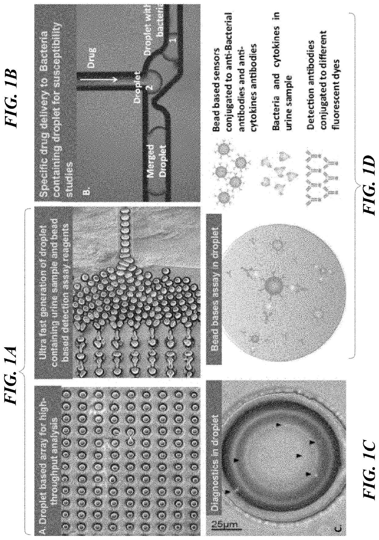
[0059]In this example, disclosed is a fully integrated droplet based microfluidic device to detect and establish antimicrobial susceptibility within intact droplets is disclosed. The technology developed herein includes: 1) bead sensors and detection assay reagents as well as urine sample co-encapsulation to generate droplet based microreactors for detection, 2) droplet microarray technology to analyze the captured bacteria and 3) droplet merging technology for timely delivery of antimicrobials for susceptibility testing.

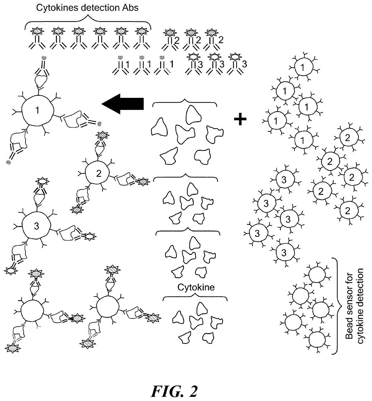
Microbead-Based Assay Using IL-6 and IL-8 Cytokine Interrogation

[0060]Two chips disclosed below are designed with variations on geometry to meet the single bacteria interrogation and co-encapsulation with assay reagents and antimicrobial agent requirements. The first design presents a simpler approach, where the polydimethylsiloxane (PDMS) chip is generated using standard soft lithograph...

example 3

c Validation

[0072]The use of a microfluidic system for bacterial detection and quantification of select pathogens in patient clinical samples and associated IL-6 / IL-8 cytokine levels is exemplified.

[0073]Clinical urine culture samples are collected in urine transport tubes that stabilize uropathogen numbers and viability, and urine samples are stored at 4° C. immediately after specimens are cultured and are therefore available for later analysis. Retrieved urine samples may be tested and correlated with clinical reports and parallel determination of CFU / mL of specific organisms. Clinically, urine cultures are plated using calibrated loops and results reported out quantitatively in log 10 concentration. More specifically, for clinical reporting, urine culture results are divided into four quantitative categories: no growth, 100,000 CFU / ml. Isolates are also speciated by the Vitek 2 (Biomerieux) automated identification system. Biotype numbers (a numerical summary of biochemical react...


PUM

PropertyMeasurementUnit
volumeaaaaaaaaaa
volumeaaaaaaaaaa
volumeaaaaaaaaaa

Abstract

Platform technology involving aqueous microdroplet reaction vessels created, arrayed, and characterized by imaging microscopy in a microfluidic device are applied to a wide variety of bioassays involving the detection and phenotypic characterization of single cells. The bioassays include the rapid and automated detection of microbial pathogens and their antibiotic sensitivity from patient samples as well as the characterization of immune responses using a patient's own cells, including the killing of tumor cells.

Description

CROSS REFERENCE TO RELATED APPLICATIONS[0001]This is a divisional application of U.S. application Ser. No. 15 / 744,198 filed 12 Jan. 2018 as the national phase of PCT / US2016 / 042677 filed 15 Jul. 2016, which claims the priority of U.S. Provisional Application No. 62 / 192,958 filed 15 Jul. 2015 and of U.S. Provisional Application No. 62 / 291,933 filed 5 Feb. 2016. Each of the aforementioned applications is hereby incorporated by reference in its entirety.BACKGROUND[0002]The rapid emergence of antibiotic resistance, and decline in discovery of new antibiotics, has created a significant gap in the effective management of microbial infections. Additionally, persistent use of antibiotics in agriculture has further increased antibiotic resistant bacteria strains, thus making antibiotic resistant microbial infections an acute public health concern. According to the WHO global surveillance report for antimicrobial resistance, very high rates of resistance have been observed in common bacterial ...

Claims


Application Information

Patent Timeline
Patent Type & Authority Applications(United States)
IPC IPC(8): G01N33/543B01L3/00B01L3/02G01N35/08G01N33/68G01N33/574G01N33/569B01F13/00
CPCG01N33/57492G01N33/54346G01N33/56966B01L2200/0668G01N33/56911B01L3/502761G01N33/533B01F13/0071G01N33/6863G01N35/08G01N33/5432B01L3/502784B01L3/0241B01F33/3021
Inventor KONRY, TANIASABHACHANDANI, POOJASARKAR, SAHELI
Owner NORTHEASTERN UNIV
Why Eureka
Try Eureka