Method of detection of DNA end(s) and its use

Pending Publication Date: 2020-08-13
INTODNA SPOLKA AKCYJNA
View PDF1 Cites 0 Cited by
  • Summary
  • Abstract
  • Description
  • Claims
  • Application Information

AI Technical Summary

Benefits of technology

The invention provides a method to directly detect and visualize DNA ends, which has not been possible with existing methods. This allows for the determination and quantification of single and double-strand DNA breaks, which are important for assessing the quality of DNA and studying mechanisms of damage induction, sensing and repair. The method is highly sensitive and specific, allowing for the detection of even a single DNA break. This knowledge can be used for diagnostic purposes and may help to better understand the causes of genetic disorders.

Problems solved by technology

However, DNA is not infallibly stable, and it is susceptible to spontaneous chemical changes occurring under physiological conditions (Lindahl T. Instability and decay of the primary structure of DNA. Nature. 1993 Apr. 22; 362(6422):709-15).
Since DNA is inherently chemically unstable, and is constantly damaged by various factors, preserving the genomic integrity requires specialised surveillance and repair mechanisms.
Only genomic degradation which escapes the correct action of the repair mechanisms may lead to somatic mutations.
An accumulation of unrepaired DNA damage compromises genome integrity and can lead to various cellular malfunctions, neoplastic transformation, or death.
Non-lethal changes in the genome may be passed on to subsequent generations, accumulate, and eventually result in serious malfunctions of the progeny.
Neoplastic transformation may eventually have serious consequences on the level of the whole organism, since a transformed cell may give rise to a clone of such cells and a growth of a malignant tumour.
In some cases, death of just a few cells, which results from DNA damage, may have serious consequences for the entire organism, for instance, a loss of irreplaceable neurons in the brain.
Many damaging factors induce DNA breaks directly, while others cause DNA lesions that may eventually lead to a discontinuity of a DNA molecule, that is, a single- and double-strand break.
While indirect approaches (γH2A.X, XRCC1, 53BP1) allow for detection of a low number of DNA breaks, currently, there are no methods of detecting individual DNA breaks directly, in live or fixed cells.
Methods based on direct labelling and detection of DNA ends are all characterised by low sensitivity, i.e., they are unable to detect individual DNA breaks in intact cells or treated biological materials.
TUNEL assay, well known to the persons skilled in the art, is able to readily detect large numbers (likely hundreds and more) of DNA breaks during apoptosis, however, it is not capable of detecting individual DNA double-strand breaks, or even groups consisting of low numbers (10 or so) of these breaks.
The existing methods are all based on traditional immunofluorescent labelling (including TUNEL), therefore, their specificity is limited and heavily dependent of the specificity of primary and secondary antibodies, while the sensitivity of these methods is limited by the presence of considerable non-specific background signals (the intensity of specific signals do not sufficiently exceed the intensity of nonspecific ones).
An indirect detection of DNA breaks by means of detecting the recruited cellular DNA damage response factors (like 53BP1 or XRCC1) has the following limitations: (i) the number of recruited protein molecules may be too low to be detected by the generally used methods like immunofluorescence, (ii) immunofluorescence is often not fully specific due to limited specificity of the antibody, (iii) sensitivity of detection is limited by the presence of a signal background which arises from nonspecific binding of the secondary antibody to various cell components, (iv) recruitment of repair factors may be activated in the absence of DNA breaks, or may be inhibited or absent under the conditions of saturation of repair capacity, or when the repair systems themselves are damaged or impaired, thus false positive or false negative results may occur.
Measurements of the number of DNA breaks by means of detecting accumulation of fluorescently tagged proteins at the site of damage in cells freshly derived from a body of a patient are not possible.
As of today, the ability to exploit this amplification is compromised, however, by the aforementioned shortcomings of the method of immunofluorescence, including nonspecific binding of the primary and secondary antibody to various cellular targets, and a resulting nonspecific signal.
Although phosphorylation of histone H2A.X on Serine139 has been commonly used as a gold standard for the detection of DSBs, it has recently become known that this method has limited specificity with respect to DSBs in DNA.
These and other published data testify to the fact that this method exhibits only limited specificity towards DSBs.
However, these known techniques have some limitations.
It is useful for the estimation of the general level of DNA damage, and the distribution of damage within a population of cells, however, it does not have sufficient sensitivity to detect individual DNA breaks.
It does not deliver any information regarding the type of DNA damage, and the localization of DNA breaks in relation to the nuclear structure, since it requires cell lysis.
None of these approaches (including immunofluorescence, as explained above) is directly applicable to detection of individual molecules, including proteins or nucleotide analogs attached to DNA ends in chromatin in cells, or in isolated chromatin.
The method has utility in detecting apotosis, DNA synthesis and / or repair, and as a general method for DNA end labeling, however is not capable of detecting individual or low numbers of DNA ends.
As demonstrated in scientific literature, its sensitivity is too low to allow for detection of individual DNA lesions.
However, there are no embodiments of using said solution, namely PLA technique, for direct detection of DNA ends that occur as a result of any types of DNA breaks.
However, this method can be used only for double-strand DNA breaks which were marked by histone H2AX phosphorylation, or to which a repair factor 53BP1 was recruited, therefore, its use is limited.
First of all, this is not a technique providing an opportunity of direct detection of DNA breaks, since it requires simultaneous detection of histone modifications or proteins involved in DNA damage repair.
Secondly, it cannot be used in detection of DNA breaks that have not been recognized by the cellular protein repair machinery, or that are subjected to malfunctioning repair machinery.
Nevertheless, only double-strand breaks can be detected by this modified PLA method.
Furthermore, this modification is based on interaction between proteins naturally occurring in the cell, which may lead to the false positive results due to the possibility of the presence of those proteins and their complexes at different sites than the sites of DNA damage.
Thus, the method presented in this publication is not specific enough, and its application is limited to only one particular type of DNA double-strand breaks, provided that they were recognized by the repair machinery.

Method used


Image

  • Method of detection of DNA end(s) and its use
  • Method of detection of DNA end(s) and its use
  • Method of detection of DNA end(s) and its use

Examples


example 1

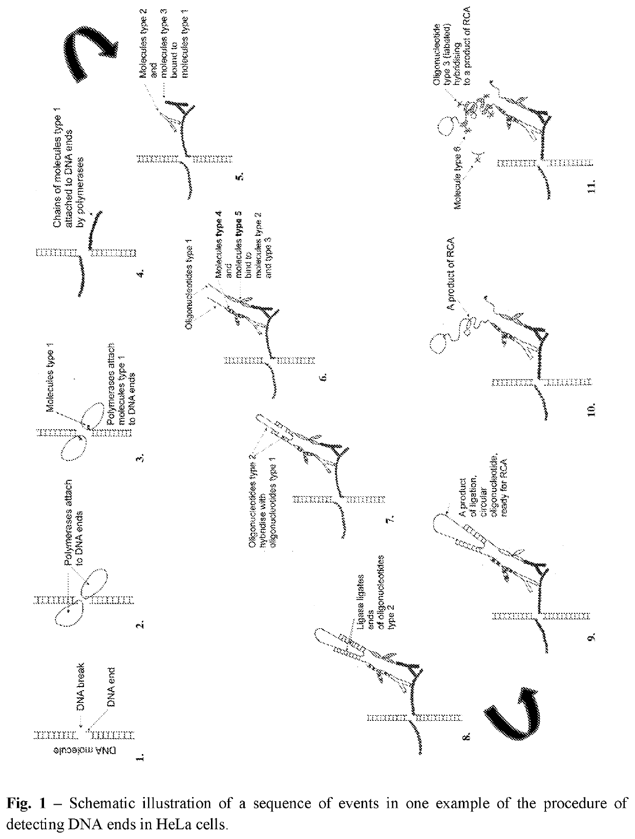
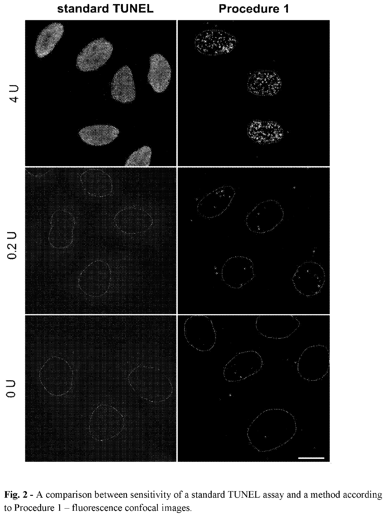
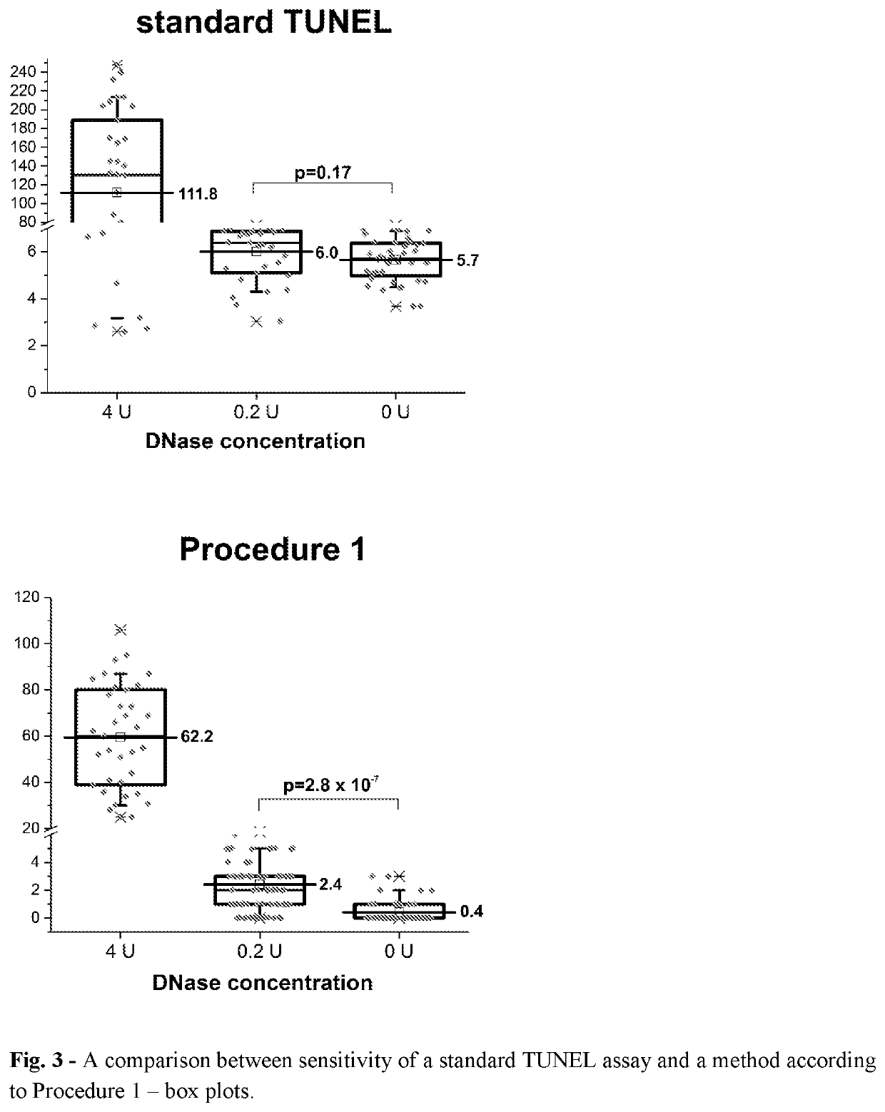
[0370]Detection of Free DNA Ends in Untreated and DNase I Treated Fixed HeLa Cells Using the Method According to Procedure 1.

[0371]In this experiment HeLa 21-4 cells (obtained from P.R. Cook, University of Oxford) were used. Cells were seeded on 18-mm coverslips (number of cells: 0.2×10⁶ per 1 coverslip) and cultured for 3 days in DMEM supplemented with 10% FBS. Subsequently, cells were incubated in 70% ethanol in water for 12 hours at −20° C. Then, cells were incubated in 0.5 mM EDTA in water for 30 minutes at room temperature. Before processing of DNA ends, some samples were treated with DNase I in order to induce DNA breaks. The reaction was performed in a droplet for 30 minutes at room temperature in a reaction mixture consisting of: 0.2 or 4 units of DNase I (Thermo Fisher Scientific, AM2222), 1× DNase I buffer (Thermo Fisher Scientific, AM8170G) and water. Subsequently, BrdU was linked to free ends of DNA using TdT enzyme using APO-BRDU kit (Phoenix Flow Systems, AU: 1001). Th...

example 2

[0381]Detection of Free DNA Ends in Untreated Fixed HeLa Cells Using the Method According to Procedure 2

[0382]In this experiment HeLa 21-4 cells (obtained from P.R. Cook, University of Oxford) were used. The cells were seeded on 18-mm coverslips (number of cells: 0.2×10⁶ per 1 coverslip), cultured for 3 days in DMEM supplemented with 10% FBS and then incubated in 70% ethanol in water for 12 hours at −20° C. After that the samples were treated according to step 3 of Procedure 2 (blocking endogenous biotin) (Molecular Probes, Endogenous Biotin Blocking Kit, E-21390). Increasing of accessibility of DNA ends (step 4) was performed with 0.5 mM EDTA in water for 30 min at RT. To incorporate unmodified and modified biotin-conjugated nucleotides, cells were dipped in UltraPure Distilled Water (Invitrogen, Thermo Fisher Scientific, 10977-035) and then incubated (1 hour incubation at 37° C. in a humid chamber) with reaction mixture consisting of 1× NEBuffer2 (NEBiolabs, B7002S), 30 μM of each...

example 3

[0385]Detection of Free DNA Ends in Fixed HeLa Cells Using the Method According to Procedure 2—Evaluation of the Effect of Blocking Endogenous Biotin.

[0386]HeLa 21-4 cells were treated as in Example 2 but without step 3 (pre-blocking) from Procedure 2.

[0387]Results:

[0388]Free DNA ends, represented as bright fluorescent foci, were readily detected in untreated HeLa cells processed according to Procedure 2 (without step 3—blocking endogenous biotin) (FIG. 4., second row, left image). The average number of foci detected per one nucleus was 128±6 (N=24).

[0389]Conclusion (Example 2 and 3):

[0390]Slightly more free DNA ends were detected in samples processed according to Procedure 2 without blocking endogenous biotin (step 3) than in samples in which this step was present (128±6 vs 116±6). However, an independent two-sample t-test yielded a p-value=0.15, indicating that the difference between these two samples is not statistically significant. Thus, one can conclude that blocking endogenou...


PUM

PropertyMeasurementUnit
Biological propertiesaaaaaaaaaa
Affinityaaaaaaaaaa

Abstract

The invention concerns the method of detection of DNA end(s) in a biological material, comprising the following steps I III and at least one of sub-steps a-h of each of steps I-III: I. PREPARATION OF THE MATERIAL comprising a fixation and / or permeabilization and / or lysis and / or isolation and / or fractionation and / or immobilization of the biological material, b. increasing accessibility of DNA end(s), c. blocking nonspecific binding site(s) for molecules type 2-6, in the biological material; H. PROCESSING OF DNA END(S) comprising d.): modification of DNA end(s) by chemical or physical processing followed by binding molecules type 1 to the DNA end(s) by catalytic or noncatalytic means; blocking nonspecific binding site(s) for molecules type 2-6 in the biological material; III RECOGNITION AND DETECTION OF THE MODIFIED DNA END(S): incubation of the biological material from step II with at least two molecules type 2 and 3 which bind to the molecules type 1 in a manner that allows steps leading to rolling circle amplification (RCA) reactions, g. detection of DNA end(s) by: i. optionally contacting suitable molecules type 4 and / or 5 with molecules type 2 and 3, wherein the molecules type 4 and / or 5 are conjugated with the oligonucleotides type 1, ii. adding oligonucleotides type 2 and enzyme ligase to allow hybridization of said added oligonucleotides type 2 to the oligonucleotides type 1 already linked to molecules type 4 and / or 5, or to molecules type 2 and 3 if they are linked to oligonucleotides type 1, and subsequently performing DNA ligation of oligonucleotides type 2, iii. performing amplification by adding enzyme polymerase and a solution of nucleotides to allow rolling circle amplification (RCA) reactions, and molecules type 6 to allow subsequent hybridization of molecules type 6 to thus obtained product of RCA reactions, h. detection of molecules type 6; wherein when more than one sub-step a-c of step I is performed then they may occur in any order. The invention concerns also use of rolling circle replication for marking the presence and position of single DNA end(s) and use above-mentioned method for detection of DNA end(s) in a biological material.

Description

FIELD OF THE INVENTION[0001]The presented invention falls within the molecular diagnostics and biomedical field. It is related to the method of detection and / or quantitative and / or qualitative analysis of DNA damage. More precisely, the present invention enables direct, highly sensitive, specific and efficient recognition and / or detection of different types of free DNA ends formed in result of induction of single- or double-strand DNA breaks in the cell nucleus, or in isolated biological materials.BACKGROUND[0002]For a life to continue from generation to generation, genomes of organisms must be passed on without any significant changes. The storage of genetic information in a form of a unique sequence of nucleobases that form deoxyribonucleic acid (DNA), and the ability to transfer it through generations, are principal features of all living organisms. Cells are the basic structural and functional units of living organisms. The sequence of bases must, therefore, be preserved during ...

Claims


Application Information

Patent Timeline
IPC IPC(8): C12Q1/6804
CPCC12Q2563/107C12Q2565/601C12Q2521/501C12Q1/6804C12Q2531/125C12Q2543/10C12Q2521/101C12Q2525/117C12Q1/6844
Inventor KORDON, MAGDALENAZAREBSKI, MIROSLAWDOBRUCKI, JERZYSOLARCZYK, KAMIL
Owner INTODNA SPOLKA AKCYJNA
Why Eureka
Try Eureka