Quantitative Imaging System and Uses Thereof
a quantitative imaging and tomography technology, applied in the field of biomedical imaging and tomography systems, can solve the problems of reduced lateral resolution, incomplete tomographic recovery of quantitative image brightness, prior art deficiency in quantitative functional anatomical imaging systems for breast screening with simultaneous, etc., to achieve the effect of making the exam comfortable and fas
- Summary
- Abstract
- Description
- Claims
- Application Information
AI Technical Summary
Benefits of technology
Problems solved by technology
Method used
Image
Examples
example 1
Functional Imaging Validation in Phantoms
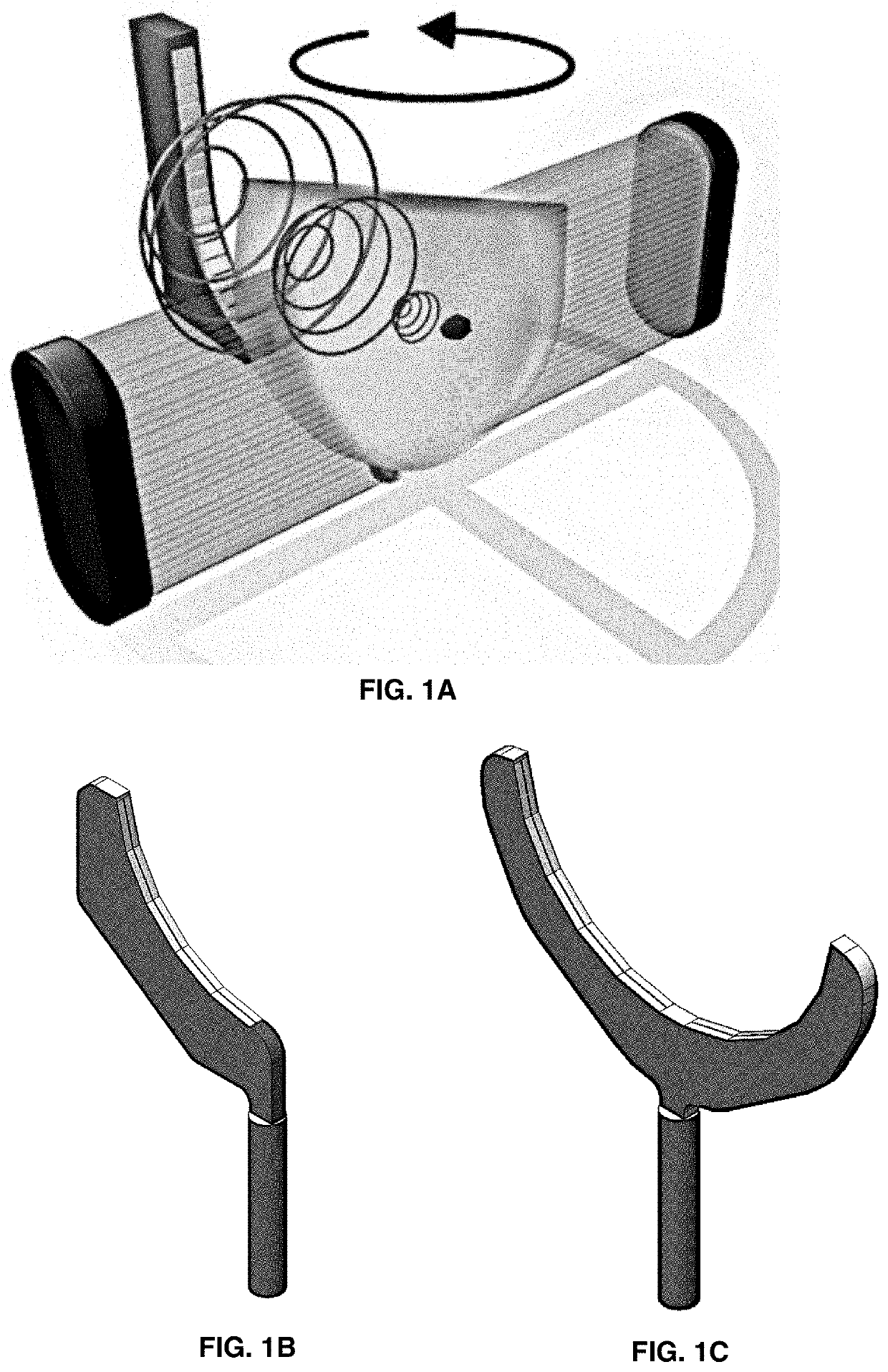
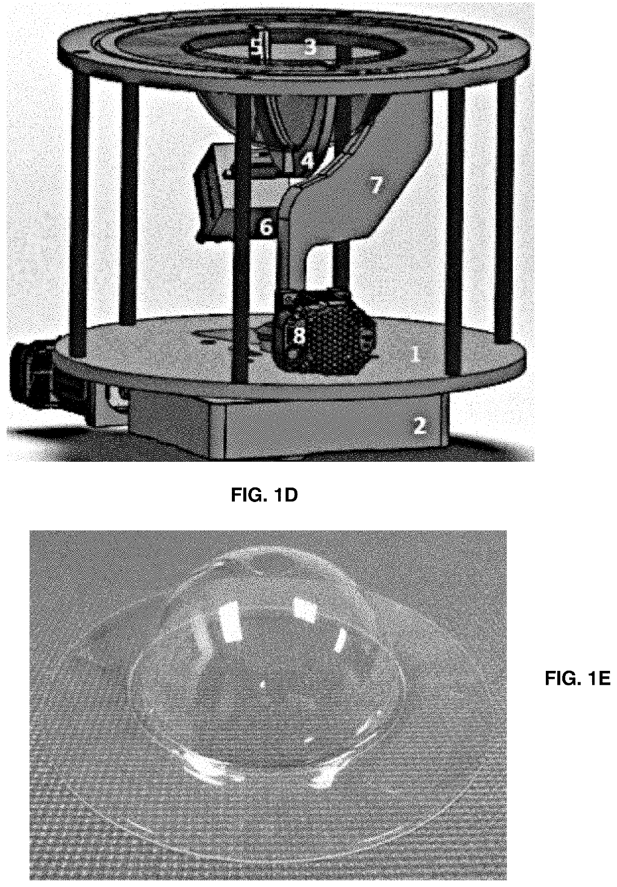
[0103]The most important advancement of this latest system design compared with previously reported systems is the full breast illumination accomplished for each rotational step of the optoacoustic transducer array using fiberoptic illuminator rotating around the breast independently from rotation of the detector probe. A pilot case study on one healthy volunteer and on patient with a suspicious small lesion in the breast are reported herein. LOUISA visualized deoxygenated veins and oxygenated arteries of a healthy volunteer, indicative of its capability to visualize hypoxic microvasculature in cancerous tumors. A small lesion detected on optoacoustic image of a patient was not visible on ultrasound, potentially indicating high system sensitivity of the optoacoustic subsystem to small but aggressively growing cancerous lesions with high density angiogenesis microvasculature. With safe level of NIR optical fluence, the main breast vasculature ...
example 2
Clinical Validation of LOUISA
[0107]The LOUISA system contains a 3D imaging module and a 2D imaging handheld probe. While LOIS-3D, the predecessor of LOUISA, employed a half-time reconstruction algorithm in the spherical coordinates (30), it was necessary to employ full time reconstruction for the hemispherical geometry of 3D image reconstruction in order to retain full view rigorous reconstruction solution for the breast imaging (31). The images of this patient were obtained at a single wavelength of 757 nm, thus blood oxygen saturation of the blood vessels and the tumor was not possible. The nature of a relatively small (3.5 mm) tumor visible in the 2 out of 3 projections (FIGS. 6A-6B, not FIG. 6C) was not conclusively ascertained. FIGS. 6A-6C are a demonstration of LOUISA sensitivity, but not of specificity of tumor differentiation.
[0108]A number of improvement in the design of the imaging module and enhancements of the signal processing and image reconstruction algorithms resulte...
example 3
3D Full View Imaging System for Preclinical Research as an Example of QOAT System
[0115]Three-dimensional laser optoacoustic ultrasonic imaging system assembly based full-view optoacoustic tomography coregistered with ultrasonic tomography is developed for applications in preclinical research using small laboratory animals, such as a mouse. FIGS. 13A-13B show the basic design and resolution of the mouse imaging system. FIG. 13A shows a basic cartoon schematic of the full view 3D optoacoustic tomography applied to the imaging of a live mouse. FIG. 13B shows a 3D optoacoustic image of 3 crossed horse hairs with diameter of 150 micron fully resolved on the image obtained with the method presented in FIG. 13A. This figure demonstrates that designs and methods applied to the breast imaging are also applicable to other volumetric regions of interest in clinical and preclinical research.
[0116]FIG. 14 shows maximum intensity projection optoacoustic images of a live mouse body presented along...
PUM
 Abstract
Description
Claims
Application Information
 - R&D
- Intellectual Property
- Life Sciences
- Materials
- Tech Scout
- Unparalleled Data Quality
- Higher Quality Content
- 60% Fewer Hallucinations
Browse by: Latest US Patents, China's latest patents, Technical Efficacy Thesaurus, Application Domain, Technology Topic, Popular Technical Reports.
© 2025 PatSnap. All rights reserved.Legal|Privacy policy|Modern Slavery Act Transparency Statement|Sitemap|About US| Contact US: help@patsnap.com


