
Marker of fetal trophoblast cell, identification method, detection kit and use thereof

a fetal trophoblast cell and marker technology, applied in the field of biological cell identification, can solve the problems of inability to passable therapy for genetic diseases with chromosomal abnormalities, risk of infection and miscarriage, and inability to meet the requirements of test tube babies, and achieve low identification accuracy and poor sensitivity

Inactive Publication Date: 2021-07-29
HANGZHOU JUNHUI BIOTECHNOLOGY CO LTD
View PDF0 Cites 1 Cited by
  • Summary
  • Abstract
  • Description
  • Claims
  • Application Information

AI Technical Summary

Benefits of technology

The present patent provides a solution for identifying fetal trophoblast cells with poor sensitivity and low accuracy. The patent uses a marker, an identification method, and a detection kit that includes a microwell array chip to disperse the sample in single cells, facilitating subsequent detection and separation. The identification method includes staining the sample with a nuclear stain and performing fluorescence detection using HK2 and nuclear staining as target cells. Overall, the patent provides a more reliable and accurate method for identifying fetaltrophoblast cells.

Problems solved by technology

At present, no passable therapy is available for genetic diseases with chromosomal abnormalities.
However, these invasive examinations cause a risk of infection and miscarriage.
Although pre-implantation prenatal diagnosis can be performed on test tube babies, it is only suitable for test tube babies and technically demanding.
The non-invasive examinations currently used in clinical practice include B-ultrasound and maternal serum biochemical examinations, but these methods are only auxiliary screening methods of prenatal genetic diseases with limited scope of application rather than prenatal diagnosis methods.
However, because the number of fetal cells in the peripheral blood of pregnant women is extremely small and volatile, it is often difficult to isolate a large number of fetal cells for reliable detection.
However, the complete genome information of the fetus is unavailable, so the types of genetic diseases that can be detected are very limited, and it is a screening method rather than a diagnostic method.
However, because the cervical exfoliated cells collected in the cervix are complex, consisting of squamous epithelial cells, cervical columnar epithelial cells, cervical glandular epithelial cells, fibroblasts, red blood cells, white blood cells, lymphocytes, and a small amount of trophoblast cells.
The markers and detection methods currently available are difficult to accurately detect fetal trophoblast cells in such a complicated cell sample.

Method used


Image

  • Marker of fetal trophoblast cell, identification method, detection kit and use thereof
  • Marker of fetal trophoblast cell, identification method, detection kit and use thereof
  • Marker of fetal trophoblast cell, identification method, detection kit and use thereof

Examples


examples

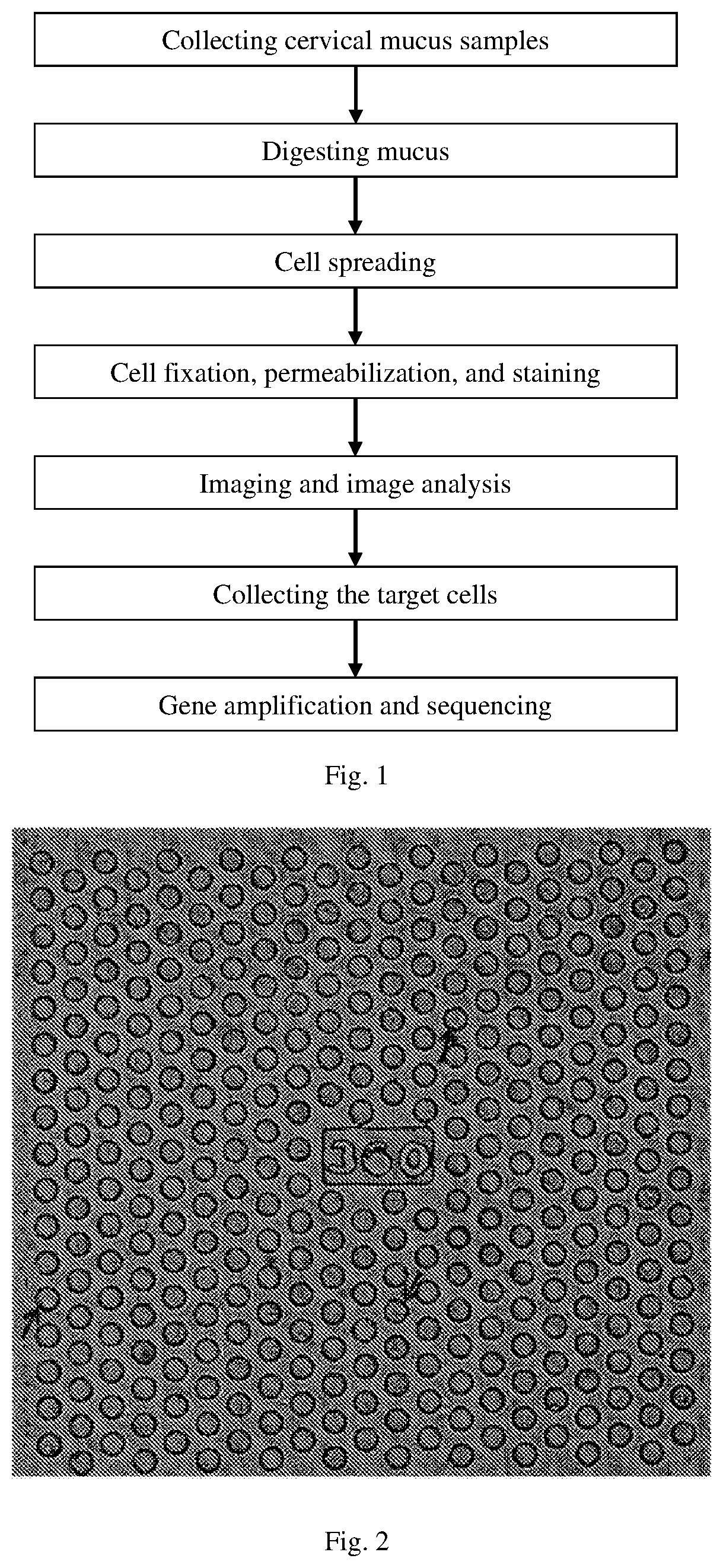
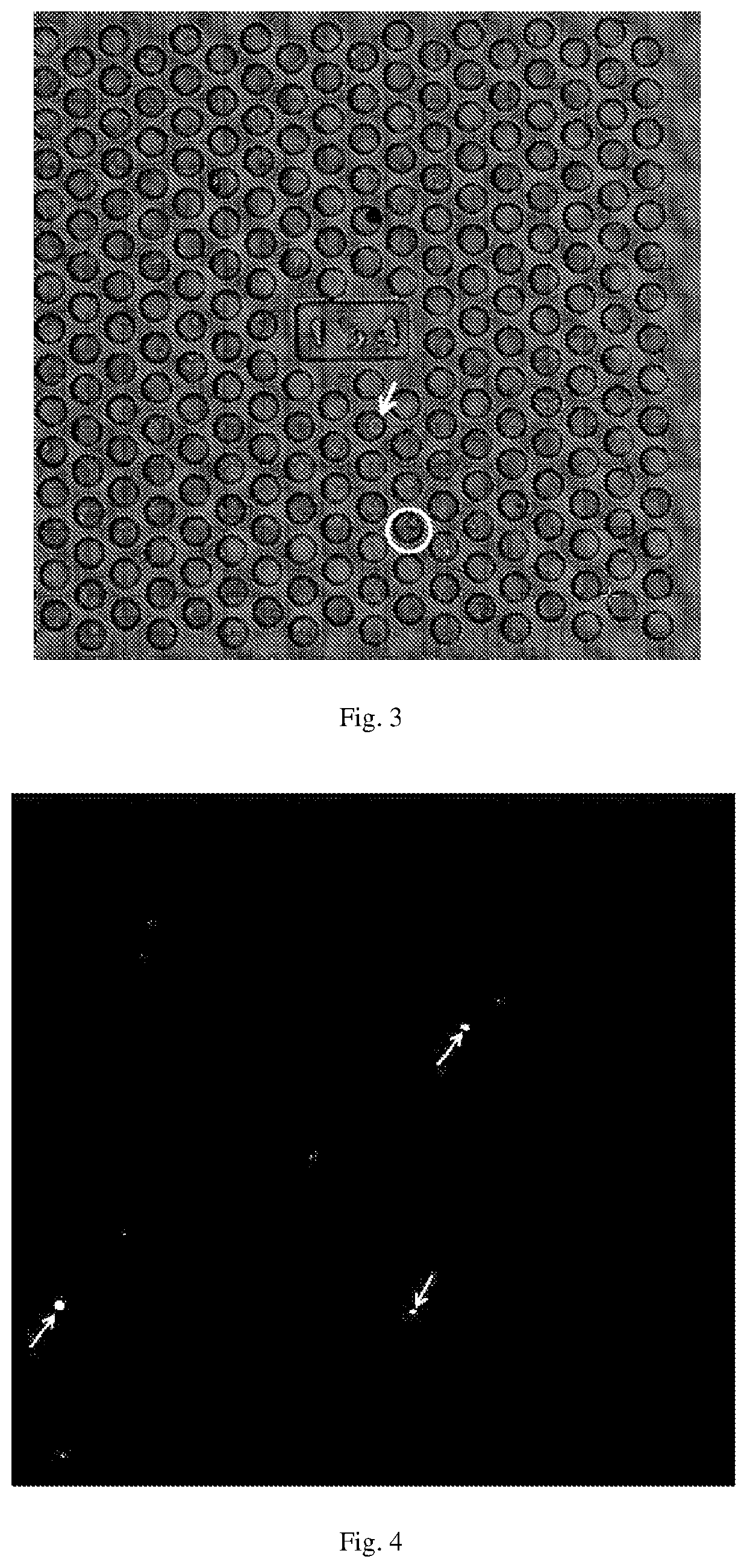
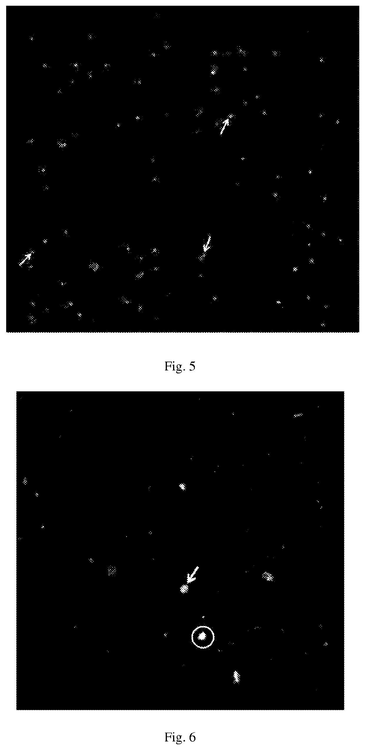
[0045]Identification and Analysis of Fetal Cells in Cervical Mucus for Prenatal Diagnosis

This embodiment includes the following steps:

(1) Using a sterile cell brush to obtain the cervical mucus from 5 women in early pregnancy (6-8 weeks of gestation) (all patients gave informed consent). The brush rotates about 10 times, and the cell brush with the mucus collected by the brush was stored in 2 ml Hank's Balanced Salt Solution (HBSS), respectively numbered as Samples 1-5, and placed in a 4-Degree ice box and transported to the laboratory.

(2) The cell brush was taken out and placed in 0.25% pancreatin (containing chelating agent) and incubated at 37° C. for 3-5 minutes to digest the mucus, and the cell dispersion was observed under a microscope. Fetal bovine serum (FBS) was used to stop the digestion of pancreatin when cells were sufficiently dispersed.

(3) The 400 g suspension obtained in the previous step was centrifuged for 5 minutes and washed with HBSS 3 times. If the cell suspensi...


PUM

PropertyMeasurementUnit
pore sizeaaaaaaaaaa
diameteraaaaaaaaaa
morphologiesaaaaaaaaaa

Abstract

A marker of fetal trophoblast cells, an identification method, a detection kit and the use thereof. The identification method comprises performing dyeing treatment on a sample containing fetal trophoblast cells with a fluorescence-labeled HK2 antibody substance, and then performing fluorescence detection, wherein HK2 positive cells are target cells. The kit comprises a fluorescence-labeled HK2 antibody substance and a cell nucleus dye. The above-mentioned technical solution can sensitively and reliably detect fetal trophoblast cells in the cervical mucus or blood samples of pregnant women in the first trimester, and especially can be used for prenatal diagnosis in early pregnancy.

Description

TECHNICAL FIELD[0001]The present application relates to the technical field of biological cell identification, and in particular to a marker, an identification method, a detection kit of fetal trophoblast cells and use thereof.BACKGROUND APPLICATION[0002]At present, no passable therapy is available for genetic diseases with chromosomal abnormalities. Therefore, first-trimester detection of abnormalities of fetal genetic information through prenatal diagnosis and active intervention can effectively reduce the rate of birth defects. Currently, prenatal diagnosis methods used in clinical practice are mainly invasive examinations, such as chorionic villus biopsy, amniocentesis, and fetal cord blood testing. However, these invasive examinations cause a risk of infection and miscarriage. Although pre-implantation prenatal diagnosis can be performed on test tube babies, it is only suitable for test tube babies and technically demanding. The non-invasive examinations currently used in clini...

Claims


Application Information

Patent Timeline
Patent Type & Authority Applications(United States)
IPC IPC(8): G01N33/68G01N33/58G01N1/30
CPCG01N33/6854G01N1/30G01N33/582G01N33/56966G01N2800/385C12Q1/68G01N2333/91215G01N33/573G01N2800/368G01N2800/387C01F1/00
Inventor SHI, QIHUIWANG, CHUNYING
Owner HANGZHOU JUNHUI BIOTECHNOLOGY CO LTD
Learn More