Methods and pharmaceutical composition for the treatment of ovarian cancer, breast cancer or pancreatic cancer
a technology for ovarian cancer and breast cancer, applied in the direction of drug compositions, antibody medical ingredients, peptides, etc., can solve the problems of -related death, poor prognosis, and limited understanding of the physiological relevance of heterogeneity and its implications for tumor developmen
- Summary
- Abstract
- Description
- Claims
- Application Information
AI Technical Summary
Benefits of technology
Problems solved by technology
Method used
Image
Examples
example 1
& Methods
[0142]Mouse Breeding and Ovarian Cancer Model
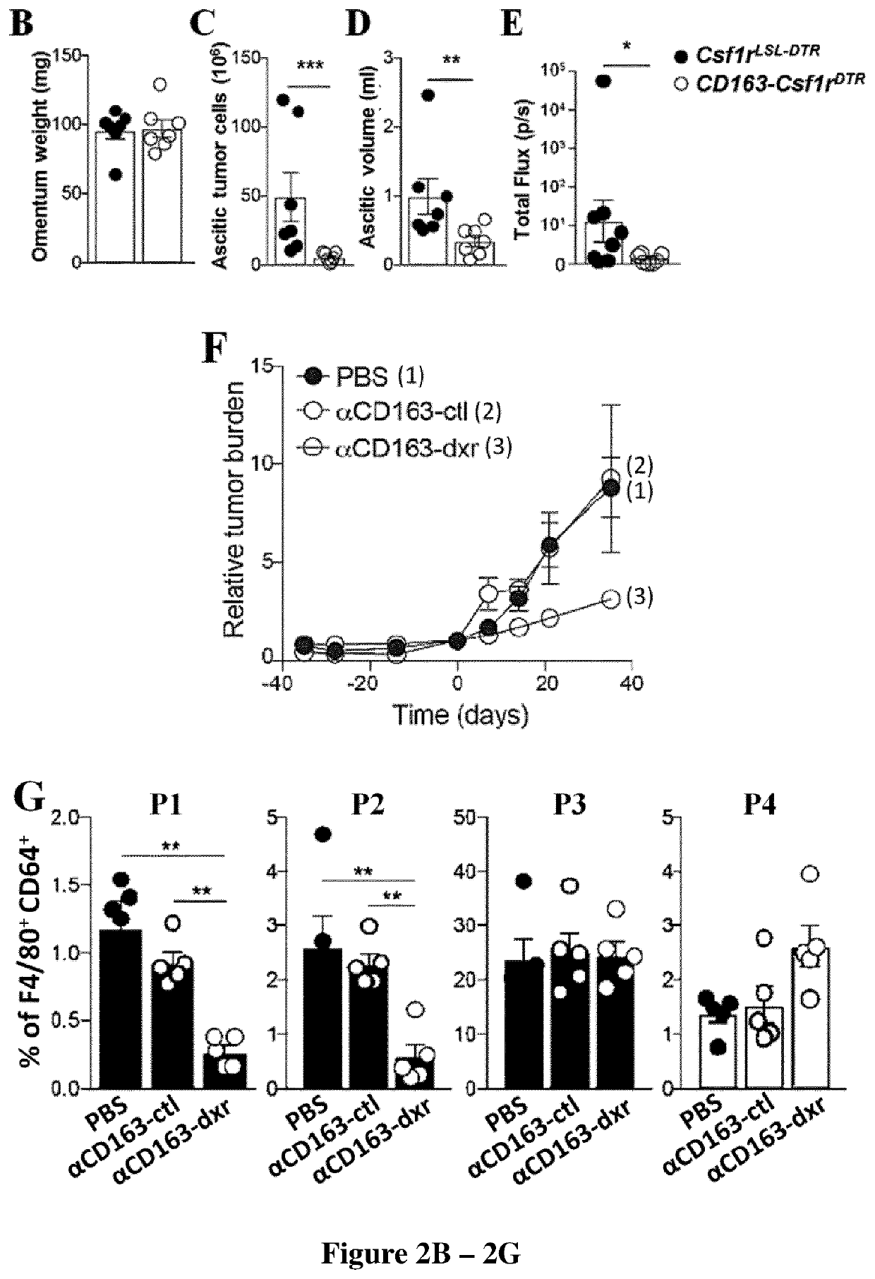
[0143]Tg(Csflr-LSL-HBEGFmCherry) (Schreiber et al., 2013), Rosa26LSL-YFP, Cx3CrlCreER and Cx3ClrGFP / + were obtained from the Jackson Laboratory (Bar Harbor, Me., US). C57BL / 6J mice were obtained from Janvier Labs (Saint-Berthevin, FR). Ccr2− / − mice and Rosa26LS-tdRFP mice were gifts from Bernard Malissen (Centre d'Immunologie Marseille Luminy, Marseille, France). Cd163iCre mice were generated from modified ES cells on a C57BL / 6 background as described (Etzerodt et al, in press). In brief; a FlpO-NeoR cassette encoding IRES-iCre was inserted in the 3′UTR of the CD163 gene using homologous recombination and used to generate chimeric mice that were subsequently crossed to Flp deleter mice to facilitate removal of NeoR cassette. To mimick peritoneal spread of epithelial ovarian cancer, 1×10⁶ ID8-Luc cells were injected i.p in 500 μl sterile PBS pH 7.4. Tumor burden was estimated weekly by injecting mice i.p. with 100 mg / kg d-Luciferi...
example 2
um is a Critical Pre-Metastatic Niche for Ovarian Cancer Cells
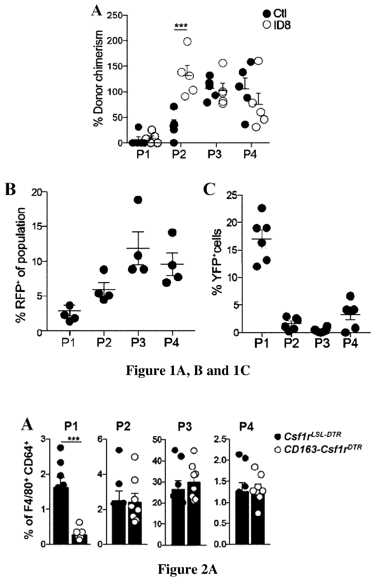
[0160]The omentum is an adipose tissue formed from a fold of the peritoneal mesothelium. In humans the greater omentum covers the majority of the abdomen, whereas in mouse the omentum is only a thin stretch of adipose tissue located between the stomach, pancreas and spleen. The peritoneal spread of ovarian cancer can be modelled using the immortalized mouse ovarian epithelial cell line ID8 (Roby et al., 2000). The intra-peritoneal (i.p.) injection of ID8 cells leads to the development of diffuse peritoneal carcinomatosis and malignant ascites with a long latency period (up to 12 weeks). We used an ID8 cell line transduced to express firefly luciferase (ID8-luc) (Hagemann et al., 2008) to monitor tumor progression after i.p. injection by non-invasive bioluminescent imaging. We observed that ID8 cells localized primarily to the omentum for up to 35 days before spreading throughout the peritoneal cavity (data not shown). Inf...
example 3
ancer Cells Colonize Fat-Associated Lymphoid Clusters in Close Contact with Omental Macrophages
[0162]The omentum has a particularly high density of fat-associated lymphoid clusters (FALC) that are thought to be important structures for capturing peritoneal antigens (Rangel-Moreno et al., 2009). Previous studies have proposed that FALC promote the colonization of omentum by ovarian cancer cells, however, neither B or T lymphocytes were shown to contribute to tumor growth (Clark et al., 2013). To visualize the localization of ID8 cells in the omentum, we labeled cells with Qdots (Qtracker®705) before i.p. injection and analyzed omentum by whole-mount confocal imaging. One day after injection, we observed that ID8 cells were located in the vicinity of FALCs, an area densely populated by omental macrophages (data not shown). To further characterize omental macrophages, we analyzed omentum by flow cytometry. Gating on the CD11b+ myeloid cell fraction (CD45.2+ Linneg (CDS, CD19, NK1.1, Ly...
PUM
 Abstract
Description
Claims
Application Information
 - R&D
- Intellectual Property
- Life Sciences
- Materials
- Tech Scout
- Unparalleled Data Quality
- Higher Quality Content
- 60% Fewer Hallucinations
Browse by: Latest US Patents, China's latest patents, Technical Efficacy Thesaurus, Application Domain, Technology Topic, Popular Technical Reports.
© 2025 PatSnap. All rights reserved.Legal|Privacy policy|Modern Slavery Act Transparency Statement|Sitemap|About US| Contact US: help@patsnap.com


