Diagnostic patch and method for diagnosis using the same

Inactive Publication Date: 2000-05-16
HISAMITSU PHARM CO INC
View PDF6 Cites 95 Cited by
  • Summary
  • Abstract
  • Description
  • Claims
  • Application Information

AI Technical Summary

Problems solved by technology

In particular, mammary cancer is known to be a cancer which is difficult to find in an early stage, though it is one of the major cancers which tend to increase.
Although the diagnosis of mammary cancer is conducted by internal examination, palpation, mammography (X ray), detection of tumor markers [such as carcinoembryonic antigen (CEA), .alpha.-fetoprotein (AFP), cancer antigen (CA15-3) and various saccharide antigens], ultrasonography or the like, these methods do not always exhibit a high diagnosis efficiency.
For example, a phyma-free cancer gives neither pain nor subjective symptoms in early stages, so that patients therewith cannot find abnormality in many cases.
However, it is very difficult to conduct the early diagnosis of primary mammary cancer by determining the concentration, because the positive rate for an early cancer is extremely low.
However, it is difficult to find an extremely small affected site by current methods easily and simply.
Therefore, the assay cannot be conducted at the site of medical services, so that the above specimen must be entrusted to an institution fitted with examination equipment or a special laboratory technician.
Accordingly, it takes many days to know the result of the assay, which is problematic particularly in cases necessitating immediate treatment.
On the other hand, atopic dermatitis is steadily increasing owing to change in the living environment, lowering in the adaptation of a living body and lowering in the natural immunity, becoming a socially significant problem.
However, the cause of the diseases has not sufficiently been elucidated as yet and the diagnosis and therapy of them depend on the macroscopic or experimental sense of a doctor in many cases.
Blood specimen collection or bi

Method used


Image

  • Diagnostic patch and method for diagnosis using the same
  • Diagnostic patch and method for diagnosis using the same
  • Diagnostic patch and method for diagnosis using the same

Examples


example 2

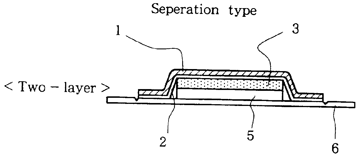
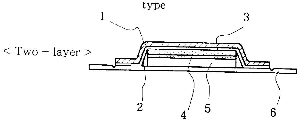
Diagnostic Patch having Two-Layer Adsorption Carrier of Separation Type

A circular pressure-sensitive adhesive tape having a diameter of 3 cm was made by spreading a pressure-sensitive adhesive comprising SIS, polyisobutylene, ester gum and liquid paraffin (at a weight ratio of 20:10:35:35) on a flexible stretch film of polyvinyl chloride as a support.

A rectangular piece (1.5 cm.times.1.8 cm) of microporous cellulose ester (pore size: 0.6 .mu.m, a product of Millipor) as a water-absorbing and adsorbent membrane was stuck on the above pressure-sensitive adhesive tape. Further, a square piece (1.8 cm.times.1.8 cm) of cellulose nitrate / cellulose acetate (pore size: 0.45 .mu.m, a product of Millipor) as an adsorption carrier was stuck on the member. The surface of the carrier was covered with a polyethylene terephthalate film to give a sticking-type diagnostic preparation.

example 3

Diagnostic Patch having One-Layer Adsorption Carrier

A circular pressure-sensitive adhesive tape having a diameter of 4 cm was made by spreading a pressure-sensitive adhesive comprising SIS, polyisobutylene, ester gum, liquid paraffin and starch acrylate (at a weight ratio of 22:8:33:35:2) on a flexible stretch film of polyvinyl chloride as a support.

A circular piece (diameter: 2 cm) of cellulose nitrate / cellulose acetate (pore size: 1 .mu.m, a product of Millipor) as an adsorption carrier was stuck on the above tape. The surface of the carrier was covered with a polyethylene terephthalate film.

example 4

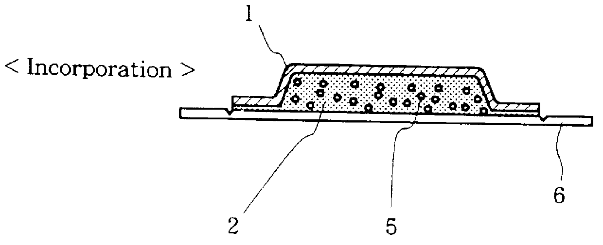
Diagnostic Patch having Incorporation-Type Adsorption Carrier

A pressure-sensitive adhesive tape having an adsorptive power was made by spreading a pressure-sensitive adhesive comprising SIS, ester gum, liquid paraffin, aluminum silicate and powdery cellulose (at a weight ratio of 24:35:35:2:4) on a circular piece (diameter: 2 cm) of a flexible stretch film of polyvinyl chloride as a support. The tape was covered with a polyethylene terephthalate film to give a diagnostic patch.


PUM


Abstract

PCT No. PCT/JP93/01737 Sec. 371 Date Apr. 29, 1996 Sec. 102(e) Date Apr. 29, 1996 PCT Filed Nov. 29, 1993 PCT Pub. No. WO94/13209 PCT Pub. Date Jun. 23, 1994A diagnostic patch comprising at least an adsorption carrier adsorbing a substance secreted from a living body, which is useful particularly for the diagnosis of mammary cancer or atopic dermatitis, and a method for the diagnosis of mammary cancer or atopic dermatitis with said preparation.

Description

The present invention relates to a diagnostic patch which has various functions and is suitably used particularly for the diagnosis of mammary cancer or atopic dermatitis, and a method for the diagnosis of mammary cancer or atopic dermatitis with said patch.BACKGROUND OF THE PRIOR ARTVarious methods have been employed in the checkup or diagnosis of diseases. For example, biochemical or biological examination is carried out by collecting blood invasively and conducting diagnosis based on the components, diagnostic markers and cells contained in the blood; pathological examination is conducted by conducting the biopsy of cancer tissue or the examination for abnormality on cellular level invasively; and the examination of urine, stool or saliva involves the collection thereof. In carrying out these method, a doctor, nurse or clinical analyst must collect blood with a syringe, must extirpate a tissue (affected site) from a living body with a sharp-edged knife or must collect an object o...

Claims


Application Information

Patent Timeline
IPC IPC(8): A61B10/00
CPCA61B10/0035A61B10/0045A61B10/0041
Inventor SAITA, MASARUSHIMOZONO, YUJIOHTA, SHIGEOYONEMURA, KEISHIMUKAI, MIZUEOKAYAMA, AKIRAIMAYAMA, SHUHEI
Owner HISAMITSU PHARM CO INC
Why Eureka
Try Eureka